Cartilage Lesion - How i Do it
•
4 likes•302 views

Cartilage Lesion is a demanding challenge for treatment , these my points of view how to treat these lesios

Recommended
More Related Content
What's hot
What's hot (20)
Similar to Cartilage Lesion - How i Do it
Similar to Cartilage Lesion - How i Do it (20)
Recently uploaded
Recently uploaded (20)
Cartilage Lesion - How i Do it
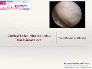
- 1. Paulo Ribeiro de Oliveira Coordenador da Unidade Funcional do Joelho Hospital de São João - Porto / F. Medicina do Porto Cartilage Lesion– what can we do ? Our Point of View ! Paulo Ribeiro de Oliveira
- 2. Paulo Ribeiro de Oliveira Coordenador da Unidade Funcional do Joelho Hospital de São João - Porto / F. Medicina do Porto Cartilage Tissue that lines the diarthrodial joints, with: 1. Low friction surface 2. Avascular 3. Permeable (water and nutrients) 4. Flexible wear resistant 5. Elasticity 6. Resistance to compressive forces 7. Ability to minimize peak loads transmitted to the subchondral bone 8. Little regenerative capacity
- 3. Paulo Ribeiro de Oliveira Coordenador da Unidade Funcional do Joelho Hospital de São João - Porto / F. Medicina do Porto Constitution : 1. Collagen fibers type II and type IX (strength and stress resistance) 2. Water 70 - 80% 3. Proteinoglicans or hydrophilic molecules "aggrecan" (20 to 40% dry weight) (elasticity and deformability) 4. Chondrocytes (10% dry weight) 5. Other collagens type V, VI, XI small amounts Cartilage
- 4. Paulo Ribeiro de Oliveira Coordenador da Unidade Funcional do Joelho Hospital de São João - Porto / F. Medicina do Porto 1. Microlesion of cells and matrix with no visible damage to the cartilage 2. Macrolesion 3. Fractures of articular cartilage and subchondral bone (osteochondral fractures) Cartilage
- 5. Paulo Ribeiro de Oliveira Coordenador da Unidade Funcional do Joelho Hospital de São João - Porto / F. Medicina do Porto Outerbridge classification: Grade 0 - Normal cartilage Grade 1 - Cartilage softened and swollen Grade 2 - Cracking not reaching the subchondral bone; less than 1.5 cm Grade 3 - Cracking reaching the subchondral bone without exposure; greater than 1.5 cm Grade 4 - subchondral bone exposure of any diameter Cartilage ICRS Classification: Normal: grade 0 Almost normal: Grade 1a- superficial lesions / softening Grade 1b - 1a and / or fissures or surface cracks Abnormal: Grade 2 - length < 50% thickness Serious injury: Grade 3 a – extension > 50% Grade 3 b - to the calcified layer Grade 3 c - to the surface of the subchondral bone (without entering) Grade 3 d - includes blisters Very serious injury: Grade 4 a - penetration of the subchondral bone but not the full diameter of the defect Grade 4b - penetration across the diameter of the defect Several classifications : Outerbridge, ICRS etc
- 6. Paulo Ribeiro de Oliveira Coordenador da Unidade Funcional do Joelho Hospital de São João - Porto / F. Medicina do Porto The primary factor in the development of a cartilage lesion is undoubtedly the relationship between the size of the lesion and the load surface, being adversely affected by: Obesity Age Axial misalignment Family history of osteoarthritis Overload activities Cartilage
- 7. Paulo Ribeiro de Oliveira Coordenador da Unidade Funcional do Joelho Hospital de São João - Porto / F. Medicina do Porto Cartilage Treatment • Individual • Effective complaints • Patient expectations • Age (physiological) • IMC • Symptomatology (mechanical pain, inflammatory or mixed) • Occupation • Ability to understand the detailed objectives and commitment to accept • Defect • Localization • Number of defects • Size • Depth • Defect geometry • The underlying subchondral space rating • Lesion contained or not • Axial deviations • Instability associated • Associated injuries (meniscal) All treatment should be individualized on the basis of these parameters
- 8. Paulo Ribeiro de Oliveira Coordenador da Unidade Funcional do Joelho Hospital de São João - Porto / F. Medicina do Porto Conservative treatment Drugs - NSAIDs Infiltration - corticosteroids, viscosupplementation, collagen ??? Orthoses Exercise, weight loss Activity modification Cartilage
- 9. Paulo Ribeiro de Oliveira Coordenador da Unidade Funcional do Joelho Hospital de São João - Porto / F. Medicina do Porto Microfractures Microfratures and augmentation Autologous osteochondral graft Osteochondral allograft Matrix autologous chondrocyte implantation Cartilage Treatment
- 10. Paulo Ribeiro de Oliveira Coordenador da Unidade Funcional do Joelho Hospital de São João - Porto / F. Medicina do Porto Microfractures ❖ Serve to facilitate the repair process with surface repopulation with undifferentiated mesenchymal cells ❖ Complete filling of the lesion with a stable clot ❖ Overcorrection of axial misalignments ❖ Role of BMP, PGF etc... Cartilage Treatment
- 11. Paulo Ribeiro de Oliveira Coordenador da Unidade Funcional do Joelho Hospital de São João - Porto / F. Medicina do Porto Cartilage Treatment
- 12. Paulo Ribeiro de Oliveira Coordenador da Unidade Funcional do Joelho Hospital de São João - Porto / F. Medicina do Porto Cartilage Treatment
- 13. Paulo Ribeiro de Oliveira Coordenador da Unidade Funcional do Joelho Hospital de São João - Porto / F. Medicina do Porto A recent systematic review of twenty-eight studies with >3000 patients found that knee function was consistently improved in the first twenty-four months after microfracture in the patients studied. After two years, knee function scores remained above preoperative levels but declined; only 67% to 85% of patients continued to report improvement in the two to five-year time frame. It was also noted that shortcomings of the microfracture technique included limited production of hyaline cartilage, unpredictable repair cartilage volume, and higher failure rates for cell transplantation surgery following failed prior microfracture compared with patients in whom similar cellular treatments were used as first-line options. Mithoefer K, McAdams T, Williams RJ, Kreuz PC, Mandelbaum BR. Clinical efficacy of the microfracture technique for articular cartilage repair in the knee: an evidence- based systematic analysis. Am J Sports Med. 2009 Oct;37(10):2053-63. There is growing evidence that modification or augmentation of microfracture may improve the quality of the repair tissue formed and ultimately the clinical outcome for patients. At the present time, it is believed that marrow stimulation techniques are best reserved as a first-line option for isolated defects of <2.5 cm2 on the femoral condyles. Biologic augmentation techniques may broaden these indications and improve longterm outcomes. Strauss EJ, Barker JU, Kercher JS, Cole BJ, Mithoefer K. Augmentation strategies following the microfracture technique for repair of focal chondral defects. Cartilage. 2010 Mar;1(2):145-52. Cartilage Microfrature and augmentation
- 14. Paulo Ribeiro de Oliveira Coordenador da Unidade Funcional do Joelho Hospital de São João - Porto / F. Medicina do Porto Axial deviations correction To correct the axial misalignment for a better distribution of the axial compressive forces Studies have shown the appearance, after osteotomy, of fibrocartilage in the medial compartment (Bergenudd H et al) This symptons improvement deteriorates with the time course (Coventry MB et al) The axial correction should never be by default and should be performed if necessary in conjunction with reconstruction techniques Cartilage Treatment
- 15. Paulo Ribeiro de Oliveira Coordenador da Unidade Funcional do Joelho Hospital de São João - Porto / F. Medicina do Porto Autologous osteochondral graft It consists in the transplant of osteochondral grafts from a non load zone to vital areas of the articular surface Outerbridge HK et al. and Hangody et al. confirmed the validity of this technique for limited defects correction, highlighting the potential morbidity of the donor area and the difficult technique of fixation of the graft Important: early mobilization , without load up to 6-8 weeks Cartilage Treatment
- 16. Paulo Ribeiro de Oliveira Coordenador da Unidade Funcional do Joelho Hospital de São João - Porto / F. Medicina do Porto Create a sufficiently smooth and sliding surface of hyaline cartilage or similar Enabling the delay of joint degeneration Cartilage Treatment Autologous osteochondral graft
- 17. Paulo Ribeiro de Oliveira Coordenador da Unidade Funcional do Joelho Hospital de São João - Porto / F. Medicina do Porto INDICATIONS Chondral or osteochondral focal lesions on loading surfaces of the joints Can be used in astragalus, head of the femur, knee, etc. Usually below the age of 50 Defects of 1 - 4 cm2 Treatement of associated pathologies: meniscal or ligamentous injuries, axial deviations correction Patient compliance to the protocol Cartilage
- 18. Paulo Ribeiro de Oliveira Coordenador da Unidade Funcional do Joelho Hospital de São João - Porto / F. Medicina do Porto Absolute: Neoplasia, infection, rheumatoid arthritis and similar pathologies Absence of a compatible donor zone 〉50 years Defect 〉8 cm2 Defect deeper than 10mm Noncompliant patient Relative: 40 - 50 years Defect size 4 - 8 cm2 Moderate degenerative changes Cartilagem CONTRAINDICATIONS
- 19. Paulo Ribeiro de Oliveira Coordenador da Unidade Funcional do Joelho Hospital de São João - Porto / F. Medicina do Porto TÉCNICA Arthroscopic Technically demanding Miniarthrotomy Best alternative for inexperienced Cartilagem Tratamento
- 20. Paulo Ribeiro de Oliveira Coordenador da Unidade Funcional do Joelho Hospital de São João - Porto / F. Medicina do Porto Pure chondral lesions - the cylinder should be 15 mm Osteochondral lesions - the cylinder should be 25 mm Cartilage Treatment
- 21. Paulo Ribeiro de Oliveira Coordenador da Unidade Funcional do Joelho Hospital de São João - Porto / F. Medicina do Porto Cartilagem Tratamento
- 22. Paulo Ribeiro de Oliveira Coordenador da Unidade Funcional do Joelho Hospital de São João - Porto / F. Medicina do Porto Cylinder perpendicular to articular surface Avoid the cylinder sinking; 1 - 2 mm above the adjacent surface No load for 3 - 5 weeks Cartilage Treatment
- 23. Paulo Ribeiro de Oliveira Coordenador da Unidade Funcional do Joelho Hospital de São João - Porto / F. Medicina do Porto MOSAICPLASTY – RMN 1 YEAR
- 24. Paulo Ribeiro de Oliveira Coordenador da Unidade Funcional do Joelho Hospital de São João - Porto / F. Medicina do Porto Bobic ( 1996) Mosaicplasty in 12 patients with ACL injury: with good or excellent results in 10/12 cases Wang ( 2002) Mosaicplasty in 16 patients with 2 to 4 years of follow-up: 80% good or excellent results Sharp et al ( 2005) Combined chondrocyte transplantation and mosaicplasty with follow up of 3 years in 13 patients with large lesions: excellent results in 10/13 patients Hangody, L. et al. JBJS . Am. 85:25-32, 2003 10 years of follow - up of 831 patients with mosaicplasty: excellent results in 92% in the femoral condyle, 87% in tibial plate, 79% in patella or trochlear region and 94% in astragalus Cartilagem Mosaicplasty Effect of Impact on Chondrocyte Viability During Insertion of Human Osteochondral Grafts BY BORIS H. BORAZJANI et al Investigation performed at University of California-San Diego, La Jolla, California Conclusions: Impact insertion of osteochondral grafts generates damaging loads that cause chondrocyte death, particularly in the superficial zone, mainly as a result of apoptosis mediated by the activation of caspases. Clinical Relevance: Chondrocyte death that occurs during impact insertion of osteochondral grafts may lead to compromised function. Understanding the mechanisms and consequences of such impact loading may provide insights into potential therapeutic interventions, or lead to changes in the insertion technique, to decrease the cell injury associated with impact loading. In conclusion : Osteochondral autograft transfer is recommended for smaller lesions, lesions in high- demand athletes, and lesions with associated bone loss, while microfracture is suited for medium-size defects with little or no bone loss in lower- demand patients and they should therefore be reserved for revision situations.
- 25. Paulo Ribeiro de Oliveira Coordenador da Unidade Funcional do Joelho Hospital de São João - Porto / F. Medicina do Porto Osteochondral allograft Use of cadaver tissue to correct larger defects Restrictions Size Height Default location Are factors that influence the adaptation of the graft to the recipient Advantages Ability to fill large defects Absence of donor morbidity Cartilage Treatment
- 26. Paulo Ribeiro de Oliveira Coordenador da Unidade Funcional do Joelho Hospital de São João - Porto / F. Medicina do Porto Restrictions Obtainment difficulties Rejection Difficulty in graft incorporation Diseases transmission Potential high cost Technical difficulty in graft conditioning Cartilage Treatment Osteochondral allograft
- 27. Paulo Ribeiro de Oliveira Coordenador da Unidade Funcional do Joelho Hospital de São João - Porto / F. Medicina do Porto Graft types Allograft in culture medium - 91% viability of chondrocytes after 14 days (Ball, ST et al.). Cryopreserved allograft - feasibility 77% of chondrocytes to one year (animal study) deterioration with time (Gole et al.) Allograft "Fresh Frozen" - The process of freezing at - 80 ° destroys the viability of chondrocytes in all graft (Enneking, WF et al.) Cartilage Treatment Osteochondral allograft Most authors concluded that the survival is directly related to the number of viable cells Transplantation of Osteochondral Allografts After Cold Storage BY THEODORE MALININ, MD, H. THOMAS TEMPLE, MD, AND BILL E. BUCK, MD Investigation performed at the Department of Orthopaedics and Rehabilitation, University of Miami School of Medicine, Miami, and the Mannheimer Foundation, Homestead, Florida Conclusions: Time-dependent loss of chondrocytes in articular cartilage stored at hypothermia, especially in specimens stored for longer than fifteen to twenty days, was observed in this study. Cartilage allografts transplanted into nonhuman primates after twenty-one days of storage underwent more severe degenerative changes than allografts that had been stored for less than twenty-one days. These findings suggest caution when transplanting cartilage stored at hypothermia for over twenty days. Clinical Relevance: Surgeons who perform fresh osteochondral allograft transplantation should be cognizant of the time- dependent changes associated with cold storage of these grafts. Full-thickness articular cartilage defects usually do not heal, presumably because of the limited regeneration capacity of the tissue. The treatment of patients with articular cartilage lesions is a challenge to orthopaedic surgeons. The problem is magnified by the high frequency of cartilage injury in the knees of young people and the relatively poor results of arthroplasty in young, active individuals. The results of treating cartilage defects have been largely disappointing. Shaving, burring, or drilling of these lesions does not predictably result in cartilage regeneration in humans or in experimental animals. Other treatments have yielded varying outcomes, and each has its inherent limitations and advantages.
- 28. Paulo Ribeiro de Oliveira Coordenador da Unidade Funcional do Joelho Hospital de São João - Porto / F. Medicina do Porto Cartilage Treatment ❖ Harvesting cartilage tissue from the host with chondrocytes, 14 - 21 days of growth and redeploy them in joint surface defects (Wakitani S, et al) ❖ Autologous chondrocytes implantation in cartilage defects can be effectively and reproducibly. We must pay attention to the possibility of the carrier agents if used may trigger synovial reactions (Brittberg, M et al) Matrix-assisted autologous chondrocyte implantation
- 29. Paulo Ribeiro de Oliveira Coordenador da Unidade Funcional do Joelho Hospital de São João - Porto / F. Medicina do Porto Cartilage Treatment
- 30. Paulo Ribeiro de Oliveira Coordenador da Unidade Funcional do Joelho Hospital de São João - Porto / F. Medicina do Porto Advantages of the 3rd generation technique The matrix with chondrocytes is subjected to mechanical stimulation, with hydrostatic pressure to chondrocytes for a minimum of seven days which will increase the production of collagen type II, aggrecan and other normal components of hyaline cartilage Easy application and adaptation to different sizes and shapes of defects Cartilage Treatment
- 31. Paulo Ribeiro de Oliveira Coordenador da Unidade Funcional do Joelho Hospital de São João - Porto / F. Medicina do Porto Cartilage Treatament
- 32. Paulo Ribeiro de Oliveira Coordenador da Unidade Funcional do Joelho Hospital de São João - Porto / F. Medicina do Porto Cathal J. Moran, MD, FRCS(Orth), Cecilia Pascual-Garrido, MD, Susan Chubinskaya, PhD, Hollis G. Potter, PhD, Russell F. Warren, MD, Brian J. Cole, MD, MBA, and Scott A. Rodeo, MD Investigation performed at the Hospital for Special Surgery, New York, NY, and Rush University Medical Center, Chicago, Illinois Cartilage
- 33. Paulo Ribeiro de Oliveira Coordenador da Unidade Funcional do Joelho Hospital de São João - Porto / F. Medicina do Porto Condylar injury Axial deformity AP instability Meniscal injury Lesion size « 2-3cm Microfractures ++ OATS ++ » 2-3 cm Inactive Microfractures +- OATS +- OCA ++ MACI ++ Active OCA ++ MACI ++ Cartilage Treatment- How I Do It
- 34. Paulo Ribeiro de Oliveira Coordenador da Unidade Funcional do Joelho Hospital de São João - Porto / F. Medicina do Porto Patellofemoral injury Misalignment of the knee extensor apparatus Lesion size « a 2-3cm Inactive Microfractures ++ MACI/AMPF +- Active ACI/AMPF ++ OATS/AMPF +- OCA/AMPF +- » a 2-3 cm Inactive Microfractures ++ MACI / AMPF +- Active OCA/AMPF ++ MACI/AMPF ++ Cartilage Treatment- How I Do It
- 35. Paulo Ribeiro de Oliveira Coordenador da Unidade Funcional do Joelho Hospital de São João - Porto / F. Medicina do Porto Thank´s