2. Neuron
204
ing the animal from a quiescent to an active egg-laying states in the model suggested the possibility that there
might be discrete behavioral states for egglaying, whichphase, a switch that requires and may be mediated
through protein kinase C–dependent (PKC–dependent) could result from distinct functional states of the neu-
rons or muscles involved in egg laying. We hoped tosignaling.
gain insight into the molecular and cellular mechanisms
that might determine these states by identifying neurons
Results and genes that affected specific parameters of egg-
laying behavior. Using the model probability density
A Three-State Stochastic Model function, we devised an algorithm that could be used to
for Egg-Laying Behavior obtain maximum likelihood (ML) estimates of the model
Because egg-laying events are relatively infrequent parameters from real experimental data (Zhou et al.,
(4–10 eggs/hr), obtaining accurate data on the timing of 1997). This made it possible to quantitate differences in
egg laying by real-time observation is both tedious and particular egg-laying parameters for different mutant
impractical. Thus, to investigate the pattern of egg-lay- and lesioned animals and thus determine how specific
ing behavior, it was necessary to use an automated nervous system defects affected specific features of the
tracking system capable of recording the behavior of egg-laying pattern.
an individual animal onvideotape over long time periods.
By analyzing the recordings generated in this fashion,
we could determine when each egg was laid, and thus Serotonin Modulates the Transition
into the Active Stateinvestigate the timing of egg-laying events. We first in-
vestigated the pattern of egg laying in wild-type animals The roles of individual neurons in controlling the timing
of egg-laying events can be determined with high preci-under favorable conditions: isotonic nematode growth
medium (NGM) seeded with abundant bacteria. We ob- sion by eliminating specific neurons by laser ablation
and assaying the effect of the ablation on behavior. Weserved that egg-laying events were often clustered, with
successive events separated by an average of 20 s. therefore eliminated the neurons with prominent synap-
tic input to the egg-laying muscles to determine howThese clustered events were interrupted by inactive pe-
riods, averaging 20 min in duration, during which egg their absence affected the timing of egg-laying events.
We first investigated the involvement of the HSNs, alaying did not occur. Quantitative analysis of these data
showed that both the duration of the inactive phases pair of serotonergic motor neurons that are required for
efficient egg laying. By tracking the behavior of animals(i.e., the long time intervals) and the intervals between
clustered egg-laying events (i.e., the short time intervals) lacking both HSNs, we found that elimination of the
HSNs did not qualitatively alter the pattern of egg laying:could be modeled as exponential random variables with
different rate constants. Neither the onset of egg-laying eggs were still laid in clusters, and the intervals between
clusters and between egg-laying events within a clusterclusters nor the laying of individual eggs within a cluster
appeared to be periodic (Figure 1a); rather, both resem- were still exponentially distributed. However, HSN abla-
tion did cause a substantial lengthening of the inactivebled stochastic Poisson processes such as in radioac-
tive decay, in which events occur at random with a fixed phase, which led to a slower overall rate of egg laying
(Figure 2a). Since loss of the HSNs decreased the fre-rate constant.
These results led us to formulate a mathematical quency of egg-laying clusters (i.e., 2 was decreased;
Table 1) but did not slow the egg-laying rate within thesemodel to describe the temporal pattern of egg laying
(Zhou et al., 1997). Our objectives were 2-fold: to devise clusters (1 was actually increased), these results sug-
gest that the HSNs stimulate egg laying by inducing thea model that accurately described the behavior we had
observed and to use this model to derive algorithms active state.
The HSNs contain at least three neurotransmitters:that would allow us to quantitatively analyze the effects
of mutations and neuronal ablations on the egg-laying serotonin, acetylcholine,and a FMRFamide-relatedneu-
ropeptide (Desai et al., 1988; Schinkmann and Li, 1992;pattern. In the model we developed (Figure 1b), animals
fluctuate between three states: an inactive state, an Rand and Nonet, 1997). Serotonin has been shown to
stimulate egg laying in nematodes (Croll, 1975; Horvitzactive state, and an egg-laying state, during which egg
laying occurs. The overall egg-laying pattern is dictated et al., 1982); hence, one possibility is that the HSNs
might induce the active phase of egg laying through theby three parameters (Figure 1c): the exponential rate
constant for the duration of the inactive phase (2), the release of serotonin. Consistent with this possibility, we
observed that serotonin-deficient mutants, like HSN-exponential rate constant for egg laying within theactive
phase (1), and the probability of remaining in the active ablated animals, exhibited an egg-laying pattern in
which egg-laying clusters were separatedby abnormallyphase after an egg-laying event (p). Simulated egg-lay-
ing data generated using this model were very similar long inactive phases (Figure 2b). Moreover, continuous
exposure of HSN-defective animals to exogenous sero-to real data (Figures 1d and 1e); thus, our formulation
appeared to provide a simple yet accurate description tonin (Figure 2c) resulted in an egg-laying pattern that
resembled a simple Poisson process, with a rate con-of the egg-laying pattern.
Although this mathematical model was devised with- stant close to the rate constant for wild-type animals in
the active state (i.e., 1). This pattern suggested that inout prior mechanistic assumptions about egg-laying be-
havior, certain features of the model had interesting the presence of exogenous serotonin, the animals were
continuously in the active phase. Taken together, thesebiological implications. In particular, the three formal
3. Control of Behavior by Serotonin in C. elegans
205
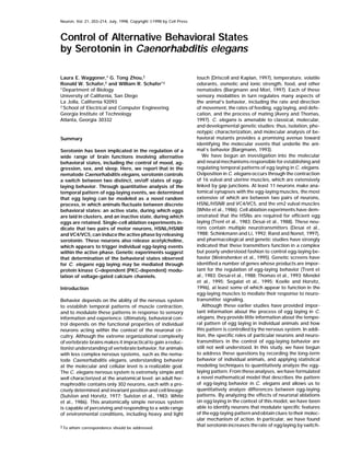
Figure 1. Egg Laying as a Three-State Point Process
Seven wild-type animals were tracked for a total of 40 hr, and the intervals between egg-laying events (contractions of the egg-laying muscles
that led to the expulsion of one or more eggs) were determined.
(a) Aperiodicity of the egg-laying pattern. The timing of egg-laying events was analyzed by computing the Fourier transform of the interval
data. If the eggs had been laid in a periodic or almost periodic fashion, then the Fourier transform would have exhibited a peak at a nonzero
frequency.
(b) Formal model for egg-laying behavior. According to the model, the animal can exist in one of three states: an inactive state (I), an active
state (A), and an egg-laying state (E). Eggs are laid upon entry into the E state; probabilities of state transitions are indicated on the branches
of the state diagram. 1 is the rate constant for the egg-laying state, and 2 is the rate constant for the inactive state (Zhou et al., 1997).
(c) Parameters of egg-laying behavior. Shown is a representative time line, with egg-laying events indicated by hash marks. According to the
model, p is the probability that after a given egg-laying event, another egg will be laid before the animal enters the inactive phase; these short
intervals resulting from a single visit to the A state are governed by the rate constant 1. Long intervals result from one or more visits to the
I (inactive) state (i.e, from E to A to I to A to E, or from E to A to I to A to I to A to E, etc.); the rate constant for these intervals is equal to p2
(for proof, see Zhou et al., 1997).
(d) Histograms of observed and simulated log intervals between egg-laying events. Shown is a histogram of the natural log of the interval
times, with the relative frequency on the y-axis. The graph shows a bimodal distribution, with one peak corresponding to the intervals between
events in a cluster and the other peak to the intervals between clusters. According to the model, if 1 and p2 are sufficiently different, the
left and right peak locations correspond to the time constants for the short and long intervals, respectively (for an exponential random variable,
the time constant equals the reciprocal of the rate constant and is the expected value of the mean interval time; for the data shown, these
are 20s and 1210s). Shown are a histogram of 216 samples of temporal egg-laying data by wild-type C. elegans and one of simulated data
based on the model (parameters: p ϭ 0.5891, 1 ϭ 0.0501, and 2 ϭ 0.0014) in the bottom graph. The dotted lines indicate the ideal distribution
based on ML analysis of the real data.
(e) Log tail distribution of intervals between egg-laying events, showing close agreement between theoretical probability and real data. The
model predicts that this distribution should be biphasic, with the steep part of the curve corresponding to the short intervals (i.e., intervals
within a cluster) and the more gradual part to the long intervals (i.e., intervals between clusters). The slope of the long intervals (i.e., the right
part of the curve) is equal to Ϫp2 (as noted, p2 is the intercluster rate constant). The solid line shows the theoretical log tail probability (p ϭ
0.5891, 1 ϭ 0.0501, and 2 ϭ 0.0014); the points correspond to the measured wild-type data from (d).
experiments argued that serotonin release from the enter an activated state in which they are more likely to
undergo contraction. According to this hypothesis, theHSNs stimulates egg laying not by directly exciting the
muscles but by modulating their activity such that they frequency of egg-laying clusters (2) is determined by
4. Neuron
206
Figure 2. Induction of Active State by Sero-
tonin
(a) Effect of HSN ablation on egg-laying pat-
tern. Shown are the log tail distributions of
the interarrival times for HSN-ablated wild-
type (N2) animals or egl-1(n986) mutant ani-
mals (in which the HSNs undergo cell death)
(Desai et al., 1988) compared to nonablated
N2 animals on NGM. The more gradual slope
(see Figure 1e legend) of the egl-1 and HSN-
ablated curves show that the intercluster in-
tervals are significantly longer in the HSN-
deficient animals. The duration of the long
intervals (i.e., Ͼ300 s) for HSN-ablated and
egl-1 animals were statistically different from
unablated wild type (level of confidence
Ͻ0.001) according to the Mann-Whitney rank
sum test.
(b) Effect of serotonin deficiency on egg-lay-
ing pattern. Shown are log tail distributions
for egg-laying intervals of bas-1 and cat-4
mutants, whichare defective in serotonin bio-
synthesis (Loer and Kenyon, 1993), compared
to the wild-type control. The more gradual
slope of the bas-1 and cat-4 tails indicates a
longer intercluster time constant. The long
intervals in the mutant animals (including
bas-1(ad446); see Table 1) were statistically
longer than in wild type (level of confidence
Ͻ0.005 for pa4 and e1141; Ͻ0.02 for ad446)
according to the Mann-Whitney rank sum test.
(c) Reversal of the effects of HSN ablation by
serotonin. Shown is the log tail distribution
of interval times for egl-1(n986), bas-1(pa4),
and wild-type hermaphrodites on 7.5 mM se-
rotonin (5-HT). The uniform linear distribu-
tions, whose slopes indicate a time constant
(1/1 ϭ 53 s for egl-1, 125 s for bas-1, and
127 s for N2) close to that for the clustered
intervals in wild-type animals, indicates that
the animals are almost exclusively in the ac-
tive state. Animals were tracked as de-
scribed, and egg-laying patterns were ob-
served until the uterus was usually essentially
empty (10 min for egl-1 and 20 min for N2
and bas-1). The number of animals and total
intervals analyzed were: N2, 9 animals, 49
intervals; egl-1, 8 animals, 117 intervals; and
bas-1, 9 animals, 49 intervals.
the rate at which serotonin is released to induce this these states, we analyzed the behavioral patterns of
active state. previously identified egg-laying–defective (Egl) mutants,
which were identified on the basis of their abnormally
slow egg-laying rate. Among the Egl mutants we sur-Genes Required to Establish the Active State
veyed, two general patterns were observed (Figure 3a).These analyses suggested that serotonin released from
Some of these mutants exhibited a pattern similar tothe HSNs controls the switch between alternative be-
that of HSN-ablated animals; they laid eggs in clustershavioral states for egg laying. Since the HSNs synapse
but had abnormally long inactivephases. These mutantsdirectly with the egg-laying muscles, one possibility is
defined genes that might affect the induction of thethat these behavioral states might correspond to differ-
active state but have little effect on egg laying withinent physiological states of the muscles. To identify mo-
lecular pathways that might be involved in determining the active phase. Interestingly, among the mutants in
5. Control of Behavior by Serotonin in C. elegans
207
Table 1. ML Estimates of Egg-Laying Parameters for Wild-Type, Mutant, and Ablated Animals
Intracluster Intercluster
Animal Type Time Constant Time Constant 1 2
(number, hours, intervals) (1/1; s) (1/p2; s) p (sϪ1
) (sϪ1
ϫ 10Ϫ3
)
N2a
18 1200 0.572 0.056 1.46
(7, 40, 216) Ϯ2 Ϯ170 Ϯ0.037 Ϯ0.008 Ϯ0.23
N2, HSNϪ 8 3400 0.534 0.125 0.55
(9, 44, 66) Ϯ2 Ϯ900 Ϯ0.069 Ϯ0.049 Ϯ0.16
egl-1(n986) 9 3630 0.685 0.106 0.40
(9, 46, 94) Ϯ2 Ϯ1020 Ϯ0.056 Ϯ0.019 Ϯ0.10
bas-1(pa4) 17 3480 0.619 0.060 0.46
(6, 38, 52) Ϯ4 Ϯ1080 Ϯ0.072 Ϯ0.014 Ϯ0.15
bas-1(ad446) 22 1870 0.446 0.046 1.22
(12, 52, 63) Ϯ6 Ϯ450 Ϯ0.072 Ϯ0.016 Ϯ0.42
cat-4(e1114) 43 4550 0.609 0.023 0.36
(12, 52, 63) Ϯ9 Ϯ1240 Ϯ0.073 Ϯ0.007 Ϯ0.13
tpa-1(k501) 33 3060 0.448 0.030 0.73
(7, 35, 66) Ϯ8 Ϯ630 Ϯ0.059 Ϯ0.016 Ϯ0.19
tpa-1(k530) 18 1970 0.508 0.055 1.04
(5, 24, 49) Ϯ6 Ϯ540 Ϯ0.083 Ϯ0.021 Ϯ0.42
CF453b
26 1770 0.539 0.038 1.05
(7, 24, 96) Ϯ6 Ϯ360 Ϯ0.062 Ϯ0.009 Ϯ0.28
CF453, VC4/5- 11 2300 0.555 0.089 0.75
(8, 53, 133) Ϯ2 Ϯ430 Ϯ0.047 Ϯ0.017 Ϯ0.16
a
N2 (a.k.a. C. elegans Bristol) is a standard wild-type strain; all mutants are in this genetic background.
b
CF453 is a strain carrying a mab-5::GFP fusion, which is expressed in VC4 and VC5 and allows identification of these cells for laser ablation.
this category were strains carrying loss-of-function al- Acetylcholine May Trigger Egg-Laying Events
within the Active Phaseleles of the gene tpa-1 (Sano et al.,1995), which encodes
the C. elegans homolog of PKC. Although tpa-1 muta- The observation that the HSNs are not required for effi-
cient egg laying within the active phase raised the ques-tions were not previously known to affect egg laying,
we found that loss-of-function PKC mutants laid eggs tion of what triggers these individual egg-laying events.
To address this question, we analyzed the effect of aat significantly lowerrates than wild type,an effect medi-
ated primarily by a lengthening of the intercluster time second pair of neurons, VC4 and VC5, on egg-laying
behavior. These cells receive synaptic input from theconstant (Figure 3b; Table 1). This result suggested that
PKC function is required for efficient induction of the HSNs and make extensive synapses with the egg-laying
muscles (White et al., 1986). Yet ablation of the VCsactive state by serotonin, but not for egg laying within
an established active state. Consistent with this hypoth- causes no gross egg-laying defect, and the role, if any,
of these cells in egg-laying behavior has up to nowesis, we found that tpa-1 mutants were highly resistant
to stimulation of egg laying by serotonin (Figure 3c). been unclear (Garriga et al., 1993). However, when we
analyzed the egg-laying patterns of VC4/VC5-ablatedThus, PKC is a candidate for a molecule that might
functiondownstream of the serotonin receptor to induce animals quantitatively, we observed that the VC neurons
had several important effects. First, VC ablation causedthe active phase.
Several Egl mutants showed a different egg-laying a small but significant increase (level of confidence
Ͻ0.02) in the intercluster time constant (Table 1). Sincepattern: they laid eggs singly rather than in clusters
(Figures 3a and 4). This pattern suggested that these VC4 and VC5 appear to be weakly serotonergic (Rand
and Nonet,1997), they may function along withthe HSNsmutants were unable to lay eggs efficiently within the
active phase, or that the active phase was unstable or to induce the active phase by releasing serotonin. In
addition, ablation of the VCs, like ablation of the HSNs,short-lived. As expected, most of these mutants laid
eggs at a higher rate when treated with serotonin, sug- caused an increased rate of egg laying within the active
phase (Table 1).gesting they were not defective in induction of the active
state. A striking exception was egl-19. Although egl- A more striking effect of the VCs on egg laying was
observed when both the HSNs and the VCs were elimi-19 hypomorphic mutants laid eggs at a reduced but
significant rate, they were completely nonresponsive to nated. Animals defective in both VC4/VC5 and HSNL/
HSNR were much more severely defective in egg layingserotonin (Figure 4 legend; see also Trent et al., 1983),
suggesting that the egl-19 gene is required to mediate than animals defective in only the HSNs (Figure 5 leg-
end). Furthermore, unlike animals defective in either thethe induction of the active state by serotonin. However,
stronger egl-19 hypomorphs showed an unclustered HSNs aloneor the VCs alone, HSN/VC-defective animals
failed to lay eggs efficiently even in the presence of highegg-laying pattern, and weaker alleles caused a signifi-
cant reduction in both the number of eggs laid in each levels of exogenous serotonin (Figures 5a and 5b). These
results suggest that a second neurotransmitter, whichcluster (i.e., p was reduced) and the rate of egg laying
within the cluster (1 was increased; Figure 4). Taken can be provided by either the HSNs or the VCs, is re-
quired in addition to serotonin for efficient egg laying.together, these data suggested that egl-19, unlike tpa-1,
is required both for induction of the active state and for One candidate for such a molecule is acetylcholine,
which is found in both the HSNs and VCs (Rand andefficient egg laying within the active state.
6. Neuron
208
Figure 3. Dependence of Active State Induction on Protein Kinase C
(a) Egg-laying patterns for wild-type and mutant animals. Shown are representative traces of egg-laying behavior for wild-type C. elegans, as
well as examples of two typical mutant egg-laying patterns. Each open box indicates an egg-laying event. The following mutants showed
clustered egg laying with long inactive phases (i.e., the middle pattern): egl-1(n986) (shown), egl-3(n150), egl-4(n478), egl-11(n587), egl-
21(n611), egl-24(n572), egl-30(n686), daf-7(e1372), and tpa-1(k501). The following mutants typically laid eggs singly rather than in clusters
(the bottom pattern): egl-7(n575), egl-10(n692), egl-12(n602), and egl-19(n582) (shown). Of the second group (in which egg laying within the
active phase appears defective), only egl-19 is serotonin nonresponsive (Trent et al., 1983).
(b) Effect of tpa-1 on egg-laying behavior. Shown are log tail distributions of egg-laying intervals for two tpa-1 mutants compared to wild-
type animals. The more gradual slope of distributions for the tpa-1 mutants (Table 1) indicates a longer intercluster time constant. The long
intervals (Ͼ300 s) in the mutant animals were statistically longer than in wild type (level of confidence Ͻ0.001 for k501 and Ͻ0.02 for k530)
according to the Mann-Whitney rank sum test.
(c) Effect of tpa-1 on serotonin response. Shown are log tail distributions of egg-laying intervals for tpa-1 (two alleles) and wild-type animals
in the presence of 7.5 mM serotonin (5-HT). The more gradual slope of the tpa-1 lines (time constant ϭ 429 s for k501, 400 s for k530, and
129 s for wild type) indicates a slower rate of egg laying. The intervals in the mutant animals were statistically longer than in wild type (level
of confidence Ͻ0.001) according to the Mann-Whitney rank sum test. For tpa-1 mutants, 45 intervals from a total of 7 animals (k530) or 67
intervals from a total of 6 animals (for k501) were analyzed.
Nonet, 1997). Acetylcholine receptor agonists stimulate laying events within those active phases. Consistent
with this hypothesis, we observed that depletion of en-egg laying in an HSN-dependent manner (Trent et al.,
1983; Weinshenker et al., 1995) and enhance thestimula- dogenous acetylcholine using conditional alleles of the
acetylcholine biosynthetic gene cha-1 caused a signifi-tion of egg laying by serotonin (D. Weinshenker and J.
Thomas, personal communication). In support of this cant decrease in the intracluster egg-laying rate (i.e., 1
was decreased; Figure 5c) but had little or no effect onhypothesis, we observed that HSN/VC-deficient animals
laid eggs efficiently when provided with both serotonin the intercluster rate (p2 was not changed).
and the nicotinic receptor agonist levamisole (Figure
5b). In contrast, levamisole alone did not stimulate egg
laying in either HSN-ablated or HSN/VC–doubly ablated Discussion
animals. Thus, the HSNs and VCs appear to release two
neurotransmitters, serotonin and acetylcholine, both of Serotonin Controls a Switch between Active
and Inactive Egg-Laying Stateswhich are required for egg laying. Since the role of sero-
tonin is to induce the active phase, a logical conjecture In summary, we have found that the complex temporal
pattern of egg laying in C. elegans can be effectivelyis that acetylcholine’s role is to trigger individual egg-
7. Control of Behavior by Serotonin in C. elegans
209
been able to identify neurons that selectively change
specific parameters of the egg-laying pattern, and we
have obtained information about the molecular mecha-
nism of action of these cells. These studies indicate that
the HSNs are not required to directly contract the egg-
laying muscles but rather to switch the egg-laying mus-
cles from a quiescent state to an active state in which
they are capable of readily undergoing contraction. At
least one mechanism through which the HSNs facilitate
the active state is likely to involve serotonin release.
Treatment of HSN-deficient animals with exogenous se-
rotonin induces egg laying in a pattern resembling a
continuous active phase. Moreover, serotonin-deficient
mutants, like HSN-ablated animals, have significantly
longer inactive periods between egg-laying clusters.
Mutations in bas-1, which cause a defect in the decar-
boxylase step of serotonin biosynthesis (Loer and Ken-
yon, 1993), cause a specific increase in the intercluster
time constant, but have little or noeffect onthe intraclus-
ter constant. In the cat-4 mutant, a large increase in the
intercluster time constant was also observed, along with
a smaller increase in the intracluster time constant.
Since the cat-4 mutation appears to be somewhat pleio-
tropic (e.g., cat-4 mutants apparently have a defective
cuticle [Loer, 1995, Soc. Neurosci., abstract]), it is un-
clear whether the effect on the intracluster rate is related
to serotonin. We identified several egg-laying–defec-
tive mutants whose phenotype resembled that of HSN-
ablated animals: active phases occurred with decreased
frequency, but egg laying within the active phase was
largely unaffected. Of these mutants, five (egl-4, egl-11,
egl-21, egl-24, and egl-30) have been shown to have
reduced serotonin response (Trent et al., 1983); thus,
these mutants define candidate genes for mediating
induction of the active state by serotonin.
Although these studies indicate that serotonin is suffi-
Figure 4. Effect of egl-19 on the Egg-Laying Pattern cient to induce the active state, recent work from other
egl-19 animals were tracked on NGM as described. Shown is the investigators indicates that serotonin is not required for
histogram of log egg-laying intervals. Loss-of-function alleles in-
active state induction. For example, C. elegans mutants
creased the time constant for egg laying in the active phase as well
that have no detectable serotonin are egg-laying com-as decreasing the number of clustered events. The degree to which
petent (Sulston et al., 1975). Moreover, C. elegans mu-these parameters were affected correlated with the severity of the
tants have been identified that are serotonin insensitiveallele. ad695, a semidominant, myotonic allele that affects inactiva-
tion kinetics (Lee et al., 1997) also decreased the intracluster egg- but can lay eggs in response to other pharmacological
laying rate. The durations of the short intervals (i.e., Ͻ300 s) in the agents (Weinshenker et al., 1995). In this study, we ob-
mutants were significantly longer than in wild type in all mutants
served that egg-laying events are still clustered in sero-
according to the Mann-Whitney rank sum test (significance Ͻ0.001
tonin-deficient mutants (Figure 2b), suggesting that afor all recessive alleles and Ͻ0.02 for ad695). ML parameter esti-
second molecule, probably released by the HSNs and/mates (p, 1 , 2 ) for each allele were: 0.171, 0.012, and 0.0037 for
or the VCs, may function redundantly with serotonin ton582; 0.246, 0.016, and 0.0024 for ad1006; 0.400, 0.019, and 0.0015
for ad1015; and 0.334, 0.033, and 0.0023 for ad695. The number of induce the active phase of egg laying. Immunocyto-
animals, hours tracked, and total intervals analyzed were: n582, 3 chemical experiments have demonstrated that the HSNs
animals, 32 hr, 53 intervals; ad1006, 5 animals, 34 hr, 47 intervals;
and VCs contain one or more FMRFamide-related neu-
ad1015, 3 animals, 20 hr, 42 intervals; and ad695, 3 animals, 24 hr,
ropeptides (FaRPs) (Schinkmann and Li, 1992); thus, an69 intervals. All recessive egl-19 mutants were completely serotonin
FaRP is a logical candidate for a second molecule thatresistant. Egg-laying rates (eggs/hr; mean Ϯ SEM) in the presence
could induce the active state.and absence of 7.5 mM serotonin were: ad1006,2.7 Ϯ 0.3(no seroto-
nin), 0.7 Ϯ 0.2 (with serotonin); ad1015, 3.9 Ϯ 0.3 (no serotonin),
10.1 Ϯ 0.3 (with serotonin); and n582, 50.0 Ϯ 0.6 (no serotonin),
1.3 Ϯ 0.4 (with serotonin). A Voltage-Gated Calcium Channel May
Determine the Active State
Our experiments also indicate that the HSNs and VCs
release another molecule,probably acetylcholine, whichmodeled as a random process involving discrete behav-
ioral states: an active, “on” state and an inactive, “off” induces individual egg-laying events within the active
period. Acetylcholine agonists have long been knownstate. By analyzing the effects of neuronal ablations and
genetic mutations in the context of this model, we have to increase egg laying in nematodes (Trent et al., 1983),
8. Neuron
210
Figure 5. Dependence of Egg Laying on Acetylcholine
(a) Effect of HSN and VC neurons on serotonin-induced egg laying. Shown are log tail distributions of egg-laying intervals of HSN-, VC-, and
HSN/VC-deficient animals on serotonin. In all cases, a monophasic, linear distribution indicating a homogenous Poisson process was observed;
the more gradual slope of the HSN/VC-ablated curve (time constant 1/1 ϭ 405 s for HSN/VCϪ, 83 s for HSNϪ, and 99 s for VCϪ) indicated
that the rate of egg laying in response to serotonin was greatly reduced (level of significance Ͻ0.001). Curves were based on analysis of 35
intervals from 6 animals (VCϪ), 70 intervals from 6 animals (HSNϪ), and 25 intervals from 12 animals (HSN/VCϪ). HSNϪ and VCϪ animals
were analyzed for 20 min, since they became depeleted of embryos after this time. To compare egg laying in the absence of serotonin, animals
were picked as larvae to individual plates and allowed to lay eggs. Both HSNϪ and HSN/VCϪ animals (but not VCϪ animals) laid eggs slowly,
such that the animals “bagged,” i.e., were consumed by hatched embryos that had been retained in the uterus. However, in HSNϪ animals,
the mean number of eggs released before bagging was 66.4 (SEM 8.6), while in HSN/VCϪ animals, the mean number of eggs released was
24.5 (SEM 4.7).
(b) Egg laying in HSN/VCϪ animals requires both serotonin and acetylcholine. HSN/VC-deficient animals laid eggs efficiently only when
provided with both serotonin and the cholinergic agonist levamisole. The histogram indicates the egg-laying rate under the indicated condition.
The indicated number of animals were tracked for 10 min as described in Experimental Procedures; solid and open boxes indicate mean and
standard error of the mean (SEM), respectively. HSN/VCϪ animals on serotonin and levamisole laid eggs at a higher rate than on serotonin
alone (level of confidence Ͻ0.005) or levamisole alone (level of significance Ͻ0.001) according to the Mann-Whitney rank sum test.
(c) Effect of acetylcholine depletion on egg laying. Depletion of endogenous acetylcholine using a conditional choline acetyltransferase mutant
allele caused a decreased rate of egg laying in the active phase. Shown are histograms of log egg laying intervals for animals carrying a
conditional allele of cha-1 (either y226 or md39) grown to adulthood at 15ЊC and shifted to 22ЊC before tracking. Estimated time constants
for long and short intervals are indicated. The short intervals (i.e., Ͻ300 s) in the mutants were statistically different from wild type (level of
confidence Ͻ0.001) according to the Mann-Whitney rank sum test. ML parameter estimates for p, 1, and 2 were 0.205, 0.016, and 0.0042
and 0.463, 0.023, and 0.0015 for y226 and md39, respectively.
and more recent work has suggested that acetylcholine serotonin specifically affected the duration of intervals
between clusters, acetylcholine affected the duration ofmight function in parallel with serotonin to stimulate
egg laying (Weinshenker et al., 1995). In support of this intervals between egg-laying events within a cluster.
Although at present we do not know the precise mecha-hypothesis, we observed that animals lacking both the
HSNs and VCs were largely resistant to stimulation of nism by which acetylcholine stimulates egg laying, the
simplest hypothesis is that it depolarizes the vulval mus-egg laying by serotonin, whereas treatment of these
animals with both serotonin and an agonist of nicotinic cle by opening nicotinic receptors, which leads to mus-
cle contraction. According to this model, the rate of egg-acetylcholine receptors (nAChRs) induced efficient egg
laying. Furthermore, analysis of acetylcholine-deficient laying events within the active phase (1) is a function
of the rate of acetylcholine release.mutants indicated that acetylcholine depletion decreases
the rate of egg laying within clusters. Thus, whereas We identified several mutants with reduced egglaying
9. Control of Behavior by Serotonin in C. elegans
211
Mechanisms for Modulation of Excitable
Cell States by Serotonin
As described above, serotonin appears to control a mo-
lecular switch between two egg-laying states, which
may correspond to functional states of the egg-laying
muscles. By investigating the genetics of egg-laying be-
havior, we have begun to obtain information about the
signaling pathways that help determine these functional
states and thus regulate the behavioral pattern. Interest-
ingly, serotonin appears to act through a similar mecha-
nism to modulate the activity of excitable cells in other
organisms. For example, recent studies of vertebrate
smooth muscle have found that serotonin can potentiate
ICa through modulation of L-type calcium channels, an
effect that appears to be mediated by Gq activation of
the PKC signaling pathway (Worley et al., 1991; Yang et
al., 1994; Hirakawa et al., 1995). In mollusks, serotonin-
dependent activation of calcium channel activity has
been directly correlated with modulation of excitation-
contraction coupling in smooth muscle cells (Nelson
Figure 6. Molecular Model for the Control of Egg Laying and Huddart, 1994). Thus, the molecular mechanisms
Serotonin, released primarily from the HSNs and to a lesser extent through which serotonin stimulates muscle activity may
from VC4 and VC5, facilitates the onset of the active phase. This be largely or completely conserved between the nema-
could occur through activation of EGL-19 calcium channels in the
todes and vertebrates. This suggests the possibility that
vulval muscles through PKC-dependent phosphorylation. TheHSNs
further genetic analysis of egg-laying behavior in theand VCs can both trigger individual muscle contractions within the
worm will provide an avenue to identify and characterizeactive phase by releasing acetylcholine; by binding to nicotinic re-
additional components of this signaling pathway (includ-ceptors and thus depolarizing the muscle cell membrane, this would
ing the candidate genes identified in our study) andlead to opening of activated EGL-19 channels and stimulation of
muscle contraction. to investigate how these molecules function in vivo to
determine the functional muscle states.
Biogenic amines, like other neuromodulators that act
through G protein–coupled serpentine receptors, havein the active phase, a pattern that suggests a defect in
been widely implicated in the control of alternative be-acetylcholine release or response. As expected, most
havioral and mental states in the brains of complex ani-of these mutants were serotonin sensitive (Trent et al.,
mals. In particular, serotonin appears to be involved in1983). The exception was egl-19, which was completely
the regulation of brain states involved in mood, sleep,serotonin resistant, and thus defective in both induction
addiction, and sexual behavior (Soubrie, 1988; Mestonof the active phase and egg laying within the active
and Gorzalka, 1992; Extein and Gold, 1993; Portas etphase. egl-19 encodes an L-type voltage-gated calcium
al., 1996). In addition, a number of studies from organ-channel homolog, which is thought, based on genetic
isms as diverse as crustaceans and humans have impli-evidence, to function in the vulval muscles (Lee et al.,
cated serotonin in the control of aggression (Olivier and1997). Thus, an appealing hypothesis is that serotonin
Mos, 1992; Brunner et al., 1993; Sandou et al., 1994;
might modulate the activity of EGL-19 calcium channels
Yeh et al., 1996). For example, in lobsters, agonistic
in the vulval muscles and thereby make them more ame-
behavior involves a series of stereotyped, increasingly
nable to contraction. In vertebrate cells, G protein–
violent behavioral patterns, between which animals
coupled receptors have been shown to facilitate PKC-
switch during the course of fights (Huber and Kravitz,
dependent phosphorylation of L-type calcium channels
1995). Serotonin does not appear to be involved in pro-
(see below), which enhances the coupling between exci-
ducing these patterns per se; rather, it appears to modu-
tation and channel opening (Boland et al., 1991). In C.
late the probability of switching from a more passive to
elegans, serotonin could induce phosphorylation of a more aggressive behavioral state (Huber et al., 1997).
EGL-19 calcium channels in the vulval muscles, making The formal parallels between the roles of serotonin in
them more likely to open in response to voltage changes crustacean aggression and nematode egg laying sug-
(Figure 6). Consistent with this hypothesis, tpa-1, cat-4, gest the possibility that the molecular mechanisms that
and bas-1 mutations all interacted genetically with egl- underlie these processes may be similar. Thus, the path-
19 hypomorphic alleles, suggesting that they may func- way through which serotonin modulates behavioral
tion in a common pathway (Table 2; see also Experimen- states in the worm may provide general insights into the
tal Procedures). Moreover, the egg-laying defects of modulatory role of serotonin in more complex behaviors
tpa-1 and cat-4 were suppressed by a gain-of-function in larger nervous systems.
allele of egl-19 (see Experimental Procedures). It is pos-
sible that modulation of other ion channels could con- Generation of Complex Response Patterns
tribute to the active state as well; for example, inhibition by Polyfunctional Neurons
of potassium channels in the egg-laying muscles or neu- A surprising conclusion of this study is that the complex
rons could increase the probability of egg-laying events pattern of C. elegans egg-laying behavior can be attrib-
uted largely to the functional versatility of the HSN andby decreasing the polarization of the membrane.
10. Neuron
212
Table 2. Genetic Interactions between egl-19 and Putative Signaling Genes
% Bag of Worms Egg-Laying Rate
Genotype (n ϭ 40; ϮSEM) (eggs/worm/hr)
wild type 0 Ϯ 0 3.5 Ϯ 0.5
egl-19(n582) 20 Ϯ 6 5.0 Ϯ 0.6
egl-19(ad1015) 3 Ϯ 2 3.9 Ϯ 0.3
tpa-1(k501) 5 Ϯ 3 3.0 Ϯ 0.6
cat-4(e1141) 5 Ϯ 3 2.4 Ϯ 0.8
bas-1(pa4) 13 Ϯ 5 2.8 Ϯ 0.1
tpa-1(k501); egl-19(n582) 62 Ϯ 8 0.4 Ϯ 0.3
tpa-1(k501); egl-19(ad1015) 8 Ϯ 4 1.5 Ϯ 0.4
cat-4(e1141); egl-19(n582) 90 Ϯ 5 0.5 Ϯ 0.3
bas-1(pa4); egl-19(ad1015) 8 Ϯ 4 1.5 Ϯ 0.6
Single animals were observed as they crawled across a standard 8VC motor neurons and to the multiplicity of signaling
cm agar plate using a stereoscopic dissecting microscope (Zeisspathways present in the muscle cells onto which they
Stemi 2000c) equipped with a Prior motorized stage. Video frames
synapse. Both the HSNs and the VCs contain at least
were captured at periodic intervals, and the stage was controlled
three neurotransmitters: a fast-acting classical transmit- based on the position of the animal using the ImagePro software
ter (acetylcholine), an amine neuromodulator (seroto- package (Media Cybernetics). A video camera (Cohu) recorded the
animal’s behavior at standard video frame rate; from these video-nin), and a peptide (or perhaps several related peptides).
tapes, the times that egg-laying events occurred were determined.Although each of these molecules is released from the
same presynaptic cells, they activate distinct signaling
ML Estimates of Model Parameters from Real Datapathways in the postsynaptic cells that affect different
For the three-state model described in Figure 1, we can prove thatparameters of the egg-laying pattern. This not only
the random variable governing the egg-laying interval times has the
allows a particular class of motor neurons to induce a following probability density function (p. d. f.) (Zhou et al., 1997):
complex output pattern in the cells onto which they
synapse, but it also opens the possibility that the differ- fx(x) ϭ k1 1 eϪ1x
ϩ k2 (p2) eϪ(p2)x
, x Ն 0,
ent response pathways in the postsynaptic cell could k1 ϭ
p(1 Ϫ 2)
1 Ϫ p2
, k2 ϭ
1(1 Ϫ p)
1 Ϫ p2
.
be differentially modified by experience,extrinsic modu-
lation, or pathway cross-talk.
Given N observations of the intervals x ϭ [x1,x2,...xN], the likelihood
Neurons that release multiple neurotransmitters are function is given by
not unique to nematodes; in fact, neurons containing a
fast-acting transmitter and a peptide, or a fast-acting f(x|) ϭ P
N
i ϭ 1
[k1 1 (eϪ1i
ϩ k2 (p2)) eϪ(p2)xi
]
transmitter and an amine, are relatively common in all
nervous systems (Kupfermann, 1991). Thus, the com-
which is a function of the parameter vector ϭ [p, 1, 2]. The
plex temporal response patterns induced by cotransmit- maximum likelihood estimate is defined as the that maximizes
ting neurons may be important for computation and f(x|) over all possible s. The bimodal peak information provided
by the histogram of the log intervals (see Figure 1d for example)behavioral control in more complex nervous systems
was used to obtain a rough estimate of and initialize the nonlinear(Marder et al., 1995). Coupled point process models,
ML algorithm, which adjusted the parameters to maximize the likeli-
such as the one we have used here to describe egg-
hood function.
laying behavior, should be widely applicable for analyz- To demonstrate the effectiveness of the ML procedure, we simu-
ing the output patterns of excitable cells in which re- lated egg-laying data using the model p. d. f. Close agreement
sponse to a fast-acting transmitter is potentiated by a between the true and estimated value was observed. The true pa-
rameters and the ML parameter estimates obtained from 100 inde-neuromodulator.
pendent realizations, each of 216 intervals (mean Ϯ standard devia-
tion), were p: true ϭ 0.5891, estimated ϭ 0.5967 Ϯ 0.0356; 1: true ϭExperimental Procedures
0.0501, estimated ϭ 0.0497 Ϯ 0.0059; and 2: true ϭ 0.0014, esti-
mated ϭ 0.0014 Ϯ 0.0002. Standard deviations in Table 1 wereAssay Conditions and Growth Media
estimated in a similar fashion.Unless otherwise noted, nematodes were grown and assayed at
For the special case of animals on serotonin (where a patternroom temperature on standard NGM seeded with E. coli strain OP50
resembling a homogeneous Poisson process is observed), the timeas a food source. For drug experiments, 5-hydroxytryptamine (creat-
constant was estimated by performing a weighted least-squaresinine sulfate complex, Sigma) was added to NGM agar at 7.5 mM,
linear regression to the log tail distribution (see below).and levamisole (hydrochloride, Sigma) was added at 6.25 M. Egg-
laying rates for egl-19 mutants were measured by counting the
number of eggs laid after 1 hr for three or more trials of 10 animals Log Tail Distribution
each on NGM in the presence or absence of 7.5 mM serotonin. It can be shown that the log tail probability (i.e., the log of the
probability thata given intervalwill belonger than time x)for intervals
Recording and Analysis of C. elegans generated by the three-state model is given by
Egg-Laying Behavior
We used a custom tracking system from Mikron Instruments (San ln Pr(X Ն x) ϭ ln(k1 eϪ1x
ϩ k2 eϪp2x
)
Diego), which tracks an animal for unlimited periods of time at up
to 60ϫ magnification (the magnification necessary to observe eggs For long intervals (x Ͼ 5/1), ln Pr(X Ն x) becomes approximately
linear with respect to x, with slope Ϫp2. Thus, the slope of thisadequately and to distinguish them from debris or clumps of food),
automatically moving the microscope stage so that the animal re- plot is inversely proportional to the average duration of the inactive
phase.mains centered in the visual field as it moves around the plate.
11. Control of Behavior by Serotonin in C. elegans
213
Histogram of Log Intervals Mary Franklin Foundation (R. W. S.). W. R. S. would especially like
to thank the Arnold andMabel Beckman Foundationfor their supportSince the intervals between eggs laid are clustered at short intervals
and sparse at long intervals, it is meaningful for reasons of better and generosity.
dynamic range to study the distribution of the log intervals (denoted
by Y ϭ ln X). Both real and simulated data exhibit a bimodal pattern Received October 31, 1997; revised May 7, 1998.
(as in Figure 1c), which can be shown to be represented by the
following p. d. f. of Y: References
fY(y) ϭ [k11 eϪ1
ev
ϩ k2(p2) eϪ(p2)ev
] ey
, Avery, L., and Thomas, J.H. (1997). Feeding and defecation. In C.
elegans II, D.L. Riddle, T. Blumenthal, B.J. Meyer, and J.R. Priess,k1 ϭ
p(1 Ϫ 2)
1 Ϫ p2
, k2 ϭ
1(1 Ϫ p)
1 Ϫ p2
.
eds. (ColdSpring Harbor, NY: ColdSpring Harbor Laboratory Press).
Bargmann, C.I. (1993). Genetic and cellular analysis of behavior in
We can show that when 1 and p2 are sufficiently different, fY(y)
C. elegans. Annu. Rev. Neurosci. 16, 47–71.
peaks at y ≈ ln(1/1) and ln(1/p2) with corresponding peak heights
Bargmann, C.I., and Mori, I. (1997). Chemotaxis and thermotaxis. Ink1/e and k2/e. When the values of 1 and p2 are close, only a single
C. elegans II, D.L. Riddle, T. Blumenthal, B.J. Meyer, and J.R. Priess,peak is observed.
eds. (Cold Spring Harbor: Cold Spring Harbor Laboratory Press).
Boland, L.M., Allen, A.C., and Dingledine, R. (1991). Inhibition byAblation of Egg-Laying Neurons
bradykinin of voltage-activated barium current in a rat dorsal rootThe two HSNs were eliminated either by introducing a mutation in
ganglion cell line: role of protein kinase C. J. Neurosci. 11, 1140–the gene egl-1, which causes the HSNs to undergo cell death in the
1149.hermaphrodite (Desai et al., 1988), or by ablating the HSN nuclei in
the first larval stage. The VC4 and VC5 neurons were ablated in early Brunner, H.G., Nelen, M., Breakfield, X.O., Ropers, H.H., and van
fourth-stage larvae of the strain CF453 (genotype: dpy-20(e1282)IV; Oost, B.A. (1993). Abnormal behavior associated with a point muta-
muIs16[dpy-20(ϩ) mab-5:GFP]; kindly provided by Craig Hunter and tion in the structural gene for monoamine oxidase A. Science 262,
Cynthia Kenyon), a strain carrying an integrated mab-5::GFP fusion. 578–580.
Expression of GFP in the developing VC4 and VC5 cells was used Croll, N.A. (1975). Indolealkylamines inthe coordinationof nematode
to identify them prior to the ablation process; cell killing was verified behavioral activities. Can. J. Zool. 53, 894–903.
the next day by scoring for the absence of VC nuclei and GFP-
Desai, C., Garriga, G., McIntire, S., and Horvitz, H.R. (1988). A genetic
expressing neurons and neuronal processes in the vulval region. For
pathway for the development of the Caenorhabditis elegans HSN
HSN/VCϪ animals, VC4 and VC5 nuclei were ablated asdescribed in
motor neurons. Nature 336, 638–646.
the strain AQ112, which carries the egl-1(n986) mutation and the
Driscoll, M., and Kaplan, J. (1997). Mechanotransduction. In C. ele-mab-5::GFP fusion; unablated AQ112 animals were used as the
gans II, D.L. Riddle, T. Blumenthal, B.J. Meyer, and J.R. Priess, eds.HSNϪ control.
(Cold Spring Harbor, NY: Cold Spring Harbor Press).
Extein, I.L., and Gold, M.S. (1993). Hypothesized neurochemicalConstruction and Characterization of Double Mutants
models for psychiatric syndromes inalchohol and drug dependence.Double mutants were generated using standard methods. Mutant
J. Addict. Dis. 12, 29–43.homozygotes were identified in the F2 generation on the basis of the
Garriga, G., Desai, C., and Horvitz, H.R. (1993). Cell interactionsfollowing phenotypes: tpa-1, resistance to tetradecanoyl phorbol
control the direction of outgrowth, branching and fasciculation ofacetate (Tabuse et al., 1989); egl-19 recessive alleles, long body
the HSN axons of Caenorhabditis elegans. Development 117, 1071–length and sluggish movement; egl-19(n2368), short body length
1087.(Lee et al., 1997); cat-4, hypochlorite hypersensitivity (Loer, 1995,
Soc. Neurosci., abstract); and bas-1, failure to slow in the presence Hirakawa, Y., Kuga, T., Kobayashi, S., Kanaide, H., and Takeshita,
of bacteria (Sawin, 1996). The pattern of egg laying in the tpa-1; A. (1995). Dual regulation of L-type Ca2ϩ
channels by serotonin
egl-19 double mutants resembled that of egl-19 single mutants. The receptor stimulation in vascular smooth muscle cells. Am. J. Physiol.
egl-19(n2368) singlemutant aswell asthe tpa-1(k501); egl-19(n2368) 268, H544–H549.
and cat-4(e1141); egl-19(n2368) double mutants all laid eggs ex-
Horvitz, H.R., Chalfie, M.,Trent, C., and Evans, P.D. (1982). Serotonin
tremely hyperactively, such that no mature (Ͼ8 cells) embryos accu-
and octopamine in the nematode Caenorhabditis elegans. Science
mulated in the uterus, and embryo production rather than vulval
216, 1012–1014.
muscle contraction was limiting for egg laying. For the doubles with
Huber, R., and Kravitz, E.A. (1995). A quantitative analysis of agonis-recessive egl-19 alleles, ML parameter estimates for p, 1, and 2
tic behavior in juvenile American lobsters. Brain Behav. Evol. 46,were 0.158, 0.020, and 0.0036 for k501; n582 (6 animals, 38 hr, 13
72–83.
intervals) and 0.255, 0.016, and 0.0030 for k501; ad1015 (4 animals,
Huber, R., Smith, K., Delago, A., Isaksson, K., and Kravitz, E.A.20 hr, 55 intervals), respectively. Egg-laying rates in Table 2 were
(1997). Serotonin and aggressive motivation in crustaceans: alteringdetermined after 1 hr incubation on seeded NGM plates (3 trials of 10
the decision to retreat. Proc. Natl. Acad. Sci. USA 94, 5939–5942.animals each). The percentage of animals forming “bags of worms”
(animals in which unlaid embryos hatch within the uterus due to an Koelle, M.R., and Horvitz, H.R. (1996). EGL-10 regulates G protein
extreme egg-laying defect) was determined at 2 days following the signaling in the C. elegans nervous system and shares a conserved
L4/adult molt. domain with many mammalian proteins. Cell 84, 115–125.
Kupfermann, I. (1991). Functional studies of cotransmission. Physiol.
Acknowledgments Rev. 71, 683–732.
Lee, R.Y.N., Lobel, L., Hengartner, M., Horvitz, H.R., and Avery, L.
We thank Barbara Meyer, Mike Nonet, Leon Avery, Craig Hunter, (1997). Mutations in the ␣1 subunit of an L-type voltage-activated
Curtis Loer, Rene Garcia, and the Caenorhabditis Genetics Center Ca2ϩ
channel cause myotonia in Caenorhabditis elegans. EMBO J.
for strains; Jon Pierce and Rick Logemann for assistance and advice 16, 6066–6076.
in setting up the tracking system; David Weinshenker for communi-
Loer, C. M., and Kenyon, C. J. (1993). Serotonin-deficient mutants
cating unpublished results; Cynthia Kenyon and members of our
and male mating behavior in the nematode Caenorhabditis elegans.
laboratories for encouragement; and Charles Zuker, Cori Bargmann,
J. Neurosci. 13, 5407–5417.
Jasper Rine, Patricia Laurenson, and Cynthia Kenyon for comments
Marder, E., Christie, A.E., and Kilman, V.L. (1995). Functional organi-on the manuscript. We would also like to thank three anonymous
zation of cotransmission systems: lessons from small nervous sys-reviewers for their suggestions. This work was supported by a NAR-
tems. Invert. Neurosci. 1, 105–112.SAD Young Investigator Award (W. R. S.), a Beckman Young Investi-
gator Award (W. R. S.), a Sloan Fellowship (W. R. S.), and grants Mendel, J.E., Korswagen, H.C., Liu, K.S., Hadju-Cronin, Y.M., Simon,
M.I., Plasterk, R.H., and Sternberg, P.W. (1995). Participation of thefrom the National Science Foundation (G. T. Z.) and the John and
12. Neuron
214
protein Go in multiple aspects of behavior in C. elegans. Science Yeh, S.R., Fricke, R.A., and Edwards, D.H. (1996). The effect of social
267, 1652–1655. experienceon serotonergic modulation of the escape circuit of cray-
fish. Science 271, 366–369.Meston, C.M., and Gorzalka, B.B. (1992). Psychoactive drugs and
human sexual behavior: the role of serotonergic activity. J. Psycho- Zhou, G.T., Schafer, R.W., and Schafer, W.R. (1997). A three-state
active Drugs 24, 1–40. biological point process model and its parameter estimation. IEEE
Trans. Signal Process., in press.Nelson, I.D., and Huddart, H. (1994). Neuromodulation in molluskan
smooth muscle: the action of 5-HT, FMRFamide and purine com-
pounds. Gen. Pharmacol. 25, 539–552.
Olivier, B., and Mos, J.(1992). Rodentmodels of aggressive behavior
and serotonergicdrugs. Prog.Neuropsychopharmacol. Biol. Psychi-
atry 16, 847–870.
Portas, C.M., Thakkar, M., Rainnie, D., and McCarley, R.W. (1996).
Microdialysis perfusion of 8-hydroxy-2-(di-n-propylamino)tetralin
(8-OH-DPAT) in the dorsal Raphe nucleus decreases serotonin re-
lease and increases rapid eye movement sleep in the freely moving
cat. J. Neurosci. 16, 2820–2828.
Rand, J.B., and Nonet, M.L. (1997). Neurotransmitter assignments
for specific neurons. In C. elegans II, D.L. Riddle, T. Blumenthal,
B.J. Meyer, and J.R. Priess, eds. (Cold Spring Harbor, NY: Cold
Spring Harbor Laboratory Press).
Sandou, F., Amara, D.A., Dierich, A., LeMeur, M., Ramboz, S., Segu,
L., Buhot, M.-C., and Hen, R. (1994). Enhanced aggressive behavior
in mice lacking 5HT1B receptor. Science 265, 1875–1878.
Sano, T., Tabuse, Y., Nishiwaki, K., and Miwa, J. (1995). The tpa-1
gene of Caenorhabditis elegans encodes two proteins similar to
Ca2ϩ
-independent protein kinase Cs: evidence by complete geno-
mic and complementary DNA sequences of the tpa-1 gene. J. Mol.
Biol. 251, 477–485.
Sawin, E.R. (1996). Genetic and cellular analysis of modulated be-
haviors in Caenorhabditis elegans. PhD thesis, Massachusetts Insti-
tute of Technology, Cambridge, MA.
Schinkmann, K., and Li, C. (1992). Localization of FMRFamide-like
peptides in Caenorhaditis elegans. J. Comp. Neurol. 316, 251–260.
Segalat, L., Elkes, D.A., and Kaplan, J.M. (1995). Modulation of sero-
tonin-controlled behaviorsby Go in Caenorhabditiselegans. Science
267, 1648–1651.
Soubrie, P. (1988). Serotonin and behavior, with special regard to
animal models of anxiety, depression, and waiting ability. In Neu-
ronal Serotonin, N.N. Osborne and M. Hamon, eds. (New York:
Wiley).
Sulston, J.E., and Horvitz, H.R. (1977). Post-embryonic cell lineages
of the nematode Caenorhabditis elegans. Dev. Biol. 56, 110–156.
Sulston, J., Dew, M., and Brenner, S. (1975). Dopaminergic neurons
in the nematode Caenorhabditis elegans. J. Comp. Neurol. 163,
215–226.
Sulston, J.E., Schierenberg, E., White, J.G., and Thomson, J.N.
(1983). The embryonic cell lineage of the nematode Caenorhabditis
elegans. Dev. Biol. 100, 64–119.
Tabuse, Y., Nishiwaki, K., and Miwa, J. (1989). Mutations in a protein
kinase C homolog confer phorbol resistance in Caenorhabditis ele-
gans. Science 243, 1713–1716.
Thomas, J.H., Birnby, D., and Vowels, J. (1993). Evidence for parallel
processing of sensory information controlling dauer formation. Ge-
netics 134, 1105–1117.
Trent, C., Tsung, N., and Horvitz, H.R. (1983). Egg-laying defective
mutants of the nematode Caenorhabditis elegans. Genetics 104,
619–647.
Weinshenker, D., Garriga, G., and Thomas, J.H. (1995). Genetic and
pharmacological analysis of neurotransmitters controlling egg-lay-
ing in C. elegans. J. Neurosci. 15, 6975–6985.
White, J., Southgate, E., Thomson, N., and Brenner, S. (1986). The
structure of the Caenorhabditis elegans nervous system. Philos.
Trans. R. Soc. Lond. B Biol. Sci. 314, 1–340.
Worley, J.F., Quayle, J.M., Standen, N.B., and Nelson, M.T. (1991).
Regulation of single calciumchannels in cerebral arteries by voltage,
serotonin, and dihydropyridines. Am. J. Physiol. 261, H1951–H1960.
Yang, M.M., Yo, Y.-L., Hseih, J.-T., and Ong, R. (1994). 5-hydroxy-
tryptamine receptor–mediated phosphoinositide hydrolysis in ca-
nine cultured tracheal smooth muscle cells. Br. J. Pharmacol. 111,
777–786.