2. of centrosomes (Wu and Palazzo, 1999; Schnackenberg et
al., 2000), meiotic apparatus (Suprenant and Rebhun, 1984;
Haimo, 1985; Kuriyama et al., 1986; Suddith et al., 2001),
nuclear envelope (Maul et al., 1987; Dessev and Goldman,
1988; Dessev et al., 1989), and PBs (Longo and Anderson,
1970; Pielak et al., 2003, 2004; Alliegro and Alliegro,
2005). One deficiency recognized in recent years, a lack of
genetic data for the species, is currently being addressed
(Marine Biological Laboratory, 2003).
In previous work on cytoskeletal events during PB for-
mation in Spisula (Pielak et al., 2003, 2004), we found that
the metaphase I peripheral aster spread onto the cortex in a
circular pattern containing relatively few central microtu-
bules. This aster disassembled during anaphase, and a ring
of thickened F-actin appeared that had essentially the same
cortical location, dimensions, and pattern as did microtu-
bules in the earlier aster. The late anaphase spindle re-
mained docked at the F-actin-poor center of the ring (the
“ring opening”), where PB extrusion then occurred. The
observed spatial correspondence immediately suggested
that microtubules of the peripheral aster are a causal agent
in generation of the PB F-actin ring.
The present studies were designed to test this hypothesis
by altering the number of peripheral asters and their cortical
contact patterns prior to PB formation. Two methods were
employed. First, exposure to lovastatin (LV), followed by
washout, induced both metaphase arrest (Turner et al.,
1995) and multiple asters under our conditions, an effect
modified and enhanced by hexylene glycol (HG; Rebhun
and Sawada, 1969; Saiki and Hamaguchi, 1997). Second,
cytochalasin D (CD) was used to block PB1 extrusion, with
extra asters being generated following washout prior to
meiosis II (Longo, 1972; Hertzler, 2002). Both approaches
gave strikingly consistent positive results, producing multi-
ple peripheral asters associated with multiple, overlapping
F-actin rings.
Materials and Methods
Ripe individuals of Spisula solidissima (Dillwyn, 1817),
maintained at 11–13 °C to inhibit spawning, were obtained
from the Aquatic Resources Division of the Marine Biolog-
ical Laboratory (MBL) in Woods Hole, Massachusetts. Go-
nadal tissue was removed by dissection and suspended in
about 150–300 ml of filtered (Whatman #2) natural seawa-
ter (F-NSW) depending on gonad size. After tissue removal
by filtration through a double cheesecloth layer, the egg
suspension passing through was allowed to settle (Ϸ20–30
min), the supernate was removed, and the eggs were washed
twice more by resuspension and settling. After determining
approximate total egg volume by gentle hand-centrifuga-
tion, final resuspension was in F-NSW refiltered at 0.45-m
(ϭ double-filtered, or DF-NSW) to a concentration of about
1% (v/v). Eggs were stored at this concentration in DF-
NSW containing 50 g/ml gentamycin (Hunt et al., 1992),
maintained in suspension at 16 °C in a rocking water bath.
Such eggs retained the capacity for normal PB formation
and (when sperm-activated) normal cleavage, but were used
for 2 days only because, beyond that, increased time to PB
formation suggested some deterioration.
Spisula oocytes were activated by using excess KCl, a
standard method for this species (Allen, 1953; Costello and
Henley, 1971). Eggs were gently hand-centrifuged and re-
suspended to 10% (v/v) in DF-NSW, and one volume of
suspension was mixed with 7.6 vol. DF-NSW and 1.4 vol.
0.5 M KCl (t ϭ 0). Eggs were monitored in phase contrast
for GVBD, sedimented gently, resuspended in DF-NSW or
experimental media to about 1% (v/v), and maintained in
suspension by gentle rocking on a nutator at 22 °C. Samples
were treated as described in particular experiments and then
prepared for fluorescence labeling and microscopy (see be-
low). The timing of stages became slightly extended with
longer-term storage of ripe animals in cold seawater, and
this was adjusted for by monitoring a range of time-point
samples in each preparation.
Fluorescence labeling and microscopy
For rapid monitoring of chromosomal stage, oocytes
were routinely fixed in seawater containing 2% or 4%
formaldehyde (equally effective), stained with DAPI (3 or 6
M), and examined and photographed using a standard
Zeiss epifluorescence microscope. For cytoskeletal labeling,
oocytes were simultaneously permeabilized and fixed for 15
min in PEM (100 mM PIPES, 5 mM EGTA, 1 mM MgCl2,
pH 6.8, using NaOH) containing 0.6% Brij-58 and 2%
formaldehyde, then washed twice in phosphate-buffered
saline (PBS). Labeling was performed in PBS using the
following agents: for F-actin, Alexa Fluor 568-phalloidin
(Molecular Probes); for microtubules, a mixture of mouse
monoclonal anti-␣ and anti- tubulins (Sigma T9026,
T-4026) at their recommended working dilutions, pre-
tagged with Alexa Fluor 488-labeled anti-mouse Fab frag-
ments (Zenon; Molecular Probes); for DNA, DAPI. Label-
ing protocols were as described in prior work, without
vitelline membrane removal (Lee et al., 2002; Pielak et al.,
2003, 2004). Samples were viewed both in a standard Zeiss
epifluorescence microscope and by confocal fluorescence
microscopy using the Zeiss Laser Scanning System LSM 5
PASCAL, with image processing via Zeiss LSM Image
Examiner (2004) and Adobe Photoshop (vers. 3.0) software.
Aster or ring diameters were measured on images of opti-
mum clarity, using Zeiss Image Examiner software. Rela-
tive pixel intensity was measured on the original 3-D re-
constructed image in face view, using the Scion Image PC
version of NIH-Image software (beta vers. 4.0.2).
Treatment with aster-perturbing agents
Following activation, Spisula oocytes proceed continu-
ously from GVBD through meiosis and the extrusion of two
22 R. M. PIELAK ET AL.
3. PBs. Therefore, we tested lovastatin (LV; Calbiochem) as a
possible means to provide a time window for addition of
aster-perturbing agents such as hexylene glycol (HG) that
require oocyte penetration. LV, a 3-hydroxy-3-methylglu-
taryl coenzyme A reductase inhibitor, induces reversible
metaphase I arrest of KCl-activated Spisula oocytes (Turner
et al., 1995). Using DF-NSW as the primary medium, as for
other experimental agents and solvent controls described
below, activated oocytes were incubated in LV at 3.5 M,
35 M, 52.5 M, and 70 M. Under our conditions, meta-
phase stability was most repeatable at 52.5 M (1.5 ϫ conc.
used by Turner et al., 1995), and this concentration was
used routinely at about 12 min post-activation, when meta-
phase spindles were still centrally located and en route to
the cortex. Solvent-only controls were exposed to 0.15%
DMSO in DF-NSW. Oocyte samples were processed for
fluorescence microscopy after various LV incubation peri-
ods before washout and during meiotic resumption after two
washes in DF-NSW.
Hexylene glycol (HG, ϭ 2-methyl-2,4-pentanediol; Sigma)
reversibly increases aster size and stability (Spisula: Reb-
hun and Sawada, 1969; starfish: Saiki and Hamaguchi,
1997). During LV-induced metaphase time windows, HG
(1% v/v) was added either “early” at t ϭ 20 min post-
activation, with t ϭ 25 min washout (5 min HG incubation),
or “late” at t ϭ 25 or 30 min, with washout at 35 or 42 min,
respectively (10 or 12 min HG incubation). Both late addi-
tion times gave the same results, so that HG use is described
below as either early or late. For all experiments with LV
plus HG, maximum oocyte exposure to LV was thus 30
min.
As in previous work (Pielak et al., 2004), cytochalasin D
(CD; Calbiochem) was used to inhibit F-actin ring assembly
prior to PB1 formation. Eggs were sedimented gently and
resuspended in 1.0 g/ml CD or 0.1% DMSO (solvent
control) at about 15 min post-activation, when metaphase
spindles were still in transit to the cortex. Various times for
initiating CD washout were then tested to determine which
would block extrusion of PB1 while permitting that of PB2.
Washout when solvent-only controls were still in metaphase
(Ϸ20 min) was too early, allowing sequential formation of
both PBs. Washout when controls were at late anaphase
(Ϸ27–30 min) was too late; no PBs formed, and oocytes
contained four pronuclei. However, washout with controls
at anaphase onset (Ϸ25 min post-activation) permitted sub-
sequent PB2 formation and was used in the experiments.
Results
F-actin ring formation in untreated oocytes
In the normal cytoskeletal sequence that precedes PB
formation, the metaphase meiotic spindle moved toward the
egg surface, the peripheral aster spread fountain-like onto
the oocyte cortex, and subsequently in anaphase, most astral
microtubules disassembled (Fig. 1A–C). The F-actin ring
appeared in the cortical location of the previously spread
aster, with similar outer diameter (Ϸ20–22 m), and with
the spindle docked at the center of the F-actin-deficient ring
opening (Fig. 1C, D). Rotated 3-D reconstructions of cor-
tical F-actin from optical section stacks revealed the circular
pattern of the ring, with chromosomes positioned in the ring
opening (Fig. 1E).
The normal pattern of F-actin deposition was also ob-
served in solvent-only controls (Fig. 1F, G), again with an
outer ring diameter of about 21–22 m, as measured in
optical sections of three rings. Thus, for comparison with
the normal pattern, altered cortical F-actin deposition is
described throughout this paper in terms of the “ring” and
“ring opening.” As examined in scans of pixel intensity
across the ring, cortical F-actin was most dense within the
ring, and the level in the ring opening resembled that of
surrounding cortex (Fig. 1H).
Lovastatin-induced metaphase arrest, followed by
washout
Lovastatin (LV) was initially tested to determine its util-
ity in providing a time window for administration of hexy-
lene glycol (HG) by means of metaphase arrest. First, we
confirmed LV-induced metaphase arrest (Fig. 2A, B), as
well as PB production in about 90% of oocytes after 30 min
arrest and washout (Fig. 2C, D). However, we were sur-
prised to discover that, under our conditions, LV-induced
metaphase arrest lasted only about 30 min, after which
anaphase began even without washout. More importantly
for our objectives, most of the LV-treated oocytes displayed
multiple asters with interconnected spindles (Fig. 2E). In a
majority of oocytes, only one of these asters was in contact
with the cortex; but in about 15%–20%, the meiotic appa-
ratus was oriented such that two or three asters touched the
cortex. In anaphase, such oocytes contained adjacent
docked spindles (Fig. 2F) that were associated either with a
single enlarged, circular or avoid F-actin ring (Ͼ30 m;
Fig. 2G) or with double F-actin rings (Fig. 2H–J). Each ring
opening was occupied by a spindle centrosomal region (e.g.,
Fig. 2H, J), and dual ring openings shared a strip of thick-
ened F-actin between them that varied in width in different
oocytes (e.g., Fig. 2H–J).
Hexylene glycol addition during lovastatin metaphase
arrest
Given the LV results, we tested HG addition during
LV-induced time windows to determine whether it would
increase astral cortical contact, enhancing or modifying the
formation of multiple rings (Fig. 3). Astral spreading oc-
curred during metaphase, as previously (Fig. 3A), and mul-
tiple spreading asters were observed (Fig. 3B). Two centro-
somes were clearly present at the docking pole of some
metaphase or early anaphase spindles (Fig. 3C). Single
enlarged rings were most evident when HG was added
23PERIPHERAL ASTER FUNCTION
4. Figure 1. Cytoskeletal features preceding polar body formation in normal oocytes and in DMSO solvent controls
(optical sections unless otherwise noted; microtubules, green; F-actin, red; chromosomes, blue-violet). (A) Docked
metaphase spindle, with spreading peripheral aster. (B) Early anaphase spindle with spread aster. (C) Anaphase
spindle abutting cortex at the ring center after most peripheral aster microtubules have disassembled. (D) Another
anaphase oocyte, microtubules omitted. One chromosome set is centered at the ring opening (arrowhead). (E) Rotated
3-D view of the F-actin ring in the oocyte shown in (D), constructed from an image stack, illustrating the
Ϸ22-m-diameter ring pattern and chromosome location in the ring opening. (F, G) Optical sections of solvent-only
control oocytes (0.15% DMSO), with same Ϸ20-m-diameter ring pattern as in oocytes without solvent (arrowheads
at ring opening). (H) Upper: face-view grayscale image of F-actin only, in rectangular area across the ring in (E);
Lower: scan of pixel intensity in upper image; level in the ring opening (ro) is similar to that in the cortex (c)
surrounding the ring (r). (A, B, D) From Pielak et al. (2003). Scale bars: 10 m (bars for A–C and E, as in D).
Figure 2. Lovastatin LV-induced formation of enlarged and multiple spreading asters, with subsequent cortical
induction of enlarged F-actin rings. (A, B) Rapid DAPI labeling of chromosome sets (arrowheads) in metaphase and
anaphase, respectively. (C, D) Optical sections showing polar body formation following lovastatin washout. (E)
Docked duplex metaphase spindle, with very large spreading aster region, prior to F-actin deposition and polar body
formation. (F) Peripheral centrosomes of two late anaphase spindles docked close together at the cortex. Note adjacent
regions of thickened cortical F-actin (arrowheads). (G) Same oocyte, rotated 3-D view showing a single enlarged,
ovoid F-actin ring and ring center (long axis of Ϸ35 m). (H) Oocyte with large region of thickened cortical F-actin,
centrosomes occupying the two ring openings, and a shared F-actin segment between them. (I) Another pattern,
similar to (H), but with a much thinner inter-ring segment (arrow). Inset: chromosomes of same egg; the two upper
chromosome groups were adjacent to the two ring openings. (J) Another oocyte with a dual ring pattern; inter-ring
segment is intermediate in thickness between that of (H) and (I). Inset: chromosomes of same egg; the two upper left
chromosome groups were adjacent to the two ring openings. NOTE: optical sectioning was restricted to the most
relevant regions to limit photobleaching; unsectioned areas appear in some 3-D reconstructions (e.g., area “x” in G).
Scale bars: 10 m (bar for B–D as in A; E and G as in F; H and I as in J).
5. “late” (Fig. 3D, E), whereas “early” HG addition produced
two or more peripheral asters and multiple rings (Fig. 3F–
K). Multiple asters and rings were observed more frequently
when HG was used in combination with LV than when LV
was used alone.
In some of these multiple-ring oocytes, F-actin stained far
more intensely in the region between ring openings than in
the surrounding area (Fig. 3G, H; confirmed by pixel inten-
sity scanning). When two cortex-associated asters were
adjacent and a third more distant, the result was one larger
Figure 4. Spindle malorientation induced by lovastatin plus hexylene
glycol: the spindle, normally perpendicular to the cortical surface, as in Fig.
1C, here is parallel. (A) Rotated 3-D view of anaphase cortical F-actin
pattern. Upper inset: anaphase chromosome sets; lower inset: centrosomes
of the two asters. (B) Composite of A ϩ A insets. (C) Optical section of
same cell, showing increased F-actin deposition between the ring openings
(arrow), and edge view of one of the cortically docked spindle poles
(arrowhead). (D) Optical section of another anaphase oocyte with spindle
parallel to the cortical surface, and pronounced F-actin deposition between
two docked spindle poles. The F-actin pattern of both of these oocytes is
similar to that of Fig. 3H, with two centrosome-associated ring centers and
increased F-actin deposition appearing principally in the segment between
them. Scale bars: 10 m (bars for B and C as in A).
Figure 3. Hexylene glycol addition during lovastatin metaphase arrest: aster and ring patterns following
washout. Optical sections: A, B, C, I; others, rotated 3-D views from image stacks. (A) Enlarged spreading
metaphase aster. (B) Two adjacent spreading metaphase asters. (C) Docked metaphase or early anaphase spindle.
Inset: higher magnification of two peripheral centrosomes (arrowheads). (D, E) Enlarged rings in late anaphase
oocytes, diameters Ͼ 30 m. (F) Pattern variation with one large ring opening containing two docked
centrosomes, and a smaller opening with one centrosome. Insets, left: astral pattern; right: associated chromo-
some sets. (G) F-actin pattern for oocyte in (F), showing major thickening only in shared region between ring
openings. (H) Another example of F-actin thickening primarily between ring openings. (I) Oocyte containing
three peripherally spread asters (two visible in this section), and three cortical regions of F-actin deposition
(arrowheads). (J) Triple ring pattern of same oocyte, with two large ring openings and a third smaller one. (K)
Composite view for (J), showing centrosomal regions of the three peripheral asters. Note that ring F-actin
deposition is not yet evident in metaphase or early anaphase oocytes (A–C). Scale bar for C inset: 5 m; others:
10 m (bar for E as in D; H as in G)
25PERIPHERAL ASTER FUNCTION
6. Figure 5. Multiple asters with interconnected spindles and two docked centrosomes, induced by cytocha-
lasin D plus washout. Images (G), (K), and (N) are 3-D views from image stacks; others are optical sections. (A)
Multiple spreading metaphase asters, prior to ring formation. (B) Same oocyte, metaphase chromosome sets. (C)
Another oocyte at later stage, with two polar bodies extruded after cytochalasin D washout. Inset: chromosome
sets, two external and one internal (slight size reduction). (D) Same oocyte as (C), F-actin distribution only. (E)
An anaphase oocyte with multiple asters and two centrosomes cortically docked (only one visible in this optical
section). (F) Same oocyte as (E), anaphase chromosome sets. (G) Same oocyte, dual cortical F-actin ring pattern.
(H) Another anaphase oocyte, with two docked centrosomes. (I) Same oocyte as (H), anaphase chromosome sets.
(J) Same oocyte, F-actin pattern only, with three cortical deposition areas (arrowheads). (K) Same oocyte,
showing dual cortical F-actin ring pattern. (L) Another anaphase oocyte, with multiple asters, interconnected
spindles, and two closely docked centrosomes. (M) Anaphase chromosome sets of same oocyte. (N) Same
oocyte, single enlarged, ovoid ring and ring opening. Scale bars: 10 m (bar for B as in A; C as in D; F and G
as in E; I–K as in H; M and N as in L).
26 R. M. PIELAK ET AL.
7. ring opening and one smaller one (Fig. 3F, G). However, if
centrosomes of three peripherally spread asters were suffi-
ciently separated, three ring openings appeared within a
very large region of cortical F-actin deposition (Fig. 3I–K).
Despite the unusual ring patterns, about 80%–90% of the
oocytes treated with LV plus HG in different preparations
ultimately produced PBs after washout, many of which
appeared to be larger than control PBs (not shown).
In a minor sub-population of LV-treated oocytes, HG
produced single anaphase spindles that were parallel to the
cortex, a highly unusual orientation for Spisula (Fig. 4).
Both centrosomal regions were docked in these oocytes, and
two ring openings appeared within a common area of F-
actin deposition (Fig. 4A). F-actin deposition was typically
intensified between the ring openings (Fig. 4A, C, D), as in
some of the multi-aster contact patterns (e.g., Fig. 3G).
Cytochalasin D inhibition of polar body formation,
followed by washout
In previous work on shrimp oocytes, cytochalasin block-
age of PB1 formation, followed by washout, led to co-
production of two meiotic spindles and two PBs at the time
of PB2 formation in controls (Hertzler, 2002). Since this
was a potential means to generate two asters in simulta-
neous cortical contact, and perhaps unusual contact patterns,
we tested the approach on Spisula oocytes. Cytochalasin
effects on Spisula PB formation had been investigated ear-
lier, but without such washout (Longo, 1972; Pielak et al.,
2004).
Washout of cytochalasin D (CD) (1.0 g/ml), with con-
trols in early anaphase, was effective in permitting the
treated oocytes to proceed into meiosis II. Subsequently,
when most controls exhibited a typical metaphase II spindle
docked at the cortex, nearly all experimental oocytes con-
tained two or more cortically spreading asters and intercon-
nected metaphase spindles (Fig. 5A, B). Nevertheless, two
PBs were ultimately produced at about the same time in
these oocytes (Fig. 5C, D). At intermediate anaphase stages,
double F-actin rings usually formed, with the two centro-
somal regions of the spindles and asters occupying the ring
openings (Fig. 5E–K). The dual spindles were sometimes
connected at an internal centrosome and docked cortically at
an angle to each other (Fig. 5H–K). In several cases, how-
ever, two docked anaphase spindles were associated with a
single, very large, ovoid F-actin ring (Fig. 5L–N), with both
centrosomes occupying the ring center.
Discussion
In the normal cytoskeletal sequence that precedes forma-
tion of the polar body (PB) in Spisula oocytes, cortical
spreading of the peripheral aster at metaphase and cortical
docking of the spindle are followed, during anaphase, by
disassembly of astral microtubules and formation of the
F-actin ring (Fig. 1). The diameters of both aster and ring
are typically about 20–22 m, and the actin-poor center of
the ring is similar in dimensions to the microtubule-deficient
central region of the earlier spreading aster (Pielak et al.,
2003, 2004). These observations suggest that spatial fea-
tures of PB ring formation are determined by the peripheral
aster.
Spatiotemporal relationship between spreading peripheral
asters and cortical F-actin rings
In the present work we asked whether altering the number
and distribution of peripheral asters would produce corre-
sponding changes in the number and pattern of cortical F-
actin rings. The answer was clear: treatment with lovastatin
(LV) alone, with LV plus hexylene glycol (HG), or with
cytochalasin D (CD), followed by washout, all produced
oocytes with multiple cortically spreading peripheral asters
(e.g., Figs. 2E; 3B, I; 5E). Subsequently, many of these
oocytes produced either a single, abnormally enlarged cir-
cular or ovoid ring (Ͼ30 m diameter; e.g., Figs. 2G; 3D,
E; 5 N), or multiple overlapping rings (Figs. 2H–J; 3F–K;
5E–K). Single enlarged rings appeared to be generated
when centrosomes were in close proximity (e.g., Fig. 2F,
G), and this was correlated with considerable increase in
astral spreading diameter (long axis of Ϸ40 m; Fig. 2E).
However, it remains to be determined whether both in-
creased microtubule density and astral spreading diameter
are critical for cortical F-actin patterning. F-actin thickening
between ring openings could be an initial stage of ring
biogenesis occurring where overlap of astral microtubules
provides the greatest density (e.g., Fig. 3G, H; Fig. 4C, D).
Alternatively, thickening could be the result of incomplete
cortical contact and spreading by the experimentally altered
asters, or a symptom of an additive or even synergistic
effect of astral overlap.
With respect to the temporal relationship, F-actin rings
were observed in normal oocytes only in anaphase (Fig. 1).
Oocytes exposed to LV, or LV plus HG, completed astral
spreading and spindle docking, but were arrested at meta-
phase due to blockage of the degradation of Spisula oocyte
cyclin A and B (Hunt et al., 1992; Turner et al., 1995).
F-actin rings were not generated unless the drugs were
washed out, permitting entry into anaphase (Fig. 2F; 4A). In
addition, after CD-washout, multiple F-actin rings were
observed only in anaphase cells (Fig. 5A, B vs. 5E, F).
Therefore, both astral contact and anaphase-triggered events
must determine biogenesis of the F-actin ring for Spisula PB
formation. This is consistent with the importance of an-
aphase entry for somatic furrowing, as evident in early work
on echinoderm zygotes whose asters had been removed or
disrupted (Cornman and Comman, 1951; Swann and
Mitchison, 1953; Mitchison, 1953; Scott, 1960), and in later
studies on the appearance of actin filament bundles in the
cortex (Mabuchi, 1994).
27PERIPHERAL ASTER FUNCTION
8. Function of asters versus centrosomal regions
The experimentally generated rings exhibited various pat-
terns, most of which can be accounted for by varying
degrees of ring overlap following a stimulus from the astral
microtubules (Fig. 6A–E). However, we observed no ring
openings “filled in” by F-actin from an adjacent ring (Fig.
6F, G). This cannot be explained simply by stimulus reduc-
tion caused by low microtubule density at adjacent astral
centers, and therefore we suggest that, in contrast to astral
microtubules, docked centrosomes may inhibit F-actin de-
position. Such a mechanism would ensure reduction of
mechanical resistance in ring openings, the cortical site of
PB extrusion. Adjacent centrosomes would then produce
one enlarged ring opening (e.g., Figs. 2G; 3G; 5N); if the
centrosomes were further apart, multiple ring openings
would be surrounded by F-actin from overlapping rings
(e.g., Fig. 2H–J; 3F–K; 5G). The centrosomes of spindles
abnormally docked parallel to the cortex would be separated
by the spindle itself, generating separate ring openings (Fig.
4A–C). While we find no published evidence for or against
such speculation, it is worth noting that centrosomes iso-
lated from Spisula oocytes, though too small (Ϸ2 m di-
ameter) for simple obstruction of ring openings, contain a
great many proteins of unknown function (Palazzo and
Vogel, 1999).
In the androgenetic bivalve Corbicula, two PBs are pro-
duced naturally after simultaneous spreading of two meta-
phase I asters. Here, the metaphase I spindle always aligns
parallel to the oocyte surface, and both asters spread corti-
cally prior to dual PB extrusion (Komaru et al., 2000). This
differs from CD-washout, in which dual anaphase II spin-
dles appear nearly end-on to the cortex (Fig. 5H), but it
resembles the parallel spindle-cortex alignment in oocytes
treated with LV plus HG, generating dual overlapping rings
(Fig. 4). The actual F-actin ring pattern in Corbicula has not
yet been determined, however.
Mechanisms involved in multiple aster and ring
generation
Multiple and enlarged asters and rings occurred after
treatment and washout using LV, LV plus HG, or CD (Figs.
2–5). Since different means produced the same general
result, multiple ring formation is aster-related, not drug-
related. Multiple asters were expected for treatment with
CD but not with LV; the latter may reflect use at a some-
what higher concentration and temperature than in earlier
work (Turner et al., 1995). Although knowledge of the
specific mechanisms that produce multiple asters is not
critical to present conclusions, we believe centrosome rep-
lication to be responsible in both cases. Typical meiotic
events continue during CD inhibition of ring and PB1
formation in Spisula (Pielak et al., 2004), and, in the case of
LV, centrosome replication and separation apparently pro-
ceed during the metaphase block or shortly after washout
(Fig. 3C).
Since astral perturbation produced marked pattern alter-
ations, the biogenetic signal or mechanism for F-actin dep-
osition must be recognized indiscriminately by the Spisula
oocyte cortex (Fig. 7). In other species, such ring induction
would account for PB formation at different cortical sites
after experimental translocation of the meiotic apparatus
(Hamaguchi et al., 2001), and for an indiscriminate furrow-
ing response in somatic cytokinesis (Kawamura, 1960; Rap-
paport, 1985). In normal Spisula oocytes, the circular cor-
tical F-actin ring and ring opening occupy nearly 40% of
oocyte diameter before PB extrusion (Fig. 1E), but we do
not yet know whether other invertebrates exhibit this ring
pattern and size (Shimizu, 1990; Hamaguchi et al., 2001).
Do astral microtubules simply provide signals marking
cortical regions, or do they deliver F-actin and other ring
components? This question remains open both for Spisula
oocytes and for PB formation. In several other systems there
is evidence that microtubules determine F-actin distribution
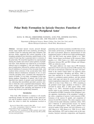
Figure 6. Diagram of pattern variations in dual rings and ring openings. (A) Ring pattern generated during
normal (control) polar body biogenesis, or enlarged in cells treated with lovastatin plus hexylene glycol (Figs.
1E; 3D, E). (B) Partial overlap of adjacent rings (Figs. 2H; 5G). (C, D) Progressively greater ring overlap
patterns (Figs. 2I, J; 3F). (E) Marked overlap, such that ring openings are joined (Figs. 2G; 5N). (F, G) Patterns
of overlap that were not observed. Absence of such patterns indicates that, whereas the earlier spreading astral
microtubules have a stimulatory effect on F-actin deposition, each docked spindle or centrosome thereof,
occupying a ring center, has an inhibitory influence. This would prevent neighboring spread asters from inducing
F-actin deposition in the adjacent ring opening. Patterns such as those in (A–E) can then be accounted for based
on the distance between spreading asters and the centrosomes of their docked spindles.
28 R. M. PIELAK ET AL.
9. and furrow position (grasshopper spermatocytes: Alsop and
Zhang, 2004), addition of new furrow membrane (sea ur-
chin zygotes: Shuster and Burgess, 2002), and furrow in-
duction via transported proteins (mammalian cells: Canman
et al., 2003); in addition, direct actin-microtubule interac-
tion has been reported in Drosophila embryos and in vitro
(Sider et al., 1999; Foe et al., 2000; Waterman-Storer et al.,
2000). With respect to PB ring signaling in Spisula oocytes,
Rho is a good candidate for study. Rho family GTPases are
believed to determine properties of F-actin rings involved in
somatic cytokinesis and oocyte wound-healing (Mabuchi et
al., 1993; Nishimura et al., 1998; Saint and Somers, 2003;
Somers and Saint, 2003; Yonemura et al., 2004; Benink and
Bement, 2005).
Regardless of molecular mechanism, peripheral asters
clearly function to induce F-actin rings associated with PB
extrusion in Spisula. Thus, at the cellular level at least, PB
biogenesis in Spisula differs from that in mammals. The
mammalian meiosis I spindle has truncated asters (Szollosi
et al., 1972), and cortical spindle positioning involves actin
filaments and formin-2 rather than astral microtubules
(Leader et al., 2002; Maro and Verlhac, 2002). Comparison
of the pathways to PB formation taken by mammalian,
non-mammalian vertebrate, and invertebrate oocytes would
be of considerable interest, and Spisula provides an excel-
lent model system for such work.
Note added in proof: Evidence for RhoA involvement in
establishing the polar body F-actin ring in Xenopus, as well
as somatic contractile rings in echinoderms, is provided in a
new paper by Bement et al. (2005).
Acknowledgments
We are indebted to Louie Kerr of the MBL and Rudi
Rottenfusser of Carl Zeiss Inc. for the excellent confocal
microscopy facilities, and to the Howard Hughes Medical
Institute Undergraduate Science Education Program in Biol-
ogy (HHMI 5200267), the Hunter College Avon-Tukman
Fund, NSF 9808368, and PSC-CUNY 65218, for support.
Literature Cited
Allen, R. D. 1953. Fertilization and artificial activation in the egg of the
surf-clam, Spisula solidissima. Biol. Bull. 105: 213–239.
Alliegro, M. C., and M. A. Alliegro. 2005. Differential expression of
tyrosinated tubulin in Spisula solidissima polar bodies. Dev. Dyn. 232:
216–220.
Alsop, G. B., and D. Zhang. 2004. Microtubules continuously dictate
distribution of actin filaments and positioning of cell cleavage in
grasshopper spermatocytes. J. Cell Sci. 117: 1591–1602.
Bement, W. M., H. A. Benink, and G. von Dassow. 2005. A microtu-
bule-dependent zone of active RhoA during cleavage plane specifica-
tion. J. Cell Biol. 170 (in press).
Benink, H. A., and W. M. Bement. 2005. Concentric zones of active
RhoA and Cdc42 around single cell wounds. J. Cell Biol. 168: 429–
439.
Canman, J. C., L. A. Cameron, P. S. Maddox, A. Straight, J. S.
Tirnauer, T. J. Mitchison, G. Fang, T. M. Kapoor, and E. D.
Salmon. 2003. Determining the position of the cell division plane.
Nature 424: 1074–1078.
Castro, A., E. Mandart, T. Lorca, and S. Galas. 2003. Involvement of
Aurora A kinase during meiosis I–II transition in Xenopus oocytes.
J. Biol. Chem. 278: 2236–2241.
Cornman, I., and M. E. Cornman. 1951. The action of podophyllin and
its fractions on marine eggs. Ann. N.Y. Acad. Sci. 51: 1443–1487.
Costello, D. P., and C. Henley. 1971. Methods for Obtaining and Han-
dling Marine Eggs and Embryos. Mollusca: Mactra solidissima, 2nd
ed. Marine Biological Laboratory, Woods Hole, MA. Also available at:
http://www.mbl.edu/BiologicalBulletin/CLASSICS/COSTELLO/
index.html [accessed 12 March 2005].
Dessev, G., and R. Goldman. 1988. Meiotic breakdown of nuclear
envelope in oocytes of Spisula solidissima involves phosphorylation
and release of nuclear lamin. Dev. Biol. 1302: 543–550.
Dessev, G., R. Palazzo, L. Rebhun, and R. Goldman. 1989. Disassem-
bly of the nuclear envelope of Spisula oocytes in a cell-free system.
Dev. Biol. 1312: 496–504.
Dube´, F., and W. R. Eckberg. 1997. Intracellular pH increase driven by
an Naϩ
/Hϩ
exchanger upon activation of surf clam oocytes. Dev. Biol.
190: 41–54.
Foe, V. E., C. M. Field, and G. M. Odell. 2000. Microtubules and
mitotic cycle phase modulate spatiotemporal distributions of F-actin
and myosin II in Drosophila syncytial blastoderm embryos. Develop-
ment 127: 1767–1787.
Haimo, L. T. 1985. Microtubule polarity in taxol-treated isolated spin-
dles. Can. J. Biochem. Cell. Biol. 63: 519–532.
Hamaguchi, Y., S. K. Satoh, T. Numata, and M. S. Hamaguchi. 2001.
Response of the cortex to the mitotic apparatus during polar body
formation in the starfish oocyte of Asterina pectinifera. Cell Struct.
Funct. 2: 627–631.
Hertzler, P. L. 2002. Twin meiosis 2 spindles form after suppression of
polar body 1 formation in oocytes of the marine shrimp Sicyonia
ingentis. Biol. Bull. 202: 100–103.
Hunt, T., F. C. Luca, and J. V. Ruderman. 1992. The requirements for
protein synthesis and degradation, and the control of destruction of
cyclins A and B in the meiotic and mitotic cell cycles of the clam
embryo. J. Cell Biol. 116: 707–724.
Katsu, Y., N. Minshall, Y. Nagahama, and N. Standart. 1999. Ca2ϩ
is
required for phosphorylation of clam p82/CPEB in vitro: implications
for dual and independent roles of MAP and Cdc2 kinases. Dev. Biol.
209: 186–199.
Kawamura, K. 1960. Studies on cytokinesis in neuroblasts of the grass-
hopper, Chortophaga viridifasciata (de Geer). II. The role of the
mitotic apparatus in cytokinesis. Exp. Cell Res. 21: 9–18.
Komaru, A., K. Ookubo, and M. Kiyomoto. 2000. All meiotic chro-
mosomes and both centrosomes at spindle pole in the zygotes discarded
as two polar bodies in clam Corbicula leana: unusual polar body
Figure 7. Schematic of observed F-actin patterns in relation to docked
spindles and their centrosomal regions. (A) Single circular ring (Figs. 1E;
3D, E). (B) Large ovoid ring (Figs. 2G; 5N). (C) Double ring (Figs. 2H–J;
3G, H; 5H–K). (D) Spindle parallel to cortex, F-actin between centrosomes
(Fig. 4B, C). The variations in size, pattern, and location demonstrate that
the cortex responds indiscriminately to the astral influence.
29PERIPHERAL ASTER FUNCTION
10. formation observed by antitubulin immunofluorescence. Dev. Genes
Evol. 210: 263–269.
Kuriyama, R., G. G. Borisy, and Y. Masui. 1986. Microtubule cycles
in oocytes of the surf clam, Spisula solidissima: an immunofluores-
cence study. Dev. Biol. 114: 151–160.
Leader, B., H. Lim, M. J. Carabatsos, A. Harrington, J. Ecsedy, D.
Pellman, R. Maas, and P. Leder. 2002. Formin-2, polyploidy, hy-
pofertility and positioning of the meiotic spindle in mouse oocytes. Nat.
Cell Biol. 4: 921–928.
Lee, K. G., A. Braun, I. Chaikhoutdinov, J. DeNobile, M. Conrad, and
W. Cohen. 2002. Rapid visualization of microtubules in blood cells
and other cell types in marine model organisms. Biol. Bull. 203:
204–206.
Longo, F. J. 1972. The effects of cytochalasin B on the events of
fertilization in the surf clam, Spisula solidissima. I. Polar body forma-
tion. J. Exp. Zool. 182: 321–344.
Longo, F. J., and E. Anderson. 1970. An ultrastructural analysis of
fertilization in the surf clam, Spisula solidissima. I. Polar body forma-
tion and development of the female pronucleus. J. Ultrastruct. Res. 33:
495–514.
Longo F. J., S. Cook, L. Mathews, and S. J. Wright. 1991. Nascent
protein requirement for completion of meiotic maturation and pro-
nuclear development: examination of fertilized and A-23187-activated
surf clam (Spisula solidissima) eggs. Dev. Biol. 148: 75–86.
Mabuchi, I. 1994. Cleavage furrow: timing of emergence of contractile
ring actin filaments and establishment of the contractile ring by fila-
ment bundling in sea urchin eggs. J. Cell Sci. 107: 1853–1862.
Mabuchi, I., Y. Hamaguchi, H. Fujimoto, N. Morii, M. Mishima, and
S. Narumiya. 1993. A rho-like protein is involved in the organisation
of the contractile ring in dividing sand dollar eggs. Zygote 1: 325–331.
Marine Biological Laboratory. 2003. Clam mini-genome project. Pp.
1–2 in LabNotes, vol. 14, no. 1. Also available at: http://www.mbl.edu/
inside/what/news/publications/labnotes/04_spring.html [accessed 12
March 2005].
Maro, B., and M. H. Verlhac. 2002. Polar body formation: new rules
for asymmetric divisions. Nat. Cell Biol. 4: 921–928.
Maul, G. G., G. Schatten, S. A. Jimenez, and A. E. Carrera. 1987.
Detection of nuclear lamin B epitopes in oocyte nuclei from mice, sea
urchins, and clams using a human autoimmune serum. Dev. Biol. 1212:
368–375.
Mitchison, J. M. 1953. Microdissection experiments on sea-urchin eggs
at cleavage. J. Exp. Biol. 30: 515–526.
Nishimura, Y., K. Nakano, and I. Mabuchi. 1998. Localization of Rho
GTPase in sea urchin eggs. FEBS. Lett. 441: 121–126.
Palazzo, R. E., and J. M. Vogel. 1999. Isolation of centrosomes from
Spisula solidissima oocytes. Methods Cell Biol. 61: 35–56.
Pielak, R. M., V. A. Gaysinskaya, and W. D. Cohen. 2003. Cytoskel-
etal events preceding polar body formation in activated Spisula eggs.
Biol. Bull. 205: 192–193.
Pielak R. M., V. A. Gaysinskaya, and W. D. Cohen. 2004. Formation
and function of the polar body contractile ring in Spisula. Dev. Biol.
269: 421–432.
Rappaport, R. 1985. Repeated furrow formation from a single mitotic
apparatus in cylindrical sand dollar eggs. J. Exp. Zool. 234: 167–171.
Rebhun, L. I., and N. Sawada. 1969. Augmentation and dispersion of
the in vivo mitotic apparatus of living marine eggs. Protoplasma 68:
1–22.
Saiki, T., and Y. Hamaguchi. 1997. Division of polar bodies induced by
their enlargement in the starfish Asterina pectinifera. Exp. Cell Res.
237: 142–148.
Saint, R., and W. G. Somers. 2003. Animal cell division: a fellowship
of the double ring? J. Cell Sci. 116: 4277–4281.
Schnackenberg, B. J., D. R. Hull, R. D. Balczon, and R. E. Palazzo.
2000. Reconstitution of microtubule nucleation potential in centro-
somes isolated from Spisula solidissima oocytes. J. Cell Sci. 113:
943–953.
Schumacher, J. M., A. Golden, and P. J. Donovan. 1998. AIR-2: An
Aurora/Ip11-related protein kinase associated with chromosomes and
midbody microtubules is required for polar body extrusion and cyto-
kinesis in Caenorhabditis elegans embryos. J. Cell Biol. 143: 1635–
1646.
Scott, A. C. 1960. Furrowing in flattened sea urchin eggs. Biol. Bull.
119: 246–259.
Shimizu, T. 1990. Polar body formation in Tubifex eggs. Pp. 260–272 in
Cytokinesis: Mechanisms of Furrow Formation During Cell Division,
G. W. Conrad and T. E. Schroeder, eds. New York Academy of
Sciences, New York.
Shuster, C. B., and D. R. Burgess. 2002. Targeted new membrane
addition in the cleavage furrow is a late, separate event in cytokinesis.
Proc. Natl. Acad. Sci. USA 99: 3633–3638.
Sider, J. R., C. A. Mandato, K. L. Weber, A. J. Zandy, D. Beach, R. J.
Finst, J. Skoble, and W. M. Bement. 1999. Direct observation of
microtubule-F-actin interaction in cell free lysates. J. Cell Sci. 112:
1947–1956.
Somers, W. G., and R. A. Saint. 2003. RhoGEF and Rho family
GTPase-activating protein complex links the contractile ring to cortical
microtubules at the onset of cytokinesis. Dev. Cell 4: 29–39.
Suddith, A. W., E. A. Vaisberg, S. A. Kuznetsov, W. Steffen, C. L.
Rieder, and R. E. Palazzo. 2001. Centriole duplication, centrosome
maturation and spindle assembly in lysates of Spisula solidissima
oocytes. Methods Mol. Biol. 161: 215–228.
Suprenant K. A., and L. I. Rebhun. 1984. Purification and character-
ization of oocyte cytoplasmic tubulin and meiotic spindle tubulin of the
surf clam Spisula solidissima. J. Cell Biol. 98: 253–266.
Swann, M. M., and J. M. Mitchison. 1953. Cleavage of sea-urchin eggs
in colchicine. J. Exp. Biol. 30: 506–514.
Swenson, K. I., N. Borgese, G. Pietrini, and J. V. Ruderman. 1987.
Three translationally regulated mRNAs are stored in the cytoplasm of
clam oocytes. Dev. Biol. 123: 10–16.
Szollosi, D., P. Calarco, and R. P. Donahue. 1972. Absence of centri-
oles in the first and second meiotic spindles of mouse oocytes. J. Cell
Sci. 11: 521–541.
Turner, J. E., C. G. Minkoff, K. H. Martin, R. Misra, and K. I.
Swenson. 1995. Oocyte activation and passage through the meta-
phase/anaphase transition of the meiotic cell cycle is blocked in clams
by inhibitors of HMG-CoA reductase activity. J. Cell Biol. 128: 1145–
1162.
Waterman-Storer, C., D. Y. Duey, K. L. Weber, J. Keech, R. E.
Cheney, E. D. Salmon, and W. M. Bement. 2000. Microtubules
remodel actomyosin networks in Xenopus egg extracts via two mech-
anisms of F-actin transport. J. Cell Biol. 150: 361–376.
Weinberg, J. R., and T. E. Helser. 1996. Growth of the Atlantic
surfclam, Spisula solidissima, from Georges Bank to the Delmarva
peninsula, USA. Mar. Biol. 126: 663–674.
Wu, X., and R. E. Palazzo. 1999. Differential regulation of maternal vs.
paternal centrosomes. Proc. Natl. Acad. Sci. USA 96: 1397–1402.
Yi, J.-H., L. Lefie`vre, C. Gagnon, M. Anctil, and F. Dube´. 2002.
Increase of cAMP upon release from prophase arrest in surf clam
oocytes. J. Cell Sci. 115: 311–320.
Yonemura, S., K. Hirao-Minakuchi, and Y. Nishimura. 2004. Rho
localization in cells and tissues. Exp. Cell Res. 295: 300–314.
30 R. M. PIELAK ET AL.