2. 1990; Zeschingk et al., 1995), chondrocytes (Oberlender
and Tuan, 1994), osteoclasts (Mbalaviele et al., 1995),
and neural cells (Doherty and Walsh, 1994).
Myogenesis is a particularly appealing system to
study the role of cadherin-mediated adhesion in cell
differentiation, because Ca2ϩ
-dependent cell adhesion,
followed by cell fusion, is an intrinsic step in the
differentiation process. The differentiation of cultured
skeletal myoblasts is commonly activated by growth
factor withdrawal and accompanied by transcriptional
activation of muscle-specific genes, growth arrest, and
fusion to form multinucleated myotubes (Olson, 1992,
1993). The muscle-specific basic helix–loop–helix tran-
scription factors, including MyoD, Myf5, myogenin,
and Mrf4, orchestrate the entire expression program of
the various muscle-specific genes.
Several lines of indirect evidence suggest that cad-
herin-mediated interactions are also involved in the
regulation of skeletal myogenesis. These include the
inhibition of fusion by calcium depletion or by anti-
N-cadherin antibodies and HAV-containing inhibitory
peptide (Knudsen et al., 1990; Mege et al., 1992). In
addition, the somites formed in N-cadherin knockout
mice are small and irregularly shaped (Radice et al.,
1997). In addition to the effect of N-cadherin on termi-
nal stages of skeletal muscle differentiation, it has been
shown to affect the expression of genes before the cell
fusion stage: injection of a dominant negative cadherin
RNA suppresses the expression of MyoD in Xenopus
embryos and affects the subsequent expression of
muscle-specific genes (Holt et al., 1994). Avian embry-
onic progenitor cells expressing only N-cadherin and
not E-cadherin differentiate into skeletal muscle, and
treatment with anti-N-cadherin antibodies inhibits the
accumulation of myosin in chick embryo cells derived
from different stages of avian embryonic development
(George-Weinstein et al., 1997). Overexpression of N-
cadherin in baby hamster kidney cells stimulates ex-
pression of sarcomeric myosin in these cells (Redfield
et al., 1997). However, although these data suggest that
cadherin-mediated interactions are involved in muscle
differentiation, they do not indicate whether they are
involved merely in the promotion of myoblast–myo-
blast adhesion per se or also induce long-range, myo-
genic signals that promote muscle gene expression.
In the present study we examined the effect of direct
long-range signaling induced by N-cadherin cluster-
ing or immobilization on myogenic differentiation. We
show here that beads conjugated to different N-cad-
herin ligands can trigger myogenesis, manifested by
accelerated myoblast adhesion, myogenin expression,
formation and assembly of various structural sarco-
meric components, and myoblast fusion. Stimulation
of myogenin expression by the N-cadherin–reactive
beads occurred irrespective of myoblast density, sug-
gesting that activation of this key step in myogenesis
is directly induced by N-cadherin signaling.
MATERIALS AND METHODS
Cell Culture
All myoblast lines examined in this study, including C2 mouse
skeletal myoblasts and L8 and L84 rat skeletal myoblasts, were
kindly provided by Dr. D. Yaffe (The Weizmann Institute of Science)
(Yaffe and Saxel, 1976, 1977). The cells were cultured in subconfluent
densities at 37°C in a humidified atmosphere containing 8% CO2 in
dishes coated with 0.1% gelatin. C2 cells were cultured in Dulbec-
co’s modified Eagle’s medium (DMEM) supplemented with 20%
heat-inactivated FCS (BioLabs, Israel), glutamine, and antibiotics. L8
and L84 cells were cultured in Waymouth’s medium containing
15% FCS. Myogenic differentiation of L8 and L84 cells was induced
by changing the growth medium to DMEM containing 2% heat-
inactivated horse serum (Biological Industries, Israel) and 4 IU/ml
insulin (Humulin R; Lilly, France). To trigger the differentiation of
C2 myoblasts, cells were either plated at high density or stimulated
by insulin and 10% horse serum in DMEM.
Preparation and Application of Cadherin-reactive
Beads
N-cadherin ectodomain (NEC) was produced as described by Lev-
enberg et al. (1998a). Briefly, 108
latex Polybead amino microsperes
(mean diameter, 6 m; Polysciences, Warrington, PA) were washed
with phosphate-buffered saline (PBS; pH 7.4), activated overnight
with 8% glutaraldehyde, washed with PBS, and incubated for 5 h
with 500 g/ml bovine serum albumin (BSA; Sigma Chemical, St.
Louis, MO), purified NEC (Levenberg et al., 1998), or anti-N-cad-
herin monoclonal antibodies (clone BE; Volk and Geiger, 1986). Free
sites were blocked with 0.5 M ethanolamine for 30 min, followed by
incubation with 10 mg/ml BSA for 30 min, and the beads were
resuspended in storage buffer (PBS containing 10 mg/ml BSA, 0.1%
sodium azide, and 5% glycerol, pH 7.4). Aliquots containing 5 ϫ 105
beads were added to cell monolayers in 35-mm-diameter culture
dishes.
Cytochemical Staining
Myoblasts were cultured on 35-mm tissue culture dishes (Falcon,
Becton Dickinson, Palo Alto, CA), coated with 0.1% gelatin, washed
twice in PBS, and fixed for 10 min with methanol at room temper-
ature. The monolayer was washed twice with PBS and stained for 25
min with 10% Giemsa solution (Fluka, Buchs, Switzerland), exten-
sively washed with water, and dry mounted for microscopic exam-
ination.
Immunochemical Reagents and Procedures
Myoblasts cultured on glass coverslips coated with 0.1% gelatin
were washed with 0.1 M 4-morpholinepropanesulfonic acid buffer
(pH 6.0), permeabilized for 2 min by 0.5% Triton X-100 in 0.1 M
4-morpholinepropanesulfonic acid buffer, and fixed for 25 min with
3% paraformaldehyde in PBS. All of these procedures were carried
out at room temperature. Anti-skeletal ␣-actin (5C5), anti-skeletal
␣-actinin (EA53), anti-skeletal myosin (MY32), anti-desmin
(DEU10), and anti-pan-cadherin (CH19) were purchased from
Sigma. Anti--catenin (94.5) was a gift from Dr. M. Wheelock (Uni-
versity of Toledo, Toledo, OH). Anti-titin (T12) and anti-myomesin
(BB78) were obtained from Dr. W. Obermann and Dr. D. Fu¨rst
(Max-Plank-Institut for Biophysical Chemistry, Gottingen, Germa-
ny). Anti-myogenin antibodies were obtained from Dr. Barbara
Winter and Dr. H. Arnold (Technical University, Braunschweig,
Germany). Anti-5-bromo-2Ј-deoxyuridine (BrdU) was purchased
from Becton Dickinson. For BrdU labeling cells were incubated for
45 min with 10 M BrdU (Sigma) in culture medium, fixed, perme-
abilized for 4 min with 0.5% Triton X-100 in 3% paraformaldehyde,
and post-fixed for 25 min with 3% paraformaldehyde. For anti-BrdU
and 4Ј,6-diamidino-2-phenylinodole (DAPI, Sigma) labeling, the
P. Goichberg and B. Geiger
Molecular Biology of the Cell3120
3. cells were treated with 2 M HCl in 0.5% Triton X-100 for an addi-
tional 15 min. The secondary antibodies were Cy-3-conjugated goat
anti-mouse immunoglobulin (Jackson ImmunoResearch Laborato-
ries, West Grove, PA). Nuclei were indirectly immunolabeled and
counterstained by 10 min incubation with 2.5 g/ml DAPI, and the
cells were mounted in Elvanol (Mowiol 4-88; Hoechst, Frankfurt,
Germany). Immunofluorescence microscopy was carried out with
an Axiophot microscope (Zeiss, Oberkochen, Germany) equipped
for multiple fluorescence examination.
Immunoblot Analysis
Whole cells were washed with PBS and extracted with Laemmli sam-
ple buffer. Proteins were separated by 10% SDS-PAGE (Laemmli, 1970)
and transferred by electroblotting to Hybond-C nitrocellulose mem-
branes (Amersham, Buckinghamshire, United Kingdom). Membranes
were blocked for 1 h with a 4% solution of dry milk in PBS and then
incubated overnight at 4°C with the primary antibodies diluted in PBS.
After washing in PBS, the membranes were incubated for 45 min at
room temperature with HRP-conjugated goat anti-mouse immuno-
globulin G (Amersham), and immunoreactive bands were visualized
using the Enhanced Chemiluminiscence system (Amersham).
Transmission Electron Microscopy
C2 cells were plated overnight on gelatin-coated 35-mm dishes.
After 48 h of treatment with beads the cells were fixed in
Kranovsky’s fixative (3% paraformaldehyde, 2% glutaraldehyde, 5
mM CaCl2, and 0.1 M sucrose in 0.1 M cacodylate buffer, pH 7.4)
and post-fixed with 1% osmium tetroxide, 0.5% potassium dichro-
mate, and 0.5% potassium hexacyanoferrate in 0.1 M cacodylate
buffer. The cells were stained en bloc with 2% aqueous uranyl
acetate, followed by ethanol dehydration. The dishes were embed-
ded in Epon 812 (Tuosimis, MD). Sections were cut using a diamond
knife (Diatome, Biel, Switzerland) and examined using a Philips
(Mahwah, NJ) CM-12 transmission electron microscope operating at
an accelerating voltage of 100 kV.
RESULTS
Interactions of Cadherin-reactive Beads with
Cultured Myoblasts
To test the effect of N-cadherin–mediated interactions
on myogenic differentiation, established myoblast cell
lines were treated with 6-m beads, coated with N-
cadherin ligands (NEC or anti-N-cadherin monoclonal
antibodies [BE]), as described by Levenberg et al.
(1998a). BSA-coated beads were used as controls.
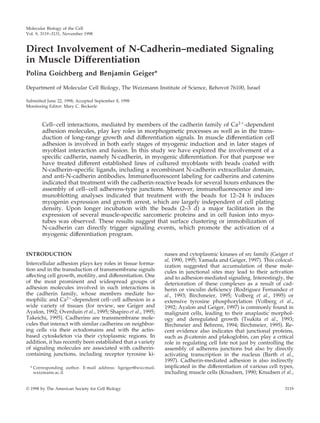
Transmission electron microscopy of C2 myoblasts
after 48 h of incubation with the beads, coated either
with NEC (Figure 1B) or with BSA (Figure 1A) indi-
cated that both types of beads attach firmly to the cell
surface. BSA-coated beads attached to the plasma
membrane via a continuos close contact area and were
engulfed by the cells after several hours of incubation,
Figure 1. Transmission electron micrographs of C2 myoblasts
treated with beads coated with BSA (A) or NEC (B and C). The cells
were incubated with the beads for 48 h in growth medium, fixed,
Figure 1 (cont). and processed for transmission electron micros-
copy. Notice that the contact region with the NEC-coated bead is
characterized by focal, foot-like adhesions (arrows), whereas the
attachment to the BSA beads is tight and uniform. Sarcomeric
filament bundles were found in the cytoplasm of the N-cadherin–
stimulated cells (C) (arrowheads point to Z lines). Bars, 1 m.
N-Cadherin in Muscle Differentiation
Vol. 9, November 1998 3121
4. whereas the NEC-coated beads, were attached to the
cells through electron-dense “foot-like processes,” re-
sembling focal contacts, and were usually not exten-
sively engulfed.
Promotion of Myotube Formation by N-Cadherin–
mediated Stimulation
To test the direct involvement of N-cadherin–medi-
ated signaling in skeletal muscle differentiation, we
have examined the effects of N-cadherin–reactive and
control beads on the rate of myotube formation by the
different myogenic cell lines under conditions that do
not favor differentiation (i.e., high serum concentra-
tion and low plating density). As demonstrated in
Figure 2, the binding of cadherin-reactive beads
(beads coated with NEC or with anti N-cadherin an-
tibodies) to the cells significantly increased the num-
ber of myotubes in these myogenic cultures from ϳ4/
Figure 2. Effect of the N-cadherin stimula-
tion on myotube formation in C2, L8, and L84
myogenic cell lines. C2, L8, and L84 myo-
blasts were treated with NEC and anti-N-
cadherin (anti N-cad) BE beads for the indi-
cated periods, fixed, and stained with
Giemsa. C2 cells were maintained in growth
medium, whereas for L8 and L84 cells the
culture medium was replaced with differen-
tiation medium simultaneously with the ad-
dition of beads. Media were changed every
second day. Notice that the number of myo-
tubes in the field is higher for the cells treated
with cadherin-reactive beads. Arrows point to
some of the multinucleated myotubes. Bar,
100 m.
P. Goichberg and B. Geiger
Molecular Biology of the Cell3122
5. mm2
(BSA-coated beads) to 7 or 8/mm2
(BE- and
NEC-coated beads, respectively). It is noteworthy that
myotubes formed after treatment with cadherin-reac-
tive beads were usually larger than those formed after
treatment with control beads. The number of nuclei
per individual tube was, however, variable, usually
displaying clusters of 5–20 nuclei, and apparently did
not depend on the type or number of bound beads.
Transmission electron microscopy of C2 cells, after
48 h treatment with cadherin-reactive beads, revealed
scattered sarcomeric structures in the cytoplasm (Fig-
ure 1C), which could be found in essentially all sec-
Figure 3. Expression of skeletal myosin in myocytes treated with beads conjugated to NEC, anti-N-cadherin BE antibodies (anti N-cad) or
BSA. C2, L8, and L84 myoblasts were treated with different beads for 48 h, permeabilized, fixed, and immunostained with anti-skeletal
myosin antibodies. C2 cells were maintained in growth medium, whereas for L8 and L84 cells growth medium was replaced with
differentiation medium simultaneously with the addition of beads. The number of cells per field was approximately equal. Notice the increase
in myosin expression in the cultures after treatment with the cadherin-reactive beads. The position of individual beads was detected by
phase-contrast microscopy, and their location is indicated by arrowheads. Bar, 10 m.
N-Cadherin in Muscle Differentiation
Vol. 9, November 1998 3123
6. tions. These sarcomers were similar to those formed
later in the course of differentiation induced by
growth factors deprivation. Such organized filaments
were not detected at that time point in Ͼ30 sections
derived from C2 cells treated with control beads.
Expression and Assembly of Sarcomeric Components
Induced by Cadherin-reactive Beads
Expression of sarcomeric constituents is an essential
element of the myogenic program and serves as a
common phenotypic marker for muscle differentiation
(Andre´s and Walsh, 1996). We have thus examined the
expression and assembly of different sarcomeric con-
stituents in C2, L8, and L84 myoblasts after treatment
with the various beads and found that cadherin stim-
ulation of the three cell lines increases the number of
cells expressing different muscle structural proteins,
including skeletal muscle myosin, myomesin, skeletal
␣-actin, titin, skeletal ␣-actinin, and M-protein. An
example showing a typical increase in the number of
cells expressing skeletal myosin in myoblasts follow-
ing such treatment is presented in Figure 3. The in-
crease in the number of C2 cells expressing several
muscle proteins was quantified after treatment with
the different beads and is summarized in Figure 4. As
shown, the various muscle proteins are first detected
in control cells ϳ48 h after plating, and the numbers
increase progressively upon longer incubation. Addi-
tion of cadherin-reactive beads to these cells nearly
doubles the number of cells expressing the various
muscle proteins at 48 h and maintains a higher num-
ber of positive cells also at 72 h (Figure 4). We further
determined the number of positive cells and overall
levels of skeletal ␣-actinin in cultures treated with the
various beads. As shown in Figure 5, the number of
␣-actinin–positive cells associated with cadherin-reac-
tive beads is significantly higher than in control cul-
tures, and, similarly, the total level of skeletal ␣-acti-
nin is elevated (Figure 5B). It is noteworthy that
because not all the cells in the culture were physically
associated with beads, the actual increase induced by
the cadherin ligands is probably even higher.
Effects of N-Cadherin Stimulation on Cell Cycle
Progression in C2 Myoblasts
Proliferation and differentiation of skeletal myoblasts
are mutually exclusive processes, and cell cycle arrest
is a prerequisite for activation of muscle-specific gene
Figure 4. Effect of cadherin-reactive beads on the expression of various sarcomeric components in C2 cells. C2 myoblasts were plated
overnight on gelatin-coated coverslips at 20% confluence, and beads coated with BSA or with the cadherin ligands were added. After
additional incubation for 48 h after application of beads, cells were permeabilized, fixed, and labeled for the various muscle proteins. The
number of positive cells in 10 randomly chosen microscopic fields (with 40ϫ objective) was determined for each sample at every time point.
P. Goichberg and B. Geiger
Molecular Biology of the Cell3124
7. expression (Andre´s and Walsh, 1996; Olson, 1992). To
examine the effect of cadherin-reactive beads on the
cell cycle, bead-treated C2 cells were pulsed with
BrdU and immunofluorescently labeled with anti-
BrdU antibodies. As shown in Figure 6, treatment of
C2 myoblasts with beads coated with anti-N-cadherin
antibodies suppresses the entry of cells into S phase
compared with control BSA-coated beads. Application
of beads coated with NEC induces a similar inhibition
of cell proliferation (our unpublished results).
Stimulation of Myogenin Expression in Myoblasts
Treated with Cadherin-reactive Beads
Skeletal muscle differentiation is driven and coordi-
nated by the expression of myogenic transcription
factors, such as MyoD, Myf5, myogenin, and Mrf4
(Olson and Klein, 1994; Yun and Wold, 1996). In the
established myoblast lines used here, MyoD and Myf5
are already present before differentiation is induced,
and myogenin transcription is up-regulated upon
myogenic induction (Olson and Klein, 1994). Because
myogenin activity is crucial for the activation of the
entire differentiation program, we have checked
whether its expression is affected by N-cadherin stim-
ulation, using both immunocytochemical (Figures 7A
and 8) and Western blotting (Figures 7B and 9A) ap-
proaches. Both assays revealed a major increase in the
expression of myogenin in cells treated with cadherin-
reactive beads. Densitometric evaluation indicated a
twofold increase in myogenin levels in cadherin bead–
treated C2 cells. In L8 and L84 cells the increase was
three- and fivefold, respectively. This increase is sim-
ilar to the increase in the incidence of myogenin-
positive nuclei.
The expression of myogenin was also elevated in
cultures of sparsely plated C2 cells, which rarely in-
teract with neighboring cells, and do not readily fuse
into myotubes. As shown in Figure 9, when such C2
cells are treated with beads for different periods and
under different growth conditions, a significant in-
crease in the myogenin level (Figure 9A) and in the
number of myogenin-positive cells (Figure 9B) is de-
tected among the cadherin-stimulated myoblasts com-
pared with the BSA controls. Myogenin-positive nu-
clei are first observed 12 h after addition of beads to
the sparsely plated C2 myoblasts cultured in growth
medium.
Enhancement of Cell–Cell Adhesion by Cadherin-
reactive Beads
To further elucidate the mechanism responsible for
N-cadherin–mediated myogenic differentiation, we
have examined the effect of the various beads on cell–
cell interactions. It was recently demonstrated that
Figure 5. Effect of cadherin-reactive beads on the expression of
skeletal ␣-actinin in C2 cells. C2 myoblasts were plated and cultured
overnight on gelatin-coated coverslips (A) or tissue culture dishes
(B) at 50% confluence. Beads were added for 48 h, and then the
samples were either fixed and immunostained or subjected to pro-
tein extraction and SDS-PAGE immunoblot analysis with antibodies
against skeletal ␣-actinin. (A) Calculation of the percentage of ␣-ac-
tinin–positive, bead-bound cells. Each column represents the
mean Ϯ SD of 4 independent experiments; 200 cells were counted in
each case. (B) Representative immunoblot analysis of the total cell
extracts prepared from C2 cells treated as in A. ␣N, anti-N-cadherin
antibodies.
Figure 6. Inhibition of S-phase entry in C2 myoblasts treated with
N-cadherin–reactive beads. C2 myoblasts were plated and cultured
overnight on gelatin-coated coverslips at 50% confluence. Cells were
incubated with the various beads for 24 or 48 h and pulsed for 45
min with BrdU, fixed, and stained with anti-BrdU antibodies. Nu-
clei were visualized by DAPI, and the percentage of BrdU-positive
nuclei was calculated. Each column represents the mean Ϯ SD of 3
independent experiments; 1500 cells were counted in each case.
Analysis of the differences between the cells treated with anti-N-
cadherin and control beads was examined using Student’s t test (one
tailed, paired) pointed to a significant decrease in the percentage of
cells in S phase after 24 h of treatment (p ϭ 0.002) and especially
after 48 h of treatment (p ϭ 0.0017).
N-Cadherin in Muscle Differentiation
Vol. 9, November 1998 3125
8. clustering of N-cadherin by bead-associated cadherin
ligands specifically enhances cell–cell adherens junc-
tion formation (Levenberg et al., 1998a). We have thus
examined the distribution of cadherin and -catenin in
cells treated with NEC- or BE-coated beads. As shown
in Figure 10, treatment with the cadherin ligands ele-
vates -catenin labeling at cell–cell junctions within a
few hours after addition of beads. Phase-contrast mi-
croscopy indicated that this increase in adherens junc-
tion formation is also accompanied by a general as-
sembly of cells into more coherent arrays (our
unpublished results). However, no significant changes
in the overall levels of -catenin or cadherin were
noted (our unpublished results).
DISCUSSION
Cadherins are important morphoregulatory molecules
that are involved in homophilic adhesion of cells. N-
cadherin is a member of the cadherin superfamily,
which plays a crucial role in embryonic morphogene-
sis, including muscle development. Previous studies
Figure 7. Effects of N-cadherin stimulation on myogenin expression in cultured myoblasts. C2, L8, or L84 myoblasts were seeded and
incubated overnight on gelatin-coated coverslips, treated with different beads, as indicated, permeabilized, fixed, and immunostained with
anti-myogenin antibodies (A). The number of cells, as determined by DAPI staining, was approximately equal in all fields, and the percentage
of bead-associated cells was ϳ25%. Notice the overall increase in the number of myogenin-positive nuclei in cells after treatment with the
cadherin-reactive beads. Bar, 20 m. (B) Immunoblot analysis of total cell extracts prepared from the three myogenic lines, treated as in A.
␣N, anti-N-cadherin antibodies.
P. Goichberg and B. Geiger
Molecular Biology of the Cell3126
9. indicated that stable adhesive interactions must be
established between fusion-competent myoblasts, as a
prerequisite for further differentiation, and that these
initial adhesions are calcium dependent (Knudsen et
al., 1990). Specific involvement of N-cadherin was also
suggested on the basis of its high levels in prefusion
myoblasts (MacCalman et al., 1992). Moreover, the
perturbation of N-cadherin–mediated adhesion in
vitro affected the rate (but not the final level) of myo-
blast fusion (Mege et al., 1992). It was suggested by
other studies that N-cadherin may not be essential for
myotube formation, because specific blocking of M-
cadherin (a muscle-specific form) by inhibitory anti-
bodies blocks the fusion of cultured L6 myocytes
(Zeschingk et al., 1995). In addition, myoblasts from
N-cadherin–null mice are able to fuse both in cul-
ture and in vivo (Charlton et al., 1997). In view of the
complexity of the various systems discussed above,
we have attempted, in the present study, to examine
Figure 8. Effect of cadherin-reactive beads on the number of myo-
genin-positive cells in C2, L8, and L84 myoblast cultures. Cells were
plated and treated with different beads as described in Figure 7. The
percentage of myogenin positive nuclei in the entire culture (A) or
in the subpopulation of bead-associated cells (B) was calculated.
Each column represents the mean Ϯ SD of 3 independent experi-
ments, each in duplicate, counting a total of 1000 cells in every case
in A and 250 cells in each case in B. ␣N-cad, anti-N-cadherin
antibodies.
Figure 9. Effect of cadherin-reactive beads on myogenin expres-
sion in sparse cultures of C2 myoblasts. C2 cells were seeded and
cultured overnight at 10% confluence, and then the medium was
replaced with fresh FCS-containing “growth medium” (FS) or with
“differentiation medium” containing horse serum and insulin (HI),
and the various beads were added. (A) Total cell extracts were
prepared at the indicated time points after addition of beads, sub-
jected to SDS-PAGE, transferred to nitrocellulose sheets, and re-
acted with anti-myogenin antibodies. The absorbance of the myo-
genin bands, determined by densitometry, is shown. (B) The
percentage of the myogenin positive nuclei after treatment was
calculated. The cells were maintained in growth medium and
treated with beads for 12 or 18 h, fixed, and immunolabeled for
myogenin. The percentage of positive cells was calculated out of the
entire population or the subpopulation of bead-associated cells.
Every column represents the mean Ϯ SD of 3 independent experi-
ments, each in duplicate. In each individual experiment a total
number of 900 cells or 200 bead-bound cells were counted. B, BSA;
␣, anti-N-cadherin antibodies; N, NEC.
N-Cadherin in Muscle Differentiation
Vol. 9, November 1998 3127
10. the direct involvement of N-cadherin in myogenesis
by its clustering with specific ligands, namely, NEC
or anti-N-cadherin antibodies.
Here we present evidence that local clustering or
immobilization of N-cadherin triggers signaling
events that activate the myogenic program in several
cultured myoblast lines. We found that the cadherin-
reactive beads activate and facilitate the myogenic
program, including myotube formation, expression of
a variety of sarcomeric components, and expression of
the myogenic transcription factor myogenin. Interest-
ingly, myotube formation depends on high cell den-
sity even after cadherin bead stimulation, whereas the
expression of the different muscle proteins, including
Figure 10. Effect of cadherin stimulation on adherens junction formation in myogenic cell lines. C2, L8, and L84 cells were sparsely plated
and further incubated for 10 h on gelatin-coated coverslips. The various beads were added to the cells for 6 h in growth medium. The cells
were then permeabilized, fixed, and immunostained for -catenin. Notice the intensive staining of the cell–cell contact sites and the apparent
increase in staining after treatment with the cadherin-reactive beads. The positions of beads are indicated by arrowheads. Bar, 10 m. anti
N-cad, anti-N-cadherin antibodies.
P. Goichberg and B. Geiger
Molecular Biology of the Cell3128
11. myogenin, was also detected in sparse cultures, ap-
parently independently of cell fusion. This is in line
with the common sequence of myogenic events trig-
gered by growth factor withdrawal, which start with
myogenin expression, induction of growth arrest, ex-
pression of structural sarcomeric components, and,
finally, fusion into myotubes (Andre´s and Walsh,
1996). It is, however, noteworthy that the growth ar-
rest induced by N-cadherin–reactive beads is not
unique to the myogenic differentiation pathway, and
treatment of a variety of mesenchymal cells with the
same types of beads inhibits proliferation and blocks
the cell cycle at the G1 phase. The mechanism under-
lying this growth inhibiting signaling process will be
described in detail elsewhere (Levenberg et al., 1998b).
Our data are consistent with the notion that growth
arrest precedes the expression of the various structural
sarcomeric components by ϳ24 h.
The crucial events in skeletal muscle differentiation
are coordinated by the expression of muscle regula-
tory proteins that act in cooperation with the MEF2
family of transcription factors to activate muscle-spe-
cific gene expression (Yun and Wold, 1996). These
proteins were also shown to interact with and be
regulated by other transcription factors and the cell
cycle regulatory system to coordinately activate the
differentiation program and to inhibit proliferation
(Olson, 1992, 1993; Rao et al., 1994; Skapek et al., 1995,
1996). The fine balance between proliferation and dif-
ferentiation appears to be critical for the induction and
progression of the myogenic program. For instance, in
committed myoblasts MyoD and Myf5 proteins are
expressed, although their activity is apparently inhib-
ited by the presence of growth-promoting factors, and
thus the progression of differentiation depends on
growth factor withdrawal, leading to myogenin ex-
pression and activation of the myogenic cascade (An-
dre´s and Walsh, 1996).
Numerous studies indicate that in the course of
myogenic differentiation inhibition of cell proliferation
and cell death are coordinately regulated, and the
inability to exit the cell cycle leads to apoptotic death
(Walsh and Perlman, 1997; Fimia et al., 1998). Cell
cycle inhibitors, such as p21 or Rb, are able to prevent
this apoptosis most probably by the induction of cell
cycle arrest (Wang and Walsh, 1996; Wang et al., 1997;
Zacksenhaus et al., 1996). As described above, treat-
ment with cadherin-reactive beads inhibits cell cycle
progression in C2 myoblasts. However, no apparent
differences in the number of apoptotic nuclei (defined
by DAPI staining) were observed after application of
the various beads (our unpublished results). Current
reports demonstrate that the decision to exit the cell
cycle and further differentiate or to die is made at the
level of myogenin-induced cell cycle arrest, i.e., at the
stages of myogenesis when cells already express myo-
genin. Because cadherin-reactive beads promote myo-
genin expression, it seems to us unlikely that stimula-
tion of cadherin-mediated adhesion directly affects the
apoptotic process.
Another aspect raised by the present study is the
specificity of the effects on myogenesis to N-cadherin.
As indicated above, additional members of the cad-
herin family are also expressed in muscle tissues, in-
cluding M- and R-cadherins (Zeschingk et al., 1995;
Rosenberg et al., 1997) and cadherin-11 (Kimura et al.,
1995), and perturbation of some of these can affect
myogenesis (Zeschingk et al., 1995). We have no direct
evidence or claim that the effect shown here for N-
cadherin stimulation is unique to this isoform and
cannot be obtained by the clustering or immobiliza-
tion of other cadherins. It is noteworthy that these
three cadherins show considerable overall homology
with N-cadherin along their cytoplasmic domains (82,
50, and 54% identity), which are presumably involved
in the transduction of N-cadherin–mediated signals.
The data presented here are in agreement with the
view that activation of cadherin-mediated signaling
leads to the expression of myogenin, which in turn
inhibits cell cycle progression, triggers the differenti-
ation program, including the expression of sarcomeric
proteins, and promotes myotube formation. The
mechanism underlying this cadherin-induced activa-
tion of myogenin expression is, however, not clear. It
was previously shown that cadherin-reactive beads
specifically activate tyrosine phosphorylation at adhe-
rens junctions and enhance cadherin-mediated cell–
cell adhesion in a variety of mesenchymal cells (Lev-
enberg et al., 1998). This is consistent with the present
results, showing that cadherin-induced stimulation
leads to a specific and generalized enhancement of
myoblast–myoblast adhesion (Figure 10). This, in turn,
could have two distinct effects that are highly relevant
to the progression of myogenic differentiation: 1) the
signals triggered by the beads might be directly in-
volved in the stimulation of myogenin expression; and
2) the apparent increase in cell adhesion, triggered by
the beads, might further promote the myogenin-in-
duced progression of differentiation.
Another possible pathway for cadherin-induced ef-
fects might involve the catenin system. -Catenin,
which is an intrinsic component of adherens junctions,
is also implicated in Wnt and Wg signaling (Willert
and Nusse, 1998) and in malignant transformation
(Korinek et al., 1997; Morin et al., 1997; Redfield et al.,
1997). In view of the capacity of extrajunctional -cate-
nin to translocate to the nucleus and to be involved in
gene transactivation, together with LEF and Tcf tran-
scription factors (Cavallo et al., 1997), it might be in-
teresting to explore the possibility that some of the
genes whose expression is regulated during myogen-
esis are under the control of -catenin, and that
changes in -catenin stability, localization, and/or ac-
N-Cadherin in Muscle Differentiation
Vol. 9, November 1998 3129
12. tivity might affect the myogenic process. Some of these
aspects are currently under study.
ACKNOWLEDGMENTS
We express our gratitude to Dr. D. Yaffe (The Weizmann Institute)
for many illuminating discussions and helpful advice, as well as for
providing the various cell lines used in this study. We are grateful
to Ilana Sabanay for her expert help with the electron microscopic
work and to Drs. B. Winter, H.H. Arnold, W. Obermann, D. Fu¨rst,
and M. Wheelock for providing antibodies used in this study. This
work was supported by the Israel Research Foundation and by the
Rita Markus Foundation. B.G. holds the Erwin Neter Chair in Cell
and Tumor Biology.
REFERENCES
Andre´s, V., and Walsh, K. (1996). Myogenin expression, cell cycle
withdrawal, and phenotypic differentiation are temporally separa-
ble events that precede cell fusion upon myogenesis. J. Cell Biol. 132,
657–666.
Ayalon, O., and Geiger, B. (1997). Cyclic changes in the organization
of cell adhesions and the associated cytoskeleton, induced by stim-
ulation of tyrosine phosphorylation on bovine aortic endothelial
cells. J. Cell Sci. 110, 547–556.
Barth, A.I., Nathke, I.S., and Nelson, W.J. (1997). Cadherins, catenins
and APC protein: interplay between cytoskeletal complexes and
signaling pathways. Curr. Opin. Cell Biol. 9, 683–690.
Birchmeier, W. (1995). E-cadherin as a tumor (invasion) suppressor
gene. Bioessays 17, 97–99.
Birchmeier, W., and Behrens, J. (1994). Cadherins expression in
carcinomas: role in the formation of cell junctions and the preven-
tion of invasiveness. Biochim. Biophys. Acta 1198, 11–26.
Cavallo, R., Rubinstein, D., and Peifer, M. (1997). Armadillo and
dTCF: a marriage made in the nucleus. Curr. Opin. Genet. Dev. 7,
459–466.
Charlton, C.A., Mohler, W.A., Radice, G.L., Hynes, R.O., and Blau,
H.M. (1997). Fusion competence of myoblasts rendered genetically
null for N-cadherin in culture. J. Cell Biol. 138, 331–336.
Doherty, P., and Walsh, F.S. (1994). Signal transduction events un-
derlying neurite outgrowth stimulated by cell adhesion molecules.
Curr. Opin. Neurobiol. 4, 49–95.
Fimia, G.M., Gottifredi, V., Belli, B., Ricciardi, M.R., Tafuri, A.,
Amati, P., and Maione, R. (1998). The activity of differentiation
factors induced apoptosis in polyoma virus large T-expressing myo-
blasts. Mol. Biol. Cell 9, 1449–1463.
Geiger, B., and Ayalon, O. (1992). Cadherins. Annu. Rev. Cell Biol.
8, 307–332.
Geiger, B., Bershadsky, A., and Yehuda-Levenberg, S. (1995). Mo-
lecular interactions in the submembrane plaque of cell-cell and
cell-matrix adhesions. Acta Anat. 154, 46–62.
Geiger, B., Ginsberg, D., Salomon, D., and Volberg, T. (1990). The
molecular basis for the assembly and modulation of adherens-type
junctions. Cell Differ. Dev. 32, 343–53.
George-Weinstein, M., Gehart, J., Blitz, J., Simak, E., and Knudsen,
K.A. (1997). N-cadherin promotes the commitment and differentia-
tion of skeletal muscle precursor cells. Dev. Biol. 185, 14–24.
Holt, C.E., Lemaire, P., and Gurdon, J.B. (1994). Cadherin-mediated
cell interactions are necessary for the activation of MyoD in Xenopus
mesoderm. Proc. Natl. Acad. Sci. USA 91, 10844–10848.
Kimura, Y., Matsunami, H., Inoue, T., Shimamura, K., Uchida, N.,
Ueno, T., Miyazaki, T., and Takeichi, M. (1995). Cadherin-11 ex-
pressed in association with mesenchymal morphogenesis in the
head, somite, and limb bud of early mouse embryos. Biol. Dev. 169,
347–358.
Knudsen, K.A. (1990). Cell adhesion molecules in myogenesis. Curr.
Opin. Cell Biol. 2, 902–906.
Knudsen, K.A., Myers, L., and McElwee, S. (1990). A role for the
Ca2ϩ
-dependent adhesion molecules, N-cadherin, in myoblast in-
teraction during myogenesis. Exp. Cell Res. 188, 175–184.
Korinek, V., Barker, N., Morin, P.J., van Wichen, D., de Weger, R.,
Kinzler, K.W., Vogelstein, B., and Clevers, H. (1997). Constitutive
transcriptional activation by a -catenin-Tcf complexes in APCϪ/Ϫ
colon carcinoma. Science 275, 1784–1787.
Laemmli, U.K. (1970). Cleavage of structural proteins during the
assembly of bacteriophage T4. Nature 227, 680–685.
Levenberg, S., Katz, B.-Z., Yamada, K., and Geiger, B. (1998a).
Long-range and selective autoregulation of cell-cell or cell-matrix
adhesions by cadherin or integrin ligands. J. Cell Sci. 111, 347–357.
Levenberg, S., Yarden, A., Kam, Z., and Geiger, B. (1998b). p27 is
involved in N-cadherin-mediated contact inhibition of cell growth
and S-phase entry. Oncogene (in press).
MacCalman, C., Bardeesy, N., Holland, P.C., and Blaschuk, O.W.
(1992). Noncoordinated developmental regulation of N-cadherin,
N-CAM, integrin, and fibronectin mRNA levels during myoblast
terminal differentiation. Dev. Dyn. 195, 127–132.
Mbalaviele, G., Chen, H., Boyce, B.F., Mundy, G.R., and Yoneda, T.
(1995). The role of cadherin in the generation of multinucleated
osteoclasts from mononucleated precursors in murine marrow.
J. Clin. Invest. 95, 2757–2765.
Mege, R.M., Goudou, D., Diaz, C., Nicolet, M., Garcia, L., Geraud,
G., and Rieger, F. (1992). N-cadherin and N-CAM in myoblast
fusion: compared localization and effect of blockage by peptides and
antibodies. J. Cell Sci. 103, 897–906.
Morin, P.J., Sparks, A., Korinek, V., Barker, N., Clevers, H., Vo-
gelstein, B., and Kinzler, K.W. (1997). Activation of -catenin-Tcf
signaling in colon cancer by mutations in -catenin or APC. Science
275, 11787–11789.
Oberlender, S.A., and Tuan, R.S. (1994). Expression and functional
involvement of N-cadherin in embryonic limb chondrogenesis. De-
velopment 120, 177–187.
Olson, E.N. (1992). Interplay between proliferation and differentia-
tion within the myogenic lineage. Dev. Biol. 154, 261–272.
Olson, E.N. (1993). Signal transduction pathways that regulate skel-
etal muscle gene expression. Mol. Endocrinol. 7, 1369–1378.
Olson, E.N., and Klein, W.H. (1994). bHLH factors in muscle devel-
opment: dead lines and commitments, what to leave in and what to
leave out. Genes Dev. 8, 1–8.
Overduin, M., Harvey, T.S., Bagby, S., Tong, K.I., Yau, P., Takeichi,
M., and Ikura, M. (1995). Solution structure of the epithelial cad-
herin domain responsible for selective cell adhesion. Science 267,
386–389.
Radice, G.L., Rayburn, H., Matsunami, H., Knudsen, K.A., Takeichi,
M., and Hynes, R.O. (1997). Developmental defects in mouse em-
bryos lacking N-cadherin. Dev. Biol. 181, 64–78.
Rao, S.S., Chu, C., and Stave Kontz, D. (1994). Ectopic expression of
cyclin D1 prevents activation of gene transcription by myogenic
basic helix-loop-helix regulators. Mol. Cell. Biol. 14, 5259–5267.
Redfield, A., Nieman, M.T., and Knudsen, K.A. (1997). Cadherins
promote skeletal muscle differentiation in three-dimensional cul-
tures. J. Cell Biol. 138, 1323–1331.
Rodrigues Fernandez, J.L., Geiger, B., Salomon, D., and Ben Ze’ev,
A. (1993). Suppression of vinculin expression by antisense transfec-
P. Goichberg and B. Geiger
Molecular Biology of the Cell3130
13. tion confers changes in cell morphology, motility, and anchorage-
dependent growth of 3T3 cells. J. Cell Biol. 122, 1285–1294.
Rosenberg, P., Esni, F., Sjodin, A., Larue, L., Carlsson, L., Gullberg,
D., Takeichi, M., Kemler, R., and Semb, H. (1997). A potential role of
R-cadherin in striated muscle formation. Dev. Biol. 187, 55–70.
Shapiro, L., Fannon, A.M., Kwong, P.D., Thompson, A., Lehman,
M.S., Grubel, G., Legrand, J.-F., Als-Neilsen, J., Colman, D.R., and
Hendrickson, W.A. (1995). Structural basis of cell-cell adhesion by
cadherins. Nature 374, 327–333.
Skapek, S.X., Rhee, J., Kim, P.S., Novitch, B.G., and Lassar, A.B.
(1996). Cyclin-mediated inhibition of muscle gene expression via a
mechanism that is independent of pRb hyperphosphorylation. Mol.
Cell. Biol. 16, 7043–7053.
Skapek, S.X., Rhee, J., Spicer, D.B., and Lassar, A.B. (1995). Inhibition
of myogenic differentiation in proliferating myoblasts by cyclin
D1-dependent kinase. Science 267, 1022–1024.
Takeichi, M. (1995). Morphogenetic roles of classic cadherins. Curr.
Opin. Cell Biol. 7, 619–627.
Tsukita, S., Itoh, M., Nagafuchi, A., Yonamura, S., and Tsukita, S.
(1993). Submembranous junctional plaque proteins include poten-
tial tumor suppressor molecules. J. Cell Biol. 123, 1049–1053.
Volberg, T., Geiger, B., Kam, Z., Pankov, R., Simcha, I., Sabanay, H.,
Coll, J.L., Adamson, E., and Ben-Ze’ev, A. (1995). Focal adhesion
formation by F9 embryonal carcinoma cells after vinculin gene
disruption. J. Cell Sci. 108, 2253–2260.
Volberg, T., Zick, Y., Dror, R., Sabanay, I., Gilon, C., Levitzki, A.,
and Geiger, B. (1992). The effect of tyrosine-specific protein phos-
phorylation on the assembly of adherens-type junctions. EMBO J.
11, 1733–1742.
Volk, T., and Geiger, B. (1986). A-CAM: a 135-kD receptor of inter-
cellular adherens junctions. II.Antibody-mediated modulation of
junction formation. J. Cell Biol. 103, 1451–1464.
Wang, J., Guo, K., Wills, K.N., Walsh, K. (1997). Rb functions to
inhibit apoptosis during myocyte differentiation. Cancer Res. 57,
351–354.
Wang, J., and Walsh, K. (1996). Resistance to apoptosis conferred by
Cdk inhibitors during myocyte differentiation. Science 273, 359–361.
Walsh, K., and Perlman, H. (1997). Cell cycle exit upon myogenic
differentiation. Curr. Opin. Genet. Dev. 7, 597–602.
Willert, K., and Nusse, R. (1998). -catenin: a key modulator of Wnt
signaling. Curr. Opin. Genet. Dev. 8, 95–102.
Yaffe, D., and Saxel, O. (1976). A myogenic cell line with altered
serum requirements for differentiation. Differentiation 7, 159–166.
Yaffe, D., and Saxel, O. (1977). Serial passaging and differentiation of
myogenic cells isolated from dystrophic mouse muscle. Nature 270,
725–727.
Yamada, K.M., and Geiger, B. (1997). Molecular interactions in cell
adhesion complexes. Curr. Opin. Cell. Biol. 9, 76–85.
Yun, K., and Wold, B. (1996). Skeletal muscle determination and
differentiation: story of a core regulatory network and its context.
Curr. Opin. Cell Biol. 8, 877–889.
Zacksenhaus, E., Jiang, Z., Chung, D., Marth J.D., Phillips, R.A., and
Gallie, B.L. (1996). pRb controls proliferation, differentiation, and
death of skeletal muscle cells and other lineages during embryogen-
esis. Genes Dev. 10, 3051–3064.
Zeschingk, M., Kozian, D., Kuch, C., Schamoll, M., and Starzinski-
Powitz, A. (1995). Involvement of M-cadherin in terminal differen-
tiation of skeletal muscle cells. J. Cell Sci. 108, 2973–2981.
N-Cadherin in Muscle Differentiation
Vol. 9, November 1998 3131