80 ĐỀ THI THỬ TUYỂN SINH TIẾNG ANH VÀO 10 SỞ GD – ĐT THÀNH PHỐ HỒ CHÍ MINH NĂ...
Specific Endocrine Glands.ppt
1. 18-1
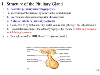
I. Structure of the Pituitary Gland
• 1. Posterior pituitary (neurohypophysis):
• a. extension of the nervous system via the infundibulum
• b. Secretes and stores neuropeptides like oxcytocin
• 2. Anterior pituitary (adenohypophysis)
• a. Connected to hypothalamus by portal vein running through the infundibulum
• b. Hypothalamus controls the adenohypophysis by release of releasing hormone
or inhibiting hormone
• c. Example would be GHRH or GHIH (somatostatin)
4. 18-4
III. Hormones of Posterior Pituitary
A. ADH
• 1. Also called vasopressin.
• 2. Osmoreceptors (specialized neurons of hypothalamus monitor
changes in intercellular osmolality (relative concentrations of
electrolytes and water).
• 3. If the concentration of electrolytes increases or if the
concentration of water decreases, then ADH secretion is stimulated.
• 4. Baroreceptors (specialized neurons found in walls of atria of
heart, large veins, carotid arteries, aortic arch) sense changes in
blood pressure (BP). If BP decreases, then ADH secretion is
stimulated.
• 5. ADH works on the collecting ducts of the kidneys to produce a
more concentrated urine as water is reabsorbed into the body
5. B. Oxcytocin
• 1. produced in the hypothalamus and stored in the posterior or
neurohypophysis
• 2. target tissues include myometrium and muscle cells around milk
ducts of mammary glands
• 3. stimuli bringing about release of oxcytocin stretching of the uterus,
cervix, sexual intercourse, and stimulation of nipple during nursing
• 4. functions:
• a. Onset of delivery
• b. Expulsion of uterine lining during menses
• c. Sperm transport during intercourse
• d. Milk letdown
• 5. special cases
• a. Nursing mothers and uterine shape
• b. Neural input to oxcytocin release
18-5
9. B. Generalized traits
• 1. controlled by releasing and inhibiting hormones from
the hypothalamus
• 2. the hormones are proteins, glycoproteins, or
polypeptides-generally too large to pass through the
membrane
• 3. bind to receptors on the target cell membrane
• 4. short half life and generally quick acting
• 5. many of the hormones are tropic hormones-stimulate
other endocrine glands to produce hormones
• 6. therefore sometimes called the master gland
18-9
10. 18-10
C. Specific Anterior Pituitary Hormones
1.Growth Hormone (GH or somatotropin)
• a. Stimulates uptake of amino acids; protein synthesis
• b. Stimulates breakdown of fats (lipolysis) to be used as an
energy source but stimulates synthesis of glycogen
(glycogenesis)
• c. glucose sparing
• d. Promotes bone, muscle and cartilage growth by bringing
about the release of somatomedins (insulin-like growth factors)
• e. Regulates blood levels of nutrients after a meal and during
periods of fasting
• f. Produced by biotech
• g. Potentials for abuse-acromegaly
12. 18-12
2. TSH (tropic hormone) and Thyroid Hormones
• a. TRH from hypothalamus causes the release of TSH
from anterior pituitary which causes secretion and storage
of hormones T3 and T4 within the thyroid gland
• b. T3 and T4 inhibit TRH and TSH secretion in a negative
feedback pattern.
13. 18-13
3. Adrenocorticotrophic Hormone (ACTH)
• a. CRH (corticotropin releasing hormone) from
hypothalamus causes release of ACTH from anterior
pituitary
• b. ACTH causes cortisol secretion from the adrenal
cortex (a glucocorticoid from the zona fasciculata)
• c. Causes aldosterone secretion from the adrenal cortex
(a mineralocorticoid from the zona glomerulosa)
• d. If adrenal cortex is diseased and malfunctional, ACTH
levels rise and
• e. Binds directly to melanocytes of the skin; causes
increase in production of melanin.
• f. ACTH, lipotropins, B endorphins and MSH are
manufactured from the same precursor
14. 18-14
4. Melanocyte Stimulating Hormone, Endorphins,
and Lipotropins
• a. ACTH, MSH, endorphins and lipotropins all derived from the
same large precursor molecule when stimulated by CRH
• b. MSH causes melanocytes to produce more melanin
• -plays a role in appetite
• -sexual behavior??
• c. Endorphins act as an analgesic; produced during times of stress.
• d. Lipotropins cause adipose cells to catabolize fat, lipolysis
15. 18-15
5. LH, FSH, Prolactin
• a. Gonadotropins: glycoprotein hormones that promote
growth and function of the gonads
• b. LH and FSH
– Both hormones regulate production of gametes and
reproductive hormones
• Testosterone in males
• Estrogen and progesterone in females
• c. GnRH from hypothalamus stimulates LH and FSH secretion
• d. Prolactin: role in milk production
– Regulation of secretion: prolactin-releasing hormone (PRH)
and prolactin-inhibiting hormones (PIH)
17. 18-17
IV. Thyroid Gland
• A. One of largest endocrine
glands;
• 1. Highly vascular.
• 2. appears dark red
• 3. Iodine enters follicular
cells by active transport.
• 4. low levels of iodine
thryroid hypertrophies called
goiter
• B. Composed of follicles:
• 1. follicular cells surrounding
thyroglobulin/thyroid hormone
• 2. Parafollicular cells:
between follicles secrete
calcitonin
• 3. calcitonin reduces blood
calcium levels by stimulating
activity of osteoblasts in bone
18. C. Thyroid hormones
• 1. two major iodine based hormones produced- triodothyronine (T3) and
thyroxine (T4)
• 2. more thyroxine is produced than the T3 which is physiologically more
active
• 3. these hormones are carried in the blood connected to globulin molecules
which protects them from breakdown-gives them a very long half life
• 4. a lot of these hormones are stored in the thyroid gland which also helps to
maintain very stable levels of these two hormones in the blood
• 5. when thyroid function is being measured by blood work, it is the TSH
which is measured
• 6. it is a more direct barometer of thyroid activity
• 7. these two hormones elevate metabolic rate
• 8. Increase rate of glucose, fat, protein metabolism in many tissues thus
increasing body temperature
• 9. Normal growth of many tissues dependent on presence of thyroid hormones
• 10. hypothyroidism vs. hyperthyroidism
18-18
20. 18-20
D. Regulation of Calcitonin Secretion
• 1. Produced by parafollicular
cells
• 2. Secretion triggered by high
Ca2+ concentration in blood;
acts to decrease Ca2+
concentration
• 3. Primary target tissue: bone.
Decreases osteoclast activity,
lengthens life span of
osteoblasts.
• 4. calcitonin concentrations
decrease with age more in
females than males
• 5. postmenopausal women
and calcitonin nasal sprays
21. 18-21
V. Parathyroid Glands
• 1. Embedded in thyroid
• 2. Historical surgery story
• 3. Two glands on each side
• 4. Secrete PTH: target tissues are bone,
kidneys and intestines.
– Increases blood calcium and phosphate
levels
– Stimulates osteoclasts
– Promotes calcium reabsorption by
kidneys and PO4 excretion because rise
of both causes bone deposition in wrong
amounts
– Increases synthesis of vitamin D which,
in turn, increases absorption of Ca and
PO4 by intestines. Net loss of PO4 under
influence of PTH.
• 5. Regulation depends on calcium levels.
• 6. inactive parathormone-hypocalcemia-
open voltage-gated sodium ion channels
23. 18-23
VI. Adrenal Glands A. Anatomy
• 1. Near superior poles of
kidneys; retroperitoneal
• 2. Inner medulla; outer
cortex
• 3. Medulla: formed from
neural crest; sympathetic.
Secretes epinephrine and
norepinephrine
• 4. Cortex: three zones
from superficial to deep
– Zona glomerulosa
– Zona fasciculata
– Zona reticularis
24. 18-24
B. Hormones of the Adrenal Medulla
• 1. Secretory products are neuropeptides: epinephrine and
norepinephrine
• 2. Combine with adrenergic membrane-bound receptors
• 3. Secretion of hormones prepares body for physical activity
• 4. Effects are short-lived; hormones rapidly metabolized; half-life is
minutes
• 5. Epinephrine
– Increases blood levels of glucose via glycogenolysis
– Increases fat breakdown in adipose tissue
– Causes dilation of blood vessels in skeletal muscles and cardiac
muscles.
• 6. Epinephrine and norepinephrine increase heart rate and force of
contraction; cause blood vessels to constrict in skin, kidneys,
gastrointestinal tract, and other viscera
25. 18-25
C. Hormones of Adrenal Cortex
• 1. Mineralocorticoids: Zona glomerulosa
– Aldosterone produced in greatest
amounts. Increases rate of sodium
reabsorption by kidneys increasing
sodium blood levels
– Stimulus for production is low blood
pressure
– Increases water reabsorption and
therefore blood volume
• 2. Glucocorticoids: Zona fasciculata
– Cortisol is major hormone. Increases
fat and protein breakdown, increases
glucose synthesis(gluconeogenesis),
promotes the increased use of fats and
proteins by muscles, decreases
inflammatory response
• 3. Androgens: Zona reticularis
– Weak androgens secreted then
converted to testosterone by peripheral
tissues. Stimulate pubic and axillary
hair growth and sexual drive in females
27. 18-27
VII. Pancreas
• A. Located along small intestine and stomach;
retroperitoneal
• B. Exocrine gland
– Produces pancreatic digestive juices
• C. Endocrine gland
– Consists of pancreatic islets
– Composed of
• Alpha cells; secrete glucagon
• Beta cells; secrete insulin
• Delta cells; secrete somatostatin
29. E. Effects of insulin
• 1. direct effect is to reduce blood glucose
• 2. acts on a number of different target tissues
• 3. muscle-causes uptake of glucose to promote glycogenesis
• 4. adipose tissue-lipogenesis
• 5. liver-glycogenesis and increased use of glucose to drive ATP
production
• 6. also acts to increase uptake of amino acids and stimulates protein
synthesis
• 7. insulin is a protein hormone-therefore binds to a membrane receptor
• 8. increases the number of glucose carrier molecules
• 9. low levels of insulin lead to a very rapid increase of blood glucose
18-29
30. G. Effects of glucagon
• 1. Affects of glucagon are antagonistic to insulin
• 2. acts to increase blood glucose levels
• 3. secreted from pancreas alpha cells and enters hepatic portal
circulation
• 4. travels directly to liver where it stimulates the liver to break down
glycogen stores-glycogenolysis
• 5. also stimulates glucose sparing and encourages the use of alternate
fuels for energy source
• 6. important to maintain adequate glucose levels as the nervous
system is an obligate consumer of glucose
• 7. promotes gluconeogenesis in liver from amino acids
• 8. glucagon does not affect many other target tissues as the liver
breaks down glucagon before it can pass on to other organs
18-30
31. H. Diabetes mellitus
• 1. normal range fasting blood sugar is 70-100 mg./deciliter
• 2. low insulin activity raises this level drastically-diabetes usually
confirmed with reading of 200mg/deciliter
• 3. two types of diabetes
• a. Type I
• -individuals don’t produce sufficient insulin
• -usually children
• - approximately 10% of diabetic population
• -insulin dependent
• b. Type II
• -individuals don’t respond to insulin normally produced
• -faulty receptors or enzymes produced after insulin combines
• -majority
• -adult onset
• -meds taken to increase insulin production
• -exercise 18-31
32. 4. Symptoms of diabetes type I
• a. Frequent urination –due to high osmotic pressures of glucose in
urine
• b. Unusual thirst –replacing fluid loss in the high urine production
• c. Extreme hunger-satiety center in hypothalamus insulin dependent
• d. Unusual weight loss- low fuel source availability
• e. Extreme fatigue and irritability
18-32
40. IX. Hormones of the Pineal Gland
A. Main hormone is melatonin
B. Melatonin inhibits GnRH and
therefore inhibits reproductive
function
C. Also may help sleep cycles by
stimulating sleep
D. Some animals pineal regulates
breeding cycles
1. light inhibits pineal secretion of
melatonin
2. dark stimulates pineal’s secretion
of melatonin
3. animals that breed in spring
experience increased day length and
decreased melatonin secretion-
reproductive structures hypertrophy
4. repurcussion-
-use as a sleep aid
-earlier onset of puberty
18-40
41. 18-41
X. Thymus gland
• A. Location-superior
mediastinum
• B. Produces hormone
thymosin
• C. Activity-
maturation and
development of the
immune system
• D. Culinary-sweet
breads