
Recommended
More Related Content
What's hot
What's hot (20)
Similar to LFTx. clinical pharmacy, PharmD 4th year
Similar to LFTx. clinical pharmacy, PharmD 4th year (20)
Recently uploaded
Recently uploaded (20)
LFTx. clinical pharmacy, PharmD 4th year
- 1. CLINICAL PHARMACY • Liver Function Tests LFT’s PharmD Fourth year • By- 2. Vaibhavi Bhatwadekar 15. Khushi Ladda • Government college of pharmacy, Aurangabad
- 2. Introduction - anatomy and physiology Functions of liver Goals of test LFT’s Drug induced liver diseases Reference
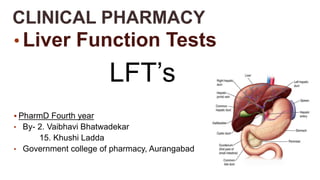
- 3. INTRODUCTION- Anatomy and physiology- Liver is the largest organ of the body, dark-reddish in colour. The liver is located in the upper right hand portion of the abdominal cavity, Beneath the diaphragm and on top of the stomach, right kidney and intestine. Weighing about 1400-1600 grams in males and 1200-1400 gram in females. Liver is divided into two lobe that is right lobe and left lobe. Each lobe contains a number of lobules. Each lobule is made of the hepatic cells the hepatic cells are the major functional cells in the liver. Hepatic cells are separated by sinusoids which form part of reticular endothelial system. Bile canaliculi present between hepatic cells empty into bile ducts.
- 4. FUNCTIONS OF THE LIVER- 1. Metabolic functions : Liver actively participates in carbohydrate, lipid, protein, mineral and vitamin metabolisms. 2. Excretory functions : Bile pigments, bile salts and cholesterol are excreted in the bile into intestine. 3. Protective functions and detoxification : Kupffer cells of liver perform phagocytosis to eliminate foreign compounds. Ammonia is detoxified to urea. Liver is responsible for the metabolism of xenobiotics (detoxification). 4. Hematological functions : Liver participates in the formation of blood (particularly in the embryo), synthesis of plasma proteins (including blood clotting factors) and destruction of erythrocytes. 5. Storage functions : Glycogen, vitamins A, D and B12 and trace element iron are stored
- 5. GOALS OF THE LFT’S- 1. To detect the presence of liver disease. 2. To distinguish among different types of liver diseases. 3. To detect the level of the disease whether the mild, moderate or severe 4. To follow the response of the treatment. 5. LFT are employed for accurate diagnosis, to assess the severity of damage, to judge prognosis and to evaluate the therapy.
- 7. LFT’S- The liver function tests are the biochemical investigations to assess the capacity of the liver to carry out any of the function its performs. The major liver function tests may be classified as follows : 1. Tests based on excretory function— Measurement of bile pigments, bile salts, bromosulphthalein. 2. Tests based on serum enzymes derived from liver—Determination of transaminases, alkaline phosphatase, 5’-nucleotidase, J-glutamyltranspeptidase. 3. Tests based on metabolic capacity— Galactose tolerance, antipyrine clearance. 4. Tests based on synthetic functions— Prothrombin time, serum albumin. 5. Tests based on detoxification—Hippuric acid synthesis.
- 8. A byproduct of RBC breakdown. Inside the liver cells , the bilirubin is conjugated with glucouronic acid to make it water soluble. High bilirubin levels are observed in gallstones, acute and chgronic hepatitis. Bilirubin pigment can be detected in serum, faeces and urine. i) Serum bilirubin estimation is based on van den Bergh diazo reaction by spectrophotometric method. Diazo reagent consists of diazotised sulfanilic acid. Water- soluble conjugated bilirubin gives direct van den Bergh reaction with diazo reagent within one minute, whereas alcohol-soluble unconjugated bilirubin is determined by indirect van den Bergh reaction. Addition of alcohol to the reaction mixture gives positive test for both conjugated and unconjugated bilirubin pigment. The unconjugated bilirubin level is then estimated by subtracting direct bilirubin value from this total value. The serum of normal adults contains less than 1 mg/ dl of total bilirubin, out of which less than 0.25 mg/dl is conjugated bilirubin. Bilirubin level rises in diseases of hepatocytes, obstruction to biliary excretion into the duodenum, in haemolysis, and defects of hepatic uptake and conjugation of bilirubin pigment such as in Gilbert’s disease.
- 9. ii) In faeces, excretion of bilirubin is assessed by inspection of stools. Clay-coloured stool due to absence of faecal excretion of the pigment indicates obstructive jaundice. iii) In urine, conjugated bilirubin can be detected by commercially available ‘dipsticks’, Fouchet’s test, foam test or ictotest tablet method. Bilirubinuria does not occur in normal subjects nor is unconjugated bilirubin excreted in the urine. Bilirubinuria occurs only when there is raised level of conjugated bilirubin (filterable). Its excretion depends upon the level of conjugated bilirubin in plasma that is not proteinbound and is therefore available for glomerular filtration. Bilirubinuria appears in patients of hepatitis before the patient becomes jaundiced. Serum bilirubin normal values:- Total 0.3-1.3 mg/dl Direct (conjugated) 0.1-0.4 mg/dl Indirect (unconjugated) 0.2-0.9 mg/dl
- 10. Urobilinogen is a clourless byproduct of bilirubin reduction. Urobilinogen is normally excreted in the urine. Its semiquantitative estimation in the urine can be done by preparing dilutions with Ehrlich’s aldehyde reagent or by ‘dipstick’ method. An increase in urobilinogen in the urine is found in hepatocellular dysfunctions such as in alcoholic liver disease, cirrhosis and malignancy of the liver. It is also raised in haemolytic disease and in pyrexia. In cholestatic jaundice due to complete biliary obstruction, urobilinogen disappears from the urine
- 11. Bromsulphalein (BSP) is a dye which is removed from circulation by the same mechanisms of binding, conjugation and excretion as bilirubin. BSP is injected intravenously and a sample of venous blood 45 minutes later is tested for percentage of injected dye remaining in the blood. The test is rarely performed nowadays because of the availability of enzyme estimations which are better indicators of hepatic dysfunction. Presently, the only value of BSP excretion test is in the diagnosis of Dubin-Johnson’s syndrome
- 12. Serum alkaline phosphatase is produced by many tissues, especially bone, liver, intestine and placenta and is excreted in the bile. Most of the normal serum alkaline phosphatase (range 33-96 U/L) is derived from bone. Elevation in activity of the enzyme can thus be found in diseases of bone, liver and in pregnancy. In the absence of bone disease and pregnancy, an elevated serum alkaline phosphatase levels generally reflect hepatobiliary disease. The greatest elevation (3 to 10 times normal) occurs in biliary tract obstruction. Slight to moderate increase is seen in parenchymal liver diseases such as in hepatitis and cirrhosis and in metastatic liver disease. It is possible to distinguish serum hepatic alkaline phosphatase from bony alkaline phosphatase by fractionation into isoenzymes but this is not routinely done.
- 13. The primary source of the enzyme, γ-GT, in serum is the liver. Its serum level parallels serum alkaline phosphatase and is used to confirm that the elevated serum alkaline phosphatase is of hepatobiliary origin. Besides its elevation in cholestasis and hepatocellular disease, the levels are high in patients with alcohol abuse even without liver disease. Normal:- 9-58 IU/L (15-85 U/L)
- 14. i) Serum aspartate transaminase or AST (formerly glutamic oxaloacetic transaminase or SGOT): AST or SGOT is a mitochondrial enzyme released from heart, liver, skeletal muscle and kidney. Its normal serum level is 0.20-0.65 μkat/L (12-38 U/L). ii) Serum alanine transaminase or ALT (formerly glutamic pyruvic transaminase or SGPT): ALT or SGPT is a cytosolic enzyme primarily present in the liver. Its normal serum level is 0.12-0.70 μkat/L (7-41 U/L). Serum levels of SGOT and SGPT are increased on damage to the tissues producing them. Thus serum estimation of SGPT (ALT) which is fairly specific for liver tissue is of greater value in liver cell injury, whereas SGOT (AST) level may rise in acute necrosis or ischaemia of other organs such as the myocardium, besides liver cell injury. Transaminase estimations are useful in the early diagnosis of viral hepatitis. Very high levels are seen in extensive acute hepatic necrosis such as in severe viral hepatitis and acute cholestasis. Alcoholic liver disease and cirrhosis are associated with mild to moderate elevation of transaminases
- 15. Albumin is solely synthesized by the liver. It has a half-life of about 20-25 days, therefore, it is a good marker to assess chronic (and not acute) liver damage. Low serum albumin is commonly observed in patients with severe liver damage. It must, however, be noted that the serum albumin concentration is also decreased due to other factors such as malnutrition. Normal serum albumin level :- 3.5-5.5 g/dl
- 16. Alpha and beta globulins are mainly synthesized by the liver. They constitute immunoglobulins . High serum gamma globulin are observed in chronic hepatitis and cirrhosis. Normal level;- 2.0-3.5 g/dl Normal Albumin to Globulin ratio(A/G) :- 1.5-3:1
- 17. The liver synthesizes all the factors concerned with blood clotting. A decrease in the concentration of plasma clotting factors is found in the impairment of liver function. This can be assessed in the laboratory by measuring prothrombin time which is prolonged in patients with liver damage, compared to normal. The half-lives of clotting factors are relatively short (5-72 hrs.), therefore, changes in prothrombin time occur quickly. Hence, this test is useful to assess acute as well as chronic liver damages; besides its help in the prognosis. Vitamin K is required for the synthesis of blood clotting factors II, VII, IX and X. Therefore, vitamin K deficiency can also cause prolonged prothrombin time which must be ruled out, before drawing conclusions on the liver functions. This is done by measuring prothrombin time before and after administration of vitamin K.
- 18. The choice of biochemical tests to measure liver functions mostly depends on the purpose of the investigation. The clinical history of the subject is often a guiding factor in this regard. A single test in isolation may have a little diagnostic value. Frequently, a combination of laboratory investigations are employed in LFT. These include serum bilirubin (conjugated and unconjugated), alanine transaminase, aspartate transaminase, alkaline phosphatase, J-glutamyl transpeptidase and proteins (albumin, globulins).
- 20. 1. Principles of Anatomy and Physiology by Gerard J.Tortora and Bryan Derrickson, 15th Edition, Wiley publication , pg no. 2. Textbook of Pathology by Harsh Mohan , 6th Edition, JAYPEE Publicatuion, pg no. 593- 595. 3. Biochemistry by U. Satyanarayana and U. Chakrapani, 4th Edition, Ellsevier publication, Pg no. 453-459