
Recommended
More Related Content
Similar to 8th thoracic Bone.pptx
Similar to 8th thoracic Bone.pptx (20)
More from ChangezKhan33
More from ChangezKhan33 (20)
Recently uploaded
Recently uploaded (20)
8th thoracic Bone.pptx
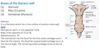
- 1. Bones of the thoracic wall (1) Sternum (2) Ribs (12 pairs) (3) Vertebrae (thoracic) Sternum It is a flat bone which lies in the midline of anterior chest wall. Parts Manubrium sterni, It Lies opposite T3,T4. Body, It lies opposite to T5 to T8. Xiphoid process, T9. The manubrium has the facet for the first costal cartilages and a facet for the clavicle. The facet for second costal cartilage occur at the sternal angle. The remaining costal cartilages fuses at sternal body.
- 2. On the basis of structure the ribs are of two types (1) Typical rib It is a long, twisted, flat bone having a rounded, smooth superior border and a sharp, thin inferior border. The inferior border overhangs (extend outwards) and forms the costal groove, which accommodates the intercostal vessels and nerve. The anterior end of each typical rib is attached to the corresponding costal cartilage. It has a head, neck, tubercle, shaft, and angle. The head has two facets for articulation with the vertebral body. The neck is a constricted portion situated between the head and the tubercle. The tubercle is a prominence on the outer surface of the rib at the junction of the neck with the shaft. The shaft is thin and flattened and twisted on its long axis. Its inferior border has the costal groove. The angle is where the shaft of the rib bends sharply forward.
- 3. (2) Atypical ribs Some ribs varies in structure than the typical ribs which are called atypical ribs. The 1st rib is an example of atypical rib. It is important clinically because of its close relationship to the lower nerves of the brachial plexus and the main vessels to the arm, namely, the subclavian artery and vein. This rib is small and flattened from above downward. The scalenus (of a triangle or having sides unequal in length) anterior muscle is attached to its upper surface and inner border. Anterior to the scalenus anterior, the subclavian vein crosses the rib, posterior to the muscle attachment, the subclavian artery and the lower trunk of the brachial plexus cross the rib and lie in contact with the bone.
- 4. Costal Cartilages Costal cartilages are bars (Rod) of cartilage connecting the upper seven ribs to the lateral edge of the sternum and the 8th, 9th, and 10th ribs to the cartilage immediately above. The cartilages of the 11th and 12th ribs end in the abdominal musculature. The costal cartilages contribute significantly to the elasticity and mobility of the thoracic walls. In old age, the costal cartilages tend to lose some of their flexibility as the result of superficial calcification
- 5. Intercostal Spaces The space between two adjacent ribs is called intercostal space. There are 9 anterior and 11 posterior intercostal spaces. Each space contains: 1- Intercostal muscles: External, Internal and innermost. 2- An Intercostal nerve. 3- Intercostal vessels: a. Intercostal arteries Anterior & Posterior. b. Intercostal veins Anterior & Posterior. The intercostal nerves, veins and arteries runs near the lower border of the rib in the following manner from above below, Vein, Artery and Nerve (VAN). The innermost intercostal muscles are covered by endothoracic fascia, which is lined internally by the parietal pleura.
- 6. Thoracic vertebrae It makes the axial part of the poster chest wall. They are 12 in number. The thoracic vertebrae may be typical and atypical. The typical thoracic vertebrae has structure of a common vertebrae while the structure of atypical vertebrae is somewhat changed from the typical.
- 7. 1st, 10th,11th and 12th T1 Has a complete facet. One very small inferior demifacet(actually half of a facet). Spine nearly horizontal has costal facet in transverse process for the tubercle of first rib. It has a small body, looks like a cervical vertebra. T10 One complete facet with the upper border Small costal facet on transverse process. T11 One complete circular facet away from upper border. No costal facet T12 Broad body & short, oblong spine. One complete facet midway between upper & lower borders. No costal facet Atypical (Non typical ) thoracic vertebrae.
- 8. Joints of the thoracic wall Joints of the Sternum Manubriosternal joint Between the manubrium and the body of the sternum. Xiphisternal joint Between xiphoid process and the body of the sternum. Joints of the Ribs and Costal Cartilages These joints are cartilaginous joints. No movement is possible.
- 9. Joints of the Costal Cartilages with the Sternum The 1st costal cartilages articulate with the manubrium through cartilaginous joints. The 2nd to 7th costal cartilages articulate with the lateral border of the sternum by synovial joints. In addition, the 6th, 7th, 8th, 9th, and 10th costal cartilages articulate with one another along their borders by small synovial joints. The cartilages of the 11th and 12th ribs are embedded in the abdominal musculature.
- 10. Joints of the Heads of the Ribs The 1st rib and the three lowest ribs have a single synovial joint with their corresponding vertebral body. For the 2nd to 9th ribs, the head articulates by means of a synovial joint with the corresponding vertebral body and that of the vertebra above it. Joints of the Tubercles of the Ribs The tubercle of a rib articulates by means of a synovial joint with the transverse process of the corresponding vertebra. This joint is absent on the 11th and 12th ribs.
- 11. Intercostal Arteries Anterior Each intercostal artery gives off branches to the muscles, skin and parietal pleura. In the region of the breast in the female, the branches to the superficial structures are particularly large.
- 12. Posterior intercostal artery Each intercostal space contains a large single posterior intercostal artery and two small anterior intercostal arteries.
- 13. Intercostal Veins 2 in each space. (1) Posterior intercostal veins It drain backward into the azygos or hemiazygos veins . And (2) Anterior intercostal veins It drain forward into the internal thoracic and the musculophrenic veins.
- 14. Azygos vein The azygos vein in right side transports deoxygenated blood from the posterior walls of the thorax and abdomen into the superior vena cava vein. Hemiazygos vein The hemiazygos vein (vena azygos minor inferior) is a vein running superiorly in the lower thoracic region, just to the left side of the vertebral column
- 16. Intercostal nerves They are the anterior branches of spinal thoracic nerves fromT1 to T11. Each nerve runs in the Intercostal space inferior to the Intercostal vessels. T3 toT6 are called Typical T12 is called Subcostal(below) The remaining nerves are called atypical. These nerves arises from the vertebrae and runs between the inner and innermost muscles of the intercostal space.
- 18. Branches of intercostal nerves Rami communicantes It connect the intercostal nerve to a ganglion(sac like swelling) of the sympathetic(expressing) trunk. Gray ramus It joins the nerve medial at the point at which the white ramus leaves it. Collateral branch It runs forward inferiorly to the main nerve on the upper border of the rib below. lateral cutaneous branch It reaches the skin on the side of the chest. It divides into an anterior and a posterior branch. Anterior cutaneous branch It is the terminal portion of the main trunk. It reaches the skin near the midline. Muscular branches These runs to the intercostal muscles. Pleural sensory branches These branches goes to the parietal pleura. Peritoneal sensory branches (7th to 11th intercostal nerves only) they run to the parietal peritoneum.
- 19. Suprapleural Membrane (Sibson fascia) On either side of the structures passing through the thoracic outlet, a dense fascial layer called the suprapleural membrane is present. It is a fibrous sheet and attached laterally to the medial border of the 1st rib and costal cartilage. It is attached at its apex to the tip of the transverse process of the seventh cervical vertebra and medially to the fascia investing the structures passing from the thorax into the neck. It protects the underlying cervical pleura and resists the changes in intrathoracic pressure occurring during respiratory movements.