
Recommended
More Related Content
Similar to intussusception.pptx
Similar to intussusception.pptx (20)
Recently uploaded
Recently uploaded (20)
intussusception.pptx
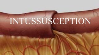
- 2. INTRODUCTION • Intussusception is a condition where the bowel “invaginates” or “telescopes” into itself. This thickens the overall size of the bowel and narrows the lumen at the folded area, leading to a palpable mass in the abdomen and obstruction to the passage of faeces through the bowel.
- 3. EPIDEMIOLOGY • It typically occurs in infants 6 months to 2 years • Affects approximately 1 in every 500 children under the age of 5 years. • more common in boys.
- 5. RISK FACTORS 1. Age: 2. Previous episodes: 3. Gastrointestinal infections:. Rotavirus, a common cause of gastroenteritis in infants and young. 4. Structural abnormalities 5. Meckel's diverticulum 6. Celiac disease 7. Intestinal polyps 8. Henoch-Schönlein purpura: 9. Family history
- 6. PATHOPHYSIOLOGY • The exact cause of intussusception is not fully understood, but it is believed to be multifactorial. One of the primary contributing factors is thought to be an abnormality in the peristaltic movements of the intestine, which could result from changes in the lymphatic tissue within the intestine, a viral infection or certain medical conditions such as polyps or tumors in the intestine. • As the invagination progresses, it leads to obstruction of the intestinal lumen, impairing the passage of food and waste material causing symptoms such as severe abdominal pain, vomiting, and bloody stools. • Furthermore, the invagination can compromise blood flow to the affected segment of the intestine, leading to ischemia and tissue necrosis. If left untreated, this can result in perforation of the intestine and peritonitis (inflammation of the abdominal cavity). The compromised blood flow can also lead to secondary complications such as infection and sepsis
- 11. CLINICAL PRESENTATION • Severe, colicky abdominal pain • Pale, lethargic and unwell child • “Redcurrant jelly stool” • Right upper quadrant mass on palpation. This is described as “sausage- shaped” • Vomiting • Intestinal obstruction
- 13. DIAGNOSIS Diagnosing intussusception involves a combination of • clinical evaluation • imaging studies: ultrasound and sometimes CT scan or X-ray • diagnostic procedures: barium enema or air enema
- 14. Clinical Evaluation - Medical History: inquire about the patient's symptoms, including abdominal pain, vomiting, bloody stools, and changes in bowel movements. - Physical Examination: perform a thorough physical examination, paying particular attention to the abdomen. a mass or signs of tenderness or distension may be noticed
- 15. Imaging Studies - Ultrasound: Ultrasonography is the primary imaging modality used to diagnose intussusception in children. It is non-invasive and does not involve exposure to ionizing radiation. Ultrasound can visualize the telescoped bowel segments and identify characteristic features such as the "target sign" or "doughnut sign." - X-ray: Abdominal X-rays may be performed to evaluate for signs of bowel obstruction or air-fluid levels. However, X-rays alone are not sufficient for confirming the diagnosis of intussusception. - Computed Tomography (CT) Scan: In certain cases where ultrasound results are inconclusive or unavailable, a CT scan may be used to aid in diagnosis
- 17. Diagnostic Procedures - Barium Enema: This procedure involves introducing a contrast material (barium) into the rectum through a small tube. X-rays are then taken to visualize the movement of the contrast material through the intestines. Barium enema can both diagnose and treat intussusception in some cases. - Air Enema: Similar to a barium enema, air enema involves introducing air instead of contrast material into the rectum. The air helps to push the telescoped bowel segments back into their normal position.
- 19. TREATMENT • Therapeutic enemas can be used to try to reduce the intussusception. Contrast, water or air are pumped into the colon to force the folded bowel out of the bowel and into the normal position. • Surgical reduction may be necessary if enemas do not work. • If the bowel becomes gangrenous (due to a disruption of the blood supply) or the bowel is perforated, then surgical resection is required.
- 20. DIFFERENTIAL DIAGNOSIS 1. Gastroenteritis 2. Appendicitis 3. Meckel's diverticulum 4. Hirschsprung's disease 5. Volvulus 6. Intestinal obstruction 7. Meconium ileus 8. Henoch-Schönlein purpura
- 21. COMPLICATIONS • Obstruction • Gangrenous bowel • Perforation • Death
- 22. REFERENCES • 1. Mayo Clinic - www.mayoclinic.org • 2. MedlinePlus - medlineplus.gov