![able to distinguish spurious results (those
due to improper specimen collection methods) from true clinical
variation due to a pathological
condition.
SECTION 1: CBC Testing
For each example, note which parameter(s) are pertinent to your
explanation. An asterisk next
to a value indicates that it has been flagged by the instrument.
Delta flags are indicated by [%D]
next to the value.
CBC #1: 57-year old Caucasian male, presented to the ED with
chest pain, diagnosed with myocardial
infarction and admitted to the ICU 2 days earlier.
CURRENT PREVIOUS
(8 hours)
REFERENCE
RANGE
UNIT
WBC 4.8 5.4 4.5 – 11.5 103/uL
RBC 2.87 4.65 4.60 – 6.00 (m) 106/uL
HGB 10.1 [%D] 14.2 14.0 – 18.0 (m) g/dL
HCT 29.1 [%D] 42.4 40.0 – 54.0 (m) %
MCV 101 [%D] 91 80 – 98 fL
MCH 35.1 30.5 26 – 32 pg
MCHC 34.7 33.4 32 – 36 g/dL
RDW 12.5 13.3 11.5 – 14.5 %
PLT 224 292 150 – 450 103/uL](data:image/gif;base64,R0lGODlhAQABAIAAAAAAAP///yH5BAEAAAAALAAAAAABAAEAAAIBRAA7)
Recommended
Recommended
More Related Content
Similar to MLSC-382A INSTRUCTOR BETH RAWSON 1 Resu
Similar to MLSC-382A INSTRUCTOR BETH RAWSON 1 Resu (20)
More from IlonaThornburg83
More from IlonaThornburg83 (20)
Recently uploaded
Recently uploaded (20)
MLSC-382A INSTRUCTOR BETH RAWSON 1 Resu
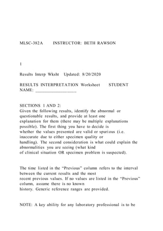
- 1. MLSC-382A INSTRUCTOR: BETH RAWSON 1 Results Interp Wksht Updated: 8/20/2020 RESULTS INTERPRETATION Worksheet STUDENT NAME: __________________ SECTIONS 1 AND 2: Given the following results, identify the abnormal or questionable results, and provide at least one explanation for them (there may be multiple explanations possible). The first thing you have to decide is whether the values presented are valid or spurious (i.e. inaccurate due to either specimen quality or handling). The second consideration is what could explain the abnormalities you are seeing (what kind of clinical situation OR specimen problem is suspected). The time listed in the “Previous” column refers to the interval between the current results and the most recent previous values. If no values are listed in the “Previous” column, assume there is no known history. Generic reference ranges are provided. NOTE: A key ability for any laboratory professional is to be
- 2. able to distinguish spurious results (those due to improper specimen collection methods) from true clinical variation due to a pathological condition. SECTION 1: CBC Testing For each example, note which parameter(s) are pertinent to your explanation. An asterisk next to a value indicates that it has been flagged by the instrument. Delta flags are indicated by [%D] next to the value. CBC #1: 57-year old Caucasian male, presented to the ED with chest pain, diagnosed with myocardial infarction and admitted to the ICU 2 days earlier. CURRENT PREVIOUS (8 hours) REFERENCE RANGE UNIT WBC 4.8 5.4 4.5 – 11.5 103/uL RBC 2.87 4.65 4.60 – 6.00 (m) 106/uL HGB 10.1 [%D] 14.2 14.0 – 18.0 (m) g/dL HCT 29.1 [%D] 42.4 40.0 – 54.0 (m) % MCV 101 [%D] 91 80 – 98 fL MCH 35.1 30.5 26 – 32 pg MCHC 34.7 33.4 32 – 36 g/dL RDW 12.5 13.3 11.5 – 14.5 % PLT 224 292 150 – 450 103/uL
- 3. MPV 7.9 8.1 6.8 – 10.2 fL MLSC-382A INSTRUCTOR: BETH RAWSON 2 Results Interp Wksht Updated: 8/20/2020 CBC #2: 27-year old Greek female, presented for prenatal evaluation (20 weeks pregnant). CURRENT PREVIOUS REFERENCE RANGE UNIT WBC 6.6 4.5 – 11.5 103/uL RBC 5.13 4.00 – 5.40 (f) 106/uL HGB 10.7 12.0 – 15.0 (f) g/dL HCT 34.9 35.0 – 49.0 (f) % MCV 68 80 – 98 fL MCH 20.9 26 – 32 pg MCHC 30.7 32 – 36 g/dL RDW 27.3 11.5 – 14.5 % PLT 309 150 – 450 103/uL MPV 7.4 6.8 – 10.2 fL
- 4. CBC #3: 19-year old African American male, presented to the outpatient draw station subsequent to his yearly physical examination; he did not have any complaints. CURRENT PREVIOUS REFERENCE RANGE UNIT WBC 5.6 4.5 – 11.5 103/uL RBC 3.58 4.60 – 6.00 (m) 106/uL HGB 11.0 14.0 – 18.0 (m) g/dL HCT 46.4* 40.0 – 54.0 (m) % MCV 130 80 – 98 fL MCH 30.7 26 – 32 pg MCHC 23.7* 32 – 36 g/dL RDW 22.4 11.5 – 14.5 % PLT 8* 150 – 450 103/uL MPV 9.7 6.8 – 10.2 fL MLSC-382A INSTRUCTOR: BETH RAWSON 3
- 5. Results Interp Wksht Updated: 8/20/2020 CBC #4: 21-year old Filipino female, diagnosed 10 days earlier with walking pneumonia due to Mycoplasma pneumoniae. CURRENT PREVIOUS (10 days) REFERENCE RANGE UNIT WBC 3.8 5.6 4.5 – 11.5 103/uL RBC 2.37 3.91 4.00 – 5.40 (f) 106/uL HGB 12.0 11.0 12.0 – 15.0 (f) g/dL HCT 22.3 [%D] 32.5 35.0 – 49.0 (f) % MCV 94.1 [%D] 83.1 80 – 98 fL MCH 50.5* 28.0 26 – 32 pg MCHC 53.6* 33.7 32 – 36 g/dL RDW 15.3 12.2 11.5 – 14.5 % PLT 306 284 150 – 450 103/uL MPV 9.9 9.6 6.8 – 10.2 fL CBC #5: 41-year old Caucasian female, presented to the ED 12 hours earlier with multiple compound fractures and major blood loss subsequent to a car accident, and was admitted to the SICU following
- 6. surgery. CURRENT PREVIOUS (8 hours) REFERENCE RANGE UNIT WBC 18.4 [%D] 10.6 4.5 – 11.5 103/uL RBC 2.85 2.00 4.00 – 5.40 (f) 106/uL HGB 8.62 [%D] 6.58 12.0 – 15.0 (f) g/dL HCT 26.1 [%D] 19.2 35.0 – 49.0 (f) % MCV 91.4 [%D] 96.3 80 – 98 fL MCH 30.2 32.9 26 – 32 pg MCHC 33.1 34.2 32 – 36 g/dL RDW 17.6 13.1 11.5 – 14.5 % PLT 124 [%D] 245 150 – 450 103/uL MPV 8.4 8.5 6.8 – 10.2 fL MLSC-382A INSTRUCTOR: BETH RAWSON 4 Results Interp Wksht Updated: 8/20/2020 SECTION 2: Coagulation Testing For each scenario the relevant screening test results are given (PT/INR, PTT, Fibrinogen, D-Dimer
- 7. values), and any anticoagulant therapy is noted. Mixing studies or additional coagulation studies are noted as appropriate. Coagulation #1: 22-month old male presented to the pediatrician for evaluation of history of easy bruising and right knee swelling after the child ran into an end table during play. CURRENT PREVIOUS (2 hours) REFERENCE RANGE UNIT PT 10.6 10.2 12.6 – 14.6 seconds INR 1.0 0.9 0.9 – 1.2 PTT 48.5 52.1 23.8 – 34.2 seconds Fibrinogen 422 160 – 455 mg/dL D-Dimer 0.21 <0.50 mg/L Anticoagulant None None None None A mixing study was performed; results are as follows: Coagulation #2: 42-year old Hispanic male presented to the ED
- 8. with lower left leg pain. Patient has a history of DVT (3 years prior). CURRENT PREVIOUS REFERENCE RANGE UNIT PT 15.8 12.6 – 14.6 seconds INR 1.5 0.9 – 1.2 PTT 23.8 23.8 – 34.2 seconds Fibrinogen 115 160 – 455 mg/dL D-Dimer 32.7 <0.50 mg/L Anticoagulant None None None PTT MIX Initial, 1:1 mix with PNP 60 minutes, 37º, 1:1 mix with PNP UNIT PATIENT 27.6 26.9 seconds CONTROL 28.5 29.1 seconds MLSC-382A INSTRUCTOR: BETH RAWSON 5 Results Interp Wksht Updated: 8/20/2020
- 9. Coagulation #3: 73-year old Asian-American female, presented to primary care physician for routine examination and anticoagulant medication management. CURRENT PREVIOUS (14 days) REFERENCE RANGE UNIT PT 72.2 20.7 12.6 – 14.6 seconds INR 6.4 2.0 0.9 – 1.2 PTT 35.4 23.8 – 34.2 seconds Fibrinogen 160 – 455 mg/dL D-Dimer <0.50 mg/L Anticoagulant Coumadin Coumadin None None Coagulation #4: 67-year old Indian male, admitted for removal of right foot abscess. CURRENT PREVIOUS (24 hours) REFERENCE RANGE UNIT
- 10. PT 9.4 13.1 12.6 – 14.6 seconds INR 0.8 1.0 0.9 – 1.2 PTT 16.4 28.7 23.8 – 34.2 seconds Fibrinogen 160 – 455 mg/dL D-Dimer <0.50 <0.50 mg/L Anticoagulant None None None None MLSC-382A INSTRUCTOR: BETH RAWSON 6 Results Interp Wksht Updated: 8/20/2020 SECTION 3: Hemoglobinopathy Testing For each of 8 patient examples you are given the citrate agar (acid pH); the cellulose acetate (alkaline pH); the hemoglobin, hematocrit, and MCV; and patient demographics. For each patient example, given the agar/acetate and CBC results, determine ALL possible hemoglobin phenotypes. If there are multiple options, which one is most likely? (HINT: look at ethnicity) Refer to the “Hgb Electrophoresis Interpretation” lecture on Canvas for more guidelines and examples.
- 11. List the hemoglobin fractions that travel with each of the 4 control fractions. Only consider A, A2, F, S, C, D, G, E, and O for this exercise. Acid agar: Alkaline acetate: F: ________________ A: _______________ A: ________________ F: _______________ S: ________________ S: _______________ C: ________________ C: _______________ Patient #1: 4-month old Caucasian male Hgb 14.2 g/dL Hct 45.1 % MCV 100 fL Final phenotype: ______________________________ (HINT: Is this normal for age?) Patient #2: 24-year old Asian-American female Hgb 12.5 g/dL Hct 38.7 % MCV 89 fL Final phenotype: ______________________________
- 12. Patient #3: 45-year old Egyptian male Hgb 13.3 g/dL Hct 39.3 % MCV 87 fL Final phenotype: ______________________________ MLSC-382A INSTRUCTOR: BETH RAWSON 7 Results Interp Wksht Updated: 8/20/2020 Patient #4: 7-year old African-American male Hgb 12.0 g/dL Hct 34.5 % MCV 68 fL Final phenotype: ______________________________ Patient #5: 29-year old African-American male Hgb 11.9 g/dL Hct 36.5 %
- 13. MCV 82 fL Final phenotype: ______________________________ Patient #6: 36-year old Thai male Hgb 12.4 g/dL Hct 37.0 % MCV 74 fL Final phenotype: ______________________________ Patient #7: 41-year old Indian female Hgb 13.1 g/dL Hct 38.6 % MCV 84 fL Final phenotype: ______________________________ Patient #8: 18-year old Israeli female Hgb 11.3 g/dL Hct 32.6 %
- 14. MCV 82 fL Final phenotype: ______________________________ MLSC-382A INSTRUCTOR: BETH RAWSON 8 Results Interp Wksht Updated: 8/20/2020 Citrate Agar – Acid pH Patient 1 Patient 2 Patient 3 Patient 4 Patient 5 Patient 6 Patient 7 Patient 8
- 15. CONTROL C S A F Cellulose Acetate – Alkaline pH Patient 1 Patient 2 Patient 3 Patient 4 Patient 5 Patient 6 Patient 7 Patient 8 CONTROL C S F A
- 16. Part 3: MLA Research Task: Locate one at least one other scholarly article from a scholarly article or essay database (other than the ones I have given (or will give you); and mark up the sections that relate to some aspect of your intentional paper. Initially, you will need to research several pdf files for your article to determine which one would be best. Find one that is interesting and easy enough to comprehend the central argument (you do not need to read the entire article, just the summary or a few pages should be enough to make a judgement about whether it will support your working thesis). The essay or article should have something to do with an idea that will fit in the topic/thesis for your paper or may provide you with ideas to put into your chosen topic/thesis. Then, complete the following steps: 1. Print out the pages of the article you will use (or the entire article if you prefer), making marginal and in-text citations in to show your comprehension of the article and how it lends itself to your paper topic. You can print off the article or page(s) and annotate, then take a picture or scan or annotate on word or another program and submit. 2. Compose one very good paragraph explaining how this article relates to your paper. 3. Compose a work cited page (with the proper “Works Cited” citation at the top) for your article (including the text you are writing about). Therefore, you should have two works listed on your work cited page. Remember, your works cited page should be in alphabetical order. Follow MLA current guidelines for works cited pages. See https://owl.purdue.edu/owl/purdue_owl.html if necessary. 4. Hand in your article, paragraph, AND your work cited page.
- 17. Make sure you put your name on the assignment! What you will turn in: 1. Annotated page(s) of a scholarly article (or entire article) for your paper 2. 3-5 or more sentence paragraph explaining how you will use the information 3. Works Cited page with scholarly article and text in MLA format Practical – Slide 10 1 MLSC-382 Updated: 9/20/2020 SLIDE 10 WBC Differential: Please individually identify all 60 cells below to the best of your ability, using the following categories as options. When recording your answers in the table, please use the indicated shorthand (see bolded abbreviations and their corresponding WBC subtype below). • Seg (segmented neutrophils) • Band (band neutrophil) • Lymph (normal lymphocyte) • Mono (monocyte) • Eo (eosinophil) • Baso (basophil) • Atyp (atypical/reactive lymphocyte) • Meta (metamyelocyte) • Myelo (myelocyte) • Pro (promyelocyte) • Blast (blast)
- 18. • Plasma (plasma cell) • Abn lymph (abnormal lymphocyte) OTHER POSSIBLE CELLS TYPES SEEN • nRBC (nucleated RBC) • Platelet (large or giant platelet) ASSUME ALL IMAGES COME FROM THE SAME SLIDE – Use context to help you decide what each cell is best identified as. Start with the cells you know, and then compare back to those. NOTE: All of the WBC subtypes listed here are NOT necessarily represented in the images included. This list represents all of the POSSIBLE options. You may end up using each cell ID once, more than once, or not at all. 1 2 3 4 5 6 7 8 9 10 11 12 13 14 15 16 17 18 19 20 21 22 23 24 Practical – Slide 10 2
- 19. MLSC-382 Updated: 9/20/2020 SLIDE 10 25 26 27 28 29 30 31 32 33 34 35 36 37 38 39 40 41 42 43 44 45 46 47 48 49 50 51 52 53 54 55 56 57 58 59 60 Practical – Slide 10 3 MLSC-382 Updated: 9/20/2020 SLIDE 10 RBC Morphology Quantitation and Platelet Estimate: Assume that each of the following images constitutes a single field, seen under the equivalent of 100x magnification. Evaluate them as a whole and report the RBC morphology based on the instructions included with the practical. NOTE: Do NOT report 4 individual answers (one for each
- 20. image); report ONE answer for the whole “slide.” Practical – Slide 10 4 MLSC-382 Updated: 9/20/2020 SLIDE 10 Practical – Slide 9 STUDENT NAME: _________________________ 1 MLSC-382 Updated: 9/20/2020 SLIDE 9 WBC Differential: Please individually identify all 60 cells below to the best of your ability, using the following categories as options. When recording your answers in the table, please use the indicated shorthand (see bolded abbreviations and their corresponding WBC subtype below). • Seg (segmented neutrophils)
- 21. • Band (band neutrophil) • Lymph (normal lymphocyte) • Mono (monocyte) • Eo (eosinophil) • Baso (basophil) • Atyp (atypical/reactive lymphocyte) • Meta (metamyelocyte) • Myelo (myelocyte) • Pro (promyelocyte) • Blast (blast) • Plasma (plasma cell) • Abn lymph (abnormal lymphocyte) OTHER POSSIBLE CELLS TYPES SEEN • nRBC (nucleated RBC) • Platelet (large or giant platelet) ASSUME ALL IMAGES COME FROM THE SAME SLIDE – Use context to help you decide what each cell is best identified as. Start with the cells you know, and then compare back to those. NOTE: All of the WBC subtypes listed here are NOT necessarily represented in the images included. This list represents all of the POSSIBLE options. You may end up using each cell ID once, more than once, or not at all. 1 2 3 4 5 6 7 8 9 10 11 12 13 14 15 16 17 18
- 22. 19 20 21 22 23 24 Practical – Slide 9 STUDENT NAME: _________________________ 2 MLSC-382 Updated: 9/20/2020 SLIDE 9 25 26 27 28 29 30 31 32 33 34 35 36 37 38 39 40 41 42 43 44 45 46 47 48 49 50 51 52 53 54 55 56 57 58 59 60
- 23. Practical – Slide 9 STUDENT NAME: _________________________ 3 MLSC-382 Updated: 9/20/2020 SLIDE 9 RBC Morphology Quantitation and Platelet Estimate: Assume that each of the following images constitutes a single field, seen under the equivalent of 100x magnification. Evaluate them as a whole and report the RBC morphology based on the instructions included with the practical. NOTE: Do NOT report 4 individual answers (one for each image); report ONE answer for the whole “slide.” Practical – Slide 9 STUDENT NAME: _________________________ 4 MLSC-382 Updated: 9/20/2020 SLIDE 9
- 24. Practical – Slide 8 1 MLSC-382 Updated: 9/20/2020 SLIDE 8 WBC Differential: Please individually identify all 60 cells below to the best of your ability, using the following categories as options. When recording your answers in the table, please use the indicated shorthand (see bolded abbreviations and their corresponding WBC subtype below). • Seg (segmented neutrophils) • Band (band neutrophil) • Lymph (normal lymphocyte) • Mono (monocyte) • Eo (eosinophil) • Baso (basophil) • Atyp (atypical/reactive lymphocyte) • Meta (metamyelocyte) • Myelo (myelocyte) • Pro (promyelocyte) • Blast (blast) • Plasma (plasma cell) • Abn lymph (abnormal lymphocyte) OTHER POSSIBLE CELLS TYPES SEEN • nRBC (nucleated RBC) • Platelet (large or giant platelet) ASSUME ALL IMAGES COME FROM THE SAME SLIDE – Use context to help you decide what each cell is best identified as. Start with the cells you know, and then compare back to those.
- 25. NOTE: All of the WBC subtypes listed here are NOT necessarily represented in the images included. This list represents all of the POSSIBLE options. You may end up using each cell ID once, more than once, or not at all. 1 2 3 4 5 6 7 8 9 10 11 12 13 14 15 16 17 18 19 20 21 22 23 24 Practical – Slide 8 2 MLSC-382 Updated: 9/20/2020 SLIDE 8 25 26 27 28 29 30 31 32 33 34 35 36 37 38 39 40 41 42
- 26. 43 44 45 46 47 48 49 50 51 52 53 54 55 56 57 58 59 60 Practical – Slide 8 3 MLSC-382 Updated: 9/20/2020 SLIDE 8 RBC Morphology Quantitation and Platelet Estimate: Assume that each of the following images constitutes a single field, seen under the equivalent of 100x magnification. Evaluate them as a whole and report the RBC morphology based on the instructions included with the practical. NOTE: Do NOT report 4 individual answers (one for each image); report ONE answer for the whole “slide.” Practical – Slide 8
- 27. 4 MLSC-382 Updated: 9/20/2020 SLIDE 8 Practical – Slide 7 1 MLSC-382 Updated: 9/20/2020 SLIDE 7 WBC Differential: Please individually identify all 60 cells below to the best of your ability, using the following categories as options. When recording your answers in the table, please use the indicated shorthand (see bolded abbreviations and their corresponding WBC subtype below). • Seg (segmented neutrophils) • Band (band neutrophil) • Lymph (normal lymphocyte) • Mono (monocyte) • Eo (eosinophil) • Baso (basophil) • Atyp (atypical/reactive lymphocyte) • Meta (metamyelocyte) • Myelo (myelocyte) • Pro (promyelocyte) • Blast (blast) • Plasma (plasma cell) • Abn lymph (abnormal lymphocyte)
- 28. OTHER POSSIBLE CELLS TYPES SEEN • nRBC (nucleated RBC) • Platelet (large or giant platelet) ASSUME ALL IMAGES COME FROM THE SAME SLIDE – Use context to help you decide what each cell is best identified as. Start with the cells you know, and then compare back to those. NOTE: All of the WBC subtypes listed here are NOT necessarily represented in the images included. This list represents all of the POSSIBLE options. You may end up using each cell ID once, more than once, or not at all. 1 2 3 4 5 6 7 8 9 10 11 12 13 14 15 16 17 18 19 20 21 22 23 24 Practical – Slide 7 2 MLSC-382 Updated: 9/20/2020 SLIDE 7
- 29. 25 26 27 28 29 30 31 32 33 34 35 36 37 38 39 40 41 42 43 44 45 46 47 48 49 50 51 52 53 54 55 56 57 58 59 60 Practical – Slide 7 3 MLSC-382 Updated: 9/20/2020 SLIDE 7 RBC Morphology Quantitation and Platelet Estimate: Assume that each of the following images constitutes a single field, seen under the equivalent of 100x magnification. Evaluate them as a whole and report the RBC morphology based on the instructions included with the practical. NOTE: Do NOT report 4 individual answers (one for each image); report ONE answer for the whole “slide.”
- 30. Practical – Slide 7 4 MLSC-382 Updated: 9/20/2020 SLIDE 7 ` Practical – Slide 6 1 MLSC-382 Updated: 9/20/2020 SLIDE 6 WBC Differential: Please individually identify all 60 cells below to the best of your ability, using the following categories as options. When recording your answers in the table, please use the indicated shorthand (see bolded abbreviations and their corresponding WBC subtype below). • Seg (segmented neutrophils) • Band (band neutrophil) • Lymph (normal lymphocyte) • Mono (monocyte)
- 31. • Eo (eosinophil) • Baso (basophil) • Atyp (atypical/reactive lymphocyte) • Meta (metamyelocyte) • Myelo (myelocyte) • Pro (promyelocyte) • Blast (blast) • Plasma (plasma cell) • Abn lymph (abnormal lymphocyte) OTHER POSSIBLE CELLS TYPES SEEN • nRBC (nucleated RBC) • Platelet (large or giant platelet) ASSUME ALL IMAGES COME FROM THE SAME SLIDE – Use context to help you decide what each cell is best identified as. Start with the cells you know, and then compare back to those. NOTE: All of the WBC subtypes listed here are NOT necessarily represented in the images included. This list represents all of the POSSIBLE options. You may end up using each cell ID once, more than once, or not at all. 1 2 3 4 5 6 7 8 9 10 11 12 13 14 15 16 17 18 19 20 21 22 23 24
- 32. Practical – Slide 6 2 MLSC-382 Updated: 9/20/2020 SLIDE 6 25 26 27 28 29 30 31 32 33 34 35 36 37 38 39 40 41 42 43 44 45 46 47 48 49 50 51 52 53 54 55 56 57 58 59 60 Practical – Slide 6 3 MLSC-382 Updated: 9/20/2020 SLIDE 6
- 33. RBC Morphology Quantitation and Platelet Estimate: Assume that each of the following images constitutes a single field, seen under the equivalent of 100x magnification. Evaluate them as a whole and report the RBC morphology based on the instructions included with the practical. NOTE: Do NOT report 4 individual answers (one for each image); report ONE answer for the whole “slide.” Practical – Slide 6 4 MLSC-382 Updated: 9/20/2020 SLIDE 6 Practical – Slide 5 1
- 34. MLSC-382 Updated: 9/20/2020 SLIDE 5 WBC Differential: Please individually identify all 60 cells below to the best of your ability, using the following categories as options. When recording your answers in the table, please use the indicated shorthand (see bolded abbreviations and their corresponding WBC subtype below). • Seg (segmented neutrophils) • Band (band neutrophil) • Lymph (normal lymphocyte) • Mono (monocyte) • Eo (eosinophil) • Baso (basophil) • Atyp (atypical/reactive lymphocyte) • Meta (metamyelocyte) • Myelo (myelocyte) • Pro (promyelocyte) • Blast (blast) • Plasma (plasma cell) • Abn lymph (abnormal lymphocyte) OTHER POSSIBLE CELLS TYPES SEEN • nRBC (nucleated RBC) • Platelet (large or giant platelet) ASSUME ALL IMAGES COME FROM THE SAME SLIDE – Use context to help you decide what each cell is best identified as. Start with the cells you know, and then compare back to those. NOTE: All of the WBC subtypes listed here are NOT necessarily represented in the images included. This list represents all of the POSSIBLE options. You may end
- 35. up using each cell ID once, more than once, or not at all. 1 2 3 4 5 6 7 8 9 10 11 12 13 14 15 16 17 18 19 20 21 22 23 24 Practical – Slide 5 2 MLSC-382 Updated: 9/20/2020 SLIDE 5 25 26 27 28 29 30 31 32 33 34 35 36 37 38 39 40 41 42 43 44 45 46 47 48
- 36. 49 50 51 52 53 54 55 56 57 58 59 60 Practical – Slide 5 3 MLSC-382 Updated: 9/20/2020 SLIDE 5 RBC Morphology Quantitation: Assume that each of the following images constitutes a single field, seen under the equivalent of 100x magnification. Evaluate them as a whole and report the RBC morphology based on the instructions included with the practical. NOTE: Do NOT report 4 individual answers (one for each image); report ONE answer for the whole “slide.” Practical – Slide 5 4 MLSC-382 Updated: 9/20/2020 SLIDE 5
- 37. Practical – Slide 4 1 MLSC-382 Updated: 9/20/2020 SLIDE 4 WBC Differential: Please individually identify all 60 cells below to the best of your ability, using the following categories as options. When recording your answers in the table, please use the indicated shorthand (see bolded abbreviations and their corresponding WBC subtype below). • Seg (segmented neutrophils) • Band (band neutrophil) • Lymph (normal lymphocyte) • Mono (monocyte) • Eo (eosinophil) • Baso (basophil) • Atyp (atypical/reactive lymphocyte) • Meta (metamyelocyte) • Myelo (myelocyte) • Pro (promyelocyte) • Blast (blast) • Plasma (plasma cell) • Abn lymph (abnormal lymphocyte) OTHER POSSIBLE CELLS TYPES SEEN • nRBC (nucleated RBC)
- 38. • Platelet (large or giant platelet) ASSUME ALL IMAGES COME FROM THE SAME SLIDE – Use context to help you decide what each cell is best identified as. Start with the cells you know, and then compare back to those. NOTE: All of the WBC subtypes listed here are NOT necessarily represented in the images included. This list represents all of the POSSIBLE options. You may end up using each cell ID once, more than once, or not at all. 1 2 3 4 5 6 7 8 9 10 11 12 13 14 15 16 17 18 19 20 21 22 23
- 39. 24 Practical – Slide 4 2 MLSC-382 Updated: 9/20/2020 SLIDE 4 25 26 27 28 29 30 31 32 33 34 35 36 37 38 39 40 41 42 43 44 45 46 47 48 49 50 51 52 53 54 55 56 57 58 59 60 Practical – Slide 4
- 40. 3 MLSC-382 Updated: 9/20/2020 SLIDE 4 RBC Morphology Quantitation and Platelet Estimate: Assume that each of the following images constitutes a single field, seen under the equivalent of 100x magnification. Evaluate them as a whole and report the RBC morphology based on the instructions included with the practical. NOTE: Do NOT report 4 individual answers (one for each image); report ONE answer for the whole “slide.” Practical – Slide 4 4 MLSC-382 Updated: 9/20/2020 SLIDE 4
- 41. Practical – Slide 3 1 MLSC-382 Updated: 9/20/2020 SLIDE 3 WBC Differential: Please individually identify all 60 cells below to the best of your ability, using the following categories as options. When recording your answers in the table, please use the indicated shorthand (see bolded abbreviations and their corresponding WBC subtype below). • Seg (segmented neutrophils) • Band (band neutrophil) • Lymph (normal lymphocyte) • Mono (monocyte) • Eo (eosinophil) • Baso (basophil) • Atyp (atypical/reactive lymphocyte) • Meta (metamyelocyte) • Myelo (myelocyte) • Pro (promyelocyte) • Blast (blast) • Plasma (plasma cell) • Abn lymph (abnormal lymphocyte) OTHER POSSIBLE CELLS TYPES SEEN • nRBC (nucleated RBC) • Platelet (large or giant platelet) ASSUME ALL IMAGES COME FROM THE SAME SLIDE – Use context to help you decide what each cell is best identified as. Start with the cells you know, and then compare back to those.
- 42. NOTE: All of the WBC subtypes listed here are NOT necessarily represented in the images included. This list represents all of the POSSIBLE options. You may end up using each cell ID once, more than once, or not at all. 1 2 3 4 5 6 7 8 9 10 11 12 13 14 15 16 17 18 19 20 21 22 23 24 Practical – Slide 3 2 MLSC-382 Updated: 9/20/2020 SLIDE 3 25 26 27 28 29 30 31 32 33 34 35 36 37 38 39 40 41 42
- 43. 43 44 45 46 47 48 49 50 51 52 53 54 55 56 57 58 59 60 Practical – Slide 3 3 MLSC-382 Updated: 9/20/2020 SLIDE 3 RBC Morphology Quantitation and Platelet Estimate: Assume that each of the following images constitutes a single field, seen under the equivalent of 100x magnification. Evaluate them as a whole and report the RBC morphology based on the instructions included with the practical. NOTE: Do NOT report 4 individual answers (one for each image); report ONE answer for the whole “slide.” Practical – Slide 3
- 44. 4 MLSC-382 Updated: 9/20/2020 SLIDE 3 Practical – Slide 2 1 MLSC-382 Updated: 9/20/2020 SLIDE 2 WBC Differential: Please individually identify all 60 cells below to the best of your ability, using the following categories as options. When recording your answers in the table, please use the indicated shorthand (see bolded abbreviations and their corresponding WBC subtype below). • Seg (segmented neutrophils) • Band (band neutrophil) • Lymph (normal lymphocyte) • Mono (monocyte) • Eo (eosinophil) • Baso (basophil) • Atyp (atypical/reactive lymphocyte) • Meta (metamyelocyte) • Myelo (myelocyte) • Pro (promyelocyte)
- 45. • Blast (blast) • Plasma (plasma cell) • Abn lymph (abnormal lymphocyte) OTHER POSSIBLE CELLS TYPES SEEN • nRBC (nucleated RBC) • Platelet (large or giant platelet) ASSUME ALL IMAGES COME FROM THE SAME SLIDE – Use context to help you decide what each cell is best identified as. Start with the cells you know, and then compare back to those. NOTE: All of the WBC subtypes listed here are NOT necessarily represented in the images included. This list represents all of the POSSIBLE options. You may end up using each cell ID once, more than once, or not at all. 1 2 3 4 5 6 7 8 9 10 11 12 13 14 15 16 17 18 19 20 21 22 23 24 Practical – Slide 2
- 46. 2 MLSC-382 Updated: 9/20/2020 SLIDE 2 25 26 27 28 29 30 31 32 33 34 35 36 37 38 39 40 41 42 43 44 45 46 47 48 49 50 51 52 53 54 55 56 57 58 59 60 Practical – Slide 2 3 MLSC-382 Updated: 9/20/2020 SLIDE 2 RBC Morphology Quantitation and Platelet Estimate: Assume that each of the following images constitutes a single field, seen under the equivalent of 100x magnification. Evaluate them as a whole and report the RBC morphology based on the instructions
- 47. included with the practical. NOTE: Do NOT report 4 individual answers (one for each image); report ONE answer for the whole “slide.” Practical – Slide 2 4 MLSC-382 Updated: 9/20/2020 SLIDE 2 Practical – Slide 1 1 MLSC-382 Updated: 9/20/2020 SLIDE 1 WBC Differential: Please individually identify all 60 cells below to the best of your ability, using the following categories as options. When recording your answers in the table, please use the indicated shorthand (see bolded abbreviations and their corresponding WBC subtype below).
- 48. • Seg (segmented neutrophils) • Band (band neutrophil) • Lymph (normal lymphocyte) • Mono (monocyte) • Eo (eosinophil) • Baso (basophil) • Atyp (atypical/reactive lymphocyte) • Meta (metamyelocyte) • Myelo (myelocyte) • Pro (promyelocyte) • Blast (blast) • Plasma (plasma cell) • Abn lymph (abnormal lymphocyte) OTHER POSSIBLE CELLS TYPES SEEN • nRBC (nucleated RBC) • Platelet (large or giant platelet) ASSUME ALL IMAGES COME FROM THE SAME SLIDE – Use context to help you decide what each cell is best identified as. Start with the cells you know, and then compare back to those. NOTE: All of the WBC subtypes listed here are NOT necessarily represented in the images included. This list represents all of the POSSIBLE options. You may end up using each cell ID once, more than once, or not at all. 1 2 3 4 5 6 7 8 9 10 11 12
- 49. 13 14 15 16 17 18 19 20 21 22 23 24 Practical – Slide 1 2 MLSC-382 Updated: 9/20/2020 SLIDE 1 25 26 27 28 29 30 31 32 33 34 35 36 37 38 39 40 41 42 43 44 45 46 47 48 49 50 51 52 53 54 55 56 57 58 59 60
- 50. Practical – Slide 1 3 MLSC-382 Updated: 9/20/2020 SLIDE 1 RBC Morphology Quantitation and Platelet Estimate: Assume that each of the following images constitutes a single field, seen under the equivalent of 100x magnification. Evaluate them as a whole and report the RBC morphology based on the instructions included with the practical. NOTE: Do NOT report 4 individual answers (one for each image); report ONE answer for the whole “slide.” Practical – Slide 1 4 MLSC-382 Updated: 9/20/2020 SLIDE 1