
Recommended
More Related Content
What's hot
What's hot (20)
Similar to Acute mesentric ischemia
Similar to Acute mesentric ischemia (20)
More from IbrahimAlbujays
Recently uploaded
Recently uploaded (20)
Acute mesentric ischemia
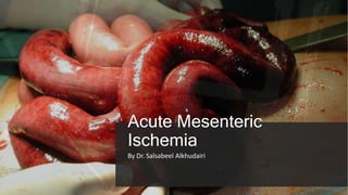
- 1. Acute Mesenteric Ischemia By Dr. Salsabeel Alkhudairi
- 2. Outline . 1. Background, Epidemiology 2. Classification & Risk factors 3. Clinical manifestations 4. Diagnosis 5. Management approach MM.DD.20XX ADD A FOOTER 2
- 3. Definition: Sudden onset of small intestinal hypoperfusion, which can be due to occlusive or nonocclusive obstruction of the arterial blood supply or obstruction of venous outflow Chronic mesenteric ischemia? accounts for less than 1 in 1000 hospital admissions High mortality 25%-80%
- 6. Etiologies of Mesenteric Ischemia
- 7. Risk factors
- 9. Clinical manifestation 1 2 3 4 Pain , NV, Fever, Abdominal distension, bloody diarrhea Hx of MI or AF Embolic AMI Abdominal angina Food fear Wt. loss , Diarrhea Ex: malnourished, bruit, diminished pulses Thrombotic AMI NOAMI HX of Hyper coagulable state Hx of DVT Settle course MVT • Hx & Ex to exclude other causes ! • Abdominal pain out of proportion to examination findings • Sick patients
- 10. Diagnosis: imaging • X-ray : not sensitive or specific • Suspicious finding In CT scan: - Bowel wall thickening - Free intraperitoneal fluid - Peri-intestinal soft tissues stranding - Abnormal bowel wall enhancement - Portal vein SMA gas • Computed tomographic angiography (CTA) best. • Other? US, duplex US, Echo.. • Lab: - Leukocytosis - Electrolyte derangements - Elevated amylase, - Lactic acidosis. - Elevated hematocrit - D- dimer!! • Additional : ECG, cardiac enzyme
- 12. Thrombotic MI
- 13. Embolic AMI
- 14. MVT
- 15. NOAMI
- 16. NOAMI
- 18. Management Aggressive resuscitation IVF, pressor “avoided”, .Electrolyte abnormalities. Anticoagulant ??? Antibiotics , pain management Arrhythmias Management Perioperative Management Occlusive vs non occlusive vs venous thrombosis Peritonitis or no peritonitis Laparotomy vs initial revascularization & post intervention look Operative vs Non operative Management Embolectomy Endovascular Approaches; Bypass Management of Ischemic Bowel Hybrid Open and Endovascular Revascularization Operative Approach
- 19. Occlusive vs non occlusive vs venous thrombosis Peritonitis or no peritonitis Operative vs Non operative Management Venous thrombosis: • Anticoagulant (95% successful) • Thrombus removal lysis “ Rare” • Intestinal stricture ? Non occlusive : • Supportive • Use of intraarterial infusion of vasodilator medication “papaverine”! • Surgery??! • No evidence for anticoagulation use
- 20. Management of Ischemic Bowel Operative Approach: Thrombotic & embolic Laparotomy, Prepping? Revascularization should precede resection of any bowel. Allow reperfusion of any threatened areas of bowel & come back for second look Don’t anastomose if questionable bowel viability Bowel resection • Assess bowel viability: - Visible& palpable pulsation - color and appearance - Peristalsis - Duplex US - Florescence IV contrast
- 21. Exposure of SMA - Anterior - Lateral
- 22. Embolectomy Operative Approach : Embolic Or thrombotic Transverse incision Embolus removal Heparinization Closure Reassessment of the bowel
- 23. Graft bypass Operative Approach : Embolic Or thrombotic Antegrade vs retrograde revascularization Aortic vs iliac inflow Autogenous vein vs prosthetic conduit Reassessment of the bowel
- 24. Hybrid Open and Endovascular Revascularization Operative Approach Open laparotomy with retrograde stenting of the SMA with access via a branch of the SMA. Success rate of patency 76%- 92% Mortality 20%-45%
- 25. Endovascular Approaches Operative Approach In case of very low suspicion for necrotic bowel In chronic mesenteric ischemia Outcome compared to Open Use of covered vs bare metal stent Assessment of bowel ?? Complication: access site thrombosis, hematomas, and infection
- 26. Lifelong anticoagulation Aspirin& Antiplatelet therapy Duplex ultrasound: detect graft stenosis. Post operative management
- 27. Summary • high index of suspicion is necessary • Use CTA to RO the diagnosis • Management depend on the etiology • Endovascular revascularization procedures may have a role with partial arterial occlusion • Post operative workup & management to prevent a sconed episode
- 28. Thank you References 1.Schwartz, Seymour I.,Brunicardi, F. Charles. (Eds.) (2011) Schwartz's principles of surgery :ABSITE and board review New York : McGraw- Hill Medical, 2.CAMERON., 2019. CURRENT SURGICAL THERAPY 13 3.Acute mesenteric ischemia: guidelines of the World Society of Emergency Surgery Bala et al. World Journal of Emergency Surgery (2017) 12:38 4.Up to date
Editor's Notes
- Splanchnic (visceral) ischemia, encompasses ischemia affecting the intestine, as well as other abdominal organs such as the liver, spleen, or kidneys. Chronic mesenteric ischemia: develops in patients with mesenteric atherosclerosis causing episodic intestinal hypoperfusion related to eating. - setting of an acute thrombosis or embolism, visceral collaterals are in sufficient to adequately perfuse the bowel in the distribution of the occluded artery, resulting in AMI. Factors associated with worse outcomes include older age, prolonged symptom duration, and the need for bowel resection. high mortality is due to challenges in diagnosis and delays in treatment. OAMI: 74%; NOAMI: 68%, MVT: 42%
- intestinal oxygen extraction is relatively low, thereby permitting sufficient oxygen to be delivered to the liver via the portal vein Collateral circulation — An extensive collateral circulation protects the intestines from transient periods of inadequate perfusion. However, prolonged reduction in splanchnic blood flow leads to vasoconstriction in the affected vascular bed and eventually reduces collateral blood flow Origin of embolic Occlusive AMI Location: Related to the angle to the Aorta Embolus 3-10 cm distal to SMA origin May have emboli on splenic artery & renal artery Origin of thrombotic Occlusive AMI Location: Origin of the SMA Origin of the celiac axis Non- occlusive AMI Relation to the hemostatic mechanism Mesenteric venous thrombosis Virchow triad Hypercoagulable state Other : IBD, Pancreatitis, sepsis
- The celiac axis and the SMA communicate principally through the junction of the superior and inferior pancreaticoduodenal arteries & gastroduodenal The SMA and IMA communicate via the marginal artery of Drummond and the meandering mesenteric artery. The marginal artery of Drummond represents the major collateral arcade and is composed of branches from the right, middle, and left colic arteries central communicating artery” the arc of Riolan” : inconstant communication between the SMA and IMA Collateralization between the IMA and systemic circulation occurs in the rectum
- Common now may be thrombosis Venous obstruction: Venous thrombosis is due to obstruction of the intestinal outflow tract, including the superior and inferior mesenteric veins and the splenic and portal veins. Embolism to the mesenteric arteries is most frequently due to a dislodged thrombus from the left atrium, left ventricle, cardiac valves, or proximal aorta. Arterial thrombosis: atherosclerotic disease, abdominal trauma, infection, thrombosed mesenteric aneurysm, and aortic or mesenteric artery dissection.
- The splanchnic circulation receives between 10 to 35 percent of cardiac output Phases of bowel ischemia Hyperactive phase: disparancy between pain & clinical finding Paralytic phase: Distended abdomen, reduced bowel sounds Septic phase: bowel leakage acute abdomen shock ischemic injury of the mesenteric circulation does not occur until perfusion pressure is reduced to approximately 30 mmHg or the mean mesenteric arterial pressure is reduced to 45 mmHg 12 hours without substantial injury,Arterial thrombosis —
- Abdominal pain described as out of proportion to examination findings Sepsis deranged vital signs resulting from inflammation and hypovolemia secondary to bowel edema. CTA can also identify other causes of mesenteric ischemia such as aortic dissection and mesenteric venous thrombosis. Pain 97%, N 44%; Vomiting: 333% diarrhea: 35%, blood per rectum: 15% Normal D-dimer levels may help to exclude acute intestinal ischemia, but elevated levels are less useful for making a diagnosis
- Finding in each category
- The anatomic site of involvement in acute mesenteric venous thrombosis is most often ileum (64 to 83 percent) or jejunum (50 to 81 percent), followed by colon (14 percent) and duodenum (4 to 8 percent) - complication of laparoscopic sleeve gastrectomy for obesity. In two retrospective surveys of more than 2900 patients, the incidence of this complication was approximately 0.7 percent
- a reduced number of mesenteric vessels (arteries and veins) and irregularity of the arterial branches of the mesenteric vasculature on vascular imaging (alternating dilation and narrowing "chain of lakes" or "string of sausages" sign).
- systemic anticoagulation to prevent thrombus formation and propagation Occlusive mesenteric ischemia: Selective catheterization of the splenic artery and SMA for catheter-directed thrombolytic therapy (used for a maximum of 48 hours)
- Non occlusive: management is largely supportive and nonoperative Selective catheterization of the SMA with intraarterial infusion of vasodilators such as papaverine “inhibition of the enzyme phosphodiesterase,” papaverine is 30 to 40 micrograms/kg/minute Mesenteric vein thrombosis: Nonoperatively, with full heparin anticoagulation and supportive care. (venous thrombectomy is not effective) Trans, hepatic or percutaneous mechanical thrombectomy Thrombolysis Surgical exploration: Open thrombectomy +\- resection In patients with cirrhosis and portal vein thrombosis, anticoagulation increased recanalization rates, reduced progression of thrombosis, and reduced variceal bleeding
- Leave the bowel in discontinuity
- Once no more thrombus is returned the vessel is gently irrigated with heparinized saline and the arteriotomy is closed with 5-0 or 6-0 polypropylene suture in a running fashion. If a longitudinal incision is made or a transverse incision converted to a longitudinal one, it should always be closed with an autogenous vein patch.
- a single bypass Graf to the SMA is adequate revascularization for AMI. Antegrade revascularization is less feasible inn critically ill patent due to need of clamping of the aorta & extensive-difficult exposure or but has superior patency results Retrograde less right common iliac artery for the inflow so the graft makes a gentle C loop and is less likely to kink Our preferred conduit is autogenous saphenous vein, although if none is available it is reasonable to use a 6-or 8-mm prosthetic. the bypass can be tunneled through the mesentery or retroperitoneally such that it lies nearly straight.
- Left brachial artery cut down for access diagnostic angiogram confirming the stenotic but patent SMA The lesion is then crossed with an appropriate wire for the selected stent covered stents have better patency. The diameter should match the diameter of the normal artery with the length extending from a millimeter or two into the aorta to just distal to the lesion. The study showed that covered stents are associated with less restenosis (18% vs. 47%), symptom recurrence (18% vs. 50%), and reintervention (9% vs. 44%) at 24 months and better primary patency at 3 years Guidelines generally include calcified ostial stenoses, high-grade eccentric stenoses, chronic occlusions, and significant residual stenosis >30% or the presence of dissection after angioplasty. Restenosis after PTA is also an indication for stent placement
- Lifelong anticoagulation: patient with arrhythmia or hypercoagulable disorders. Aspirin: After revascularization using bypass grafting. Antiplatelet therapy: Dual( least the first 6 weeks) Single-agent therapy should be continued for life. Duplex ultrasound: Q 6\m for a year after bypass then annually then after to detect graft stenosis.
- open surgical techniques have achieved an immediate clinical success rate that approaches 100%, a surgical mortality rate of 0% to 17% The freedom from recurrent stenosis rates at 1, 2, 3, and 4 years were 65%, 47%, 39%, and 13%, respectively Endovascular: The long-term clinical relief without reintervention was 82%; overall technical success rate of 91%, angioplasty and stenting demonstrate an inferior technical and clinical success rate