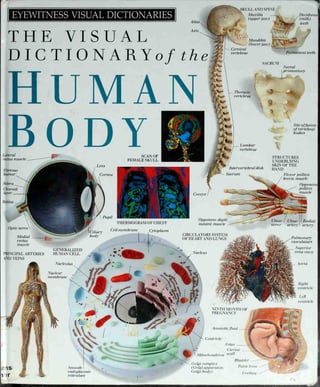
5. EYEWITNESS VISUAL DICTIONARIES
THE VISUAL
DICTIONARY of the
HUMAN
BODY
Frontalis
Brachialis
Deltoid
Rectus abdominis
Rectus fern oris
Gastrocnemius
SUPERFICIAL SKELETAL MUSCLES
6. Frontal bone
temporal
bone
i
Nasal _
septum
Mentalforamen
Fetus
Fallopian
tube
Uterine wall
Zygomatic
bone
Cervix
THE DEVELOPING FETUS
Presynaptic
axon
Mandible
(lower jaw)
Mitochondrion
Microtubule
Parietal lobe
Occipital lobe
Cerebellum
FRONT VIEW OF SKULL
- .y>
Frontal lobe
Temporal
lobe Synaptic
vesicle STRUCTURE OF A
SYNAPTIC KNOR
EXTERNAL VIEW OF RRAIN
>**JJJ
tlns Ixis
T
* % S 9
SPINE
7. EYEWITNESS VISUAL DICTIONARIES
THE VISUAL
DICTIONARY of the
HUMAN
BODYLateral rectus muscle
Vitreous humor
Optic nerve
Cornea
Conjunctiva
Lens
SECTION THROUGH LEFT EYE
DK PUBLISHING, INC
WWW.DK.COM
9. Metatarsal
BONES OF FOOT
Calcaneus]
Middle
phalanx
Larynx
Bladder
CHEST AND ABDOMINAL CAVITIES
WITH SOME OBGANS BEMOVED
Medulla
Contents
The Human Body 6
Head 8
Body Organs 10
Body Cells 12
Skeleton 14
Skull 16
Spine 18
Bones and Joints 20
Muscles 22
Hands 26
Feet 28
Skin and Hair 30
Brain 32
Nervous System 34
Eye 36
Ear 38
Nose, Mouth, and Throat 40
Teeth 42
Digestive System 44
Heart 48
Circulatory System 50
Respiratory System 52
Urinary System 54
Reproductive System 56
Development of a Baby 58
Index 60
Acknowledgments 64
Mucosa
Villus
INTEBNAL SUBFACE OF JEJUNUM
Fetal skull
Primary teeth
in maxilla
DEVELOPMENT OF
TEETH IN A FETUS
Branch of
pulmonary
vein
Branch of _
pulmonary
artery
£^
Cuticle
SECTION OF HAIB BBONCHIOLE WITH LOBULE
10. Back
The human body
Although there is enormous
variation between the external
appearances of humans, all bodies
contain the same basic features.
The outward form of the
human body depends on the
size of the skeleton, the shape
of the muscles,the thickness
of the fat layer beneath
the skin, the elasticity or
sagginess of the skin, and
the person's age and
gender. Males tend to be taller
than females, with broader
shoulders, more body hair,
and a different pattern of fat
deposits underthe skin; the
female body tends to be
less muscular and has
a shallower and wider
pelvis to allow
for childbirth.
BACK VIEW S OF
MALE AND FEMALE
Nape of neck
Shoulder
Scapula
(shoulder blade)
Arm
Hand
Upper arm
fffl
Forearm
Natal
cleft
Buttock
y
Leg.
Foot
Glutealfold
Poplitealfossa
Ca{f
Heel
'L
11. FRONT VIEWS OF
MALE AND FEMALE
Forehead
Face.
Eye
Nose
Lips
Chin
Thorax
(chest)
Abdomen
Scrotum
Knuckles-
Thigh
Head
I
'
i ^_i
Clavicle (collarbone)
Suprasternal notch
Breast
Nipple
_ Cubitalfossa
Umbilicus
(navel)
Wrist
Knee
Thumb
Finger
Palm
Pudenda
Shin
*
Toe
Instep
Ankle
12. Head
SIDE VIEW OF EXTERNAL FEATURES OF HEAD
In NEWBORN BABY, the head accounts for
one-quarter of the total body length; by
adulthood, the proportion has reduced to
one-eighth. Contained in the head are the
body's main sense organs: eyes, ears,
olfactory nerves that detect smells, and the
taste buds of the tongue. Signals from these
organs pass to the body's great coordination
center: the brain, housed in the protective,
bony dome of the skull. Hair on the head
insulates against heat loss, and adult males
also grow thick facial hair. The face has
three important openings: two nostrils
through which air passes, and the mouth,
which takes in nourishment and helps form
speech. Although all heads are basically
similar, differences in the size, shape, and
color of features produce an infinite variety
of appearances.
_ Crown
(vertex)
2=
Forehead
Eyebrow
Eyelash
Eye
_ Nose
Ear
Throat
SECTION THROUGH HEAD
Skull
Pineal body
Pituitary gland
Cerebellum
Pons
MeduIla oblonga la
Pharynx
Cervical vertebra
Spinal cord
Intervertebral dish
Superior sagittal sinus
Cerebrum
Frontal sinus
Sphenoidal sinus
Superior concha
Middle concha
Inferior concha
Vestibule
Maxilla (upperjaw)
Hard palate
Soft palate
Tongue
Uvula
Mandible (lower ja w)
Palatine tonsil
Epiglottis
Trachea
snphagus
^L
13. FRONT VIEW OF EXTERNAL
FEATURES OF HEAD
Frontal
bone
Glabella
Upper
eyelid
Iris
Pupil
Sclera
(white)
Lower
eyelid
Caruncle
Root
of nose
Dorsum
of nose
Ala
ofnose
Nasal
septum
Lateral angle of mouth
Frontal
notch
Supraorbital
notch
Supraorbital
margin
Lateral angle
of eye
Infraorbital
margin
Zygomatic
arch
Auricle (pinna)
of ear
Alar groove
Naris (nostril)
Philtral ridge
Philtrum
I ermilion border
of lip
Mentolabial sulcus
14. Body organs
All the vital body organs except for
the brain are enclosed within the trunk
or torso (the body apart from the head
and limbs). The trunk contains two
large cavities separated by a muscular
sheet called the diaphragm. The upper
cavity, known as the thorax or chest cavity, contains
the heart and lungs. The lower cavity, called the
abdominal cavity, contains the stomach, intestines,
liver, and pancreas, which all play a role in digesting
food. Also within the trunk are the kidneys and
bladder, which are part of the urinary system, and the
reproductive organs, which hold the seeds of new
human life. Modern imaging techniques, such as
contrast X-rays and different types of scans, make it
possible to see and study body organs without the
need to cut through their protective coverings of skin,
fat, muscle, and bone.
IMAGING THE BODY
S< IMK.HWI OF
IIEVRT CHVMBERS
ANGIOGRAM OF
RIGHT LING
CONTRAST X-RAY OF
GALLBLADDER
doi mi: CONTRAST
X-RA1 OF COLON
ULTRASOUND SCN
OF TWINS IN UTERUS
MAJOR INTERNAL
STRUCTURES
Thyroid gland
Greater
omentum
WGIOGRAM OF
KIDNEYS
SCINTIGRAM OF
NERVOl S SYSTEM
WCMM.KWl OF
K1 FRIES OF HEAD
CI SCW TIIROI (.11
FEMALE CHEST
THERMOGRAM OF
CHEST REGION
ANGIOGRAM OF
R'I FRIES OF HEART
MR] S< IAIN TIIROI (.11
HEM) IT EYE LEVEL
10
_
15. CHEST AND ABDOMINAL
CAVITIES WITH SOME
ORGANS REMOVED
Right common carotid artery
Right jugular vein
Right
subclavian
artery-
Right
lung
Upper
lobe
Middle
lobe
Lower
lobe
Heart
Left atrium
Right atrium
Left ventricle
Right ventricle
Right adrenal gland
Right ureter
Inferior vena cava
Common iliac vein
Rectum
External iliac vein
^_ -
Left lung
Primary bronchus
Secondary bronchus
Tertiary bronchus
Diaphragm
Esophagus
Spleen
Left adrenal gland
Pancreas
Left kidney
Left ureter
Abdominal aorta
Common iliac artery
Internal iliac artery
External iliac artery
Colon
Rladder
Adipose
(fat) tissue
11
16. Body cells
Microvillus
Everyone is made up of billions of CELLS, which are the basic structural units
of the body. Bones, muscles, nerves, skin, blood, and all other body tissues are
formed from different types of cells. Each cell has a specific function but
works with other types of cells to perform the enormous number of tasks
needed to sustain life. Most body cells have a similar basic structure.
Each cell has an outer layer (called the cell membrane) and
contains a fluid material (cytoplasm). Within the cytoplasm are
man) specialized structures (organelles). The most important
organelle is the nucleus, which contains
ital genetic material and acts as
the cell's control center.
(iitaninv
2
THE DOl ISLE HELIX
Diagrammatic representation of l)N , which is structured like a
spiral ladder. I) contains all the vital genetic information and
instruction codes necessarj for the maintenance and
continuation of life.
17. GENERALIZED HUMAN CELL
Cytoplasm
Lysosome
TYPES OF CELLS
Cell membrane
Mitochondrial
crista
BONE-FORMING
CELL
NERVE CELLS IN
SPINAL CORD
Nucleus
Rough
endoplasmic
reticulum
Microfilament
SPERM CELLS IN
SEMEN
SECRETORY THYROID
GLAND CELLS
Pore of
nuclear membrane
Ribosome
Centriole
Mitochondrion
Microtubule
ACID-SECRETING
STOMACH CELLS
CONNECTD7E TISSUE
CELLS
is«a£«fc
oxisome
MUCUS-SECRETING
DUODENAL CELLS
RED AND TWO
WHITE BLOOD CELLS
Pinocytotic vesicle
Golgi complex (Golgi apparatus;
Golgi body)
^ggm
FAT CELLS IN
ADIPOSE TISSUE
EPITHELIAL CELLS IN
CHEEK
13
18. Skeleton Metacarpal
Carpus
The SKELETON IS a MOBILE framework made up of 206 bones, approximately
half of which are in the hands and feet. Although individual bones are rigid,
the skeleton as a whole is remarkably flexible and allows the human body a
huge range of movement. The skeleton serves as an anchorage for the skeletal
muscles, and as a protective cage for the body's internal organs.
Female bones are usually smaller and lighter
than male bones, and the female pelvis
is shallower and has a wider cavity.
/ Ina
Radius
Humerus
Shoulder joint
Elbow joint
Wristjoint
Hip joint
Rib cage
Sternum
(breastbone) '5-T/MftV»
Intervertebral
disk
** **i
Shall
"True" ribs
ft si to 7th)
Clavicle
(collarbone)
"False" ribs
(8th to 10th)
Ischium
I
Ilium
"Floating" ribs
(1 1th and 12th)
Scapula
(shoulder blade)
% Humerus Radius I Ina Carpus
14
1_
20. Skull
The SKULL is the most complicated bony structure
of the body—but every feature serves a purpose.
Internally, the main hollow chamber of the skull
has three levels that support the brain, with
every bump and hollow corresponding to the
shape of the brain. Underneath and toward
the back of the skull is a large round hole, called
the foramen magnum, through which the spinal
cord passes. To the front of this are many smaller openings
through which nerves, arteries, and veins pass to and from the
brain. The roof of the skull is formed from four thin, curved bones
that are firmly fixed together from the age of about two years. At
the front of the skull are two orbits, which contain the eyeballs,
and a central hole for the airway of the nose. The jawbone hinges
on either side of the skull at ear level.
RIGHT SIDE VIEW OF A
FETAL SKULL
Anteriorfontanelle
Parietal
bone Coronal suture
Frontal
bone
RIGHT SIDE VIEW OF SKULL
Coronal suture
Greater wing of
sphenoid bone
Parietal bone
Squamous
suture
Lambdoidl
suture
Frontal bone
Frontozygomatie
suture
Occipital bone
Nasal bone
Mental
symphysis
Supraorbital margin
Sphenoidal
Mastoid fontanelle
fontanelle External
auditory
meatus
Orbital
cavity VIEW OF SKULL FROM BELOW
External occipital crest
Lambdaid
suture
Occipital bone
Temporal bone
External
auditory meatus
Mastoid process
Nasal bone
Anterior nasal spine
Maxilla
(upper jaw)
I Mandible
M (lower
jaw)
Styloid process
Coronoid
process
Mentalforamen!
Zygomatic arch
Posterior border,
of vomer
Concha
Mandible (lowerjaw)
Foramen magnum
Occipital condyle
Carotid canal
Mastoid process
Pharyngeal
tubercle
Pterygoid plate
Pterygoid hamulus
Creater palatine
foramen
Posterior nasal aperture
16
21. FRONT VIEW OF SKULL
Nasion Frontal bone
Glabella
Parietal bone
Temporal bone
Lesser wing of
sphenoid bone
Greater wing of
sphenoid bone
Frontal process
of maxilla
Nasal septum
Middle nasal concha
Inferior nasal concha
Anterior nasal spine
Mentalforamen
Nasal bone
Supraorbital
foramen
Supraorbital
margin
Superior
orbital
fissure
Lacrimal bone
Zygomatic
bone
Inferior orbital
fissure
Infraorbital
margin
Infraorbitalforamen
Maxilla
(upperjaw)
Mandible
(lowerjaw)
Mental
protuberance
17
22. Spine
SPINE DIVIDED INTO frontal view
VERTEBRAL SECTIONS
Cervical vertebrae
The spine (or SPINAL COLUMN) has two main functions: it
serves as a protective surrounding for the delicate spinal cord
and forms the supporting backbone of the skeleton. The spine
consists of 24 separate, differently shaped bones (vertebrae)
with a curved, triangular bone (the sacrum) at the bottom. The
sacrum is made up of fused vertebrae; at its lower end is a
small tail-like structure made up of tiny bones collectively
called the coccyx. Between each pair of vertebrae is a disk of
cartilage that cushions the bones during movement. The top
two vertebrae differ in appearance from the others and work as
a pair: the first, called the atlas, rotates around a stout vertical
peg on the second, called the axis. This arrangement allows the
skull to move freely up and clown, and from side to side.
Thoracic vertebrae _
TYPES OF VERTEBRAE
1TLAS WIS
Lumbar vertebrae _
Sacral vertebrae
Coccygeal vertebrae
CERVICAL VERTEBRA
interior
arch
interioi
tubercle
I ertebral
foramen
Transverse
process
Skull
Lateral mass with
superior articularfacet
Posterior
arch
Posterior
/ tubercle
Transverse
foramen
Vertebralforamen
Spinous
process
Lamina
Transverse process
andforamen
Body
Interior
tubercle
Posterior
tubercle
Superior
articular-
process
Spinous
process
Vertebral
foramen
Transverse
foramen
23. RIGHT SIDE VIEW CERVICAL VERTEBRA AND
SECTION OF SPINAL CORD
Vertebral artery
Vertebral body.
Superior articular
process
Anterior horn
Posterior horn
Anterior median
fissure
Spinous process of
cervical vertebra
Spinal cord
Anterior root
Spinal ganglion
Anterior branch
of spinal nerve
Posterior column
Dura mater
Lateral column
Posterior root
Posterior branch
ofspinal nerve
SACRUM COCCYX
Ala
THORACIC VERTERRA
Superior articular
process
Body
LUMRAR VERTERRA
Pedicle
.
Vertebral
foramen Body
Spinous
process
Lamina
Transverse
process
_
^.Costal
facet Vertebral
foramen
Superior
articular
process
andfacet
Lateral part
of sacrum
Cornu ,
Spinous
process
Lamina
Inferior
articular
process
Transverse
process Sacral
promontory
Lumbar vertebrae
Transverse
process
Facetfor
coccyx
Site offusion of
vertebral bodies
Sacralforamen
Auricular
surface
Intervertebral disk
Sacrum
Coccyx
19
24. Bones and
joints
Bonks form the body's hard, strong
skeletal framework. Each bone has a
hard, compact exterior surrounding a
spongy, lighter interior. The long bones
of the arms and legs, such as the femur
(thigh bone), have a central cavity
containing bone marrow. Bones are
composed chiefly of calcium,
phosphorus, and a fibrous substance
known as collagen. Bones meet at joints,
which are of several different types. For
example, the hip is a ball-and-socket
joint that allows the femur a wide range
of movement, whereas finger joints are
simple hinge joints that allow only
bending and straightening. Joints are
held in place by bands of tissue called
ligaments. Movement of joints is
facilitated by the smooth hyaline
cartilage that covers the bone ends and
by the synovial membrane that lines and
lubricates the joint.
LIGAMENTS SURROUNDING HIP JOINT
Iliac
crest. Iliacfossa
Iliac
spine
Greater trochanter
offemur
Iliofemoral
ligament
i
Intertrochanteric
line
Lesser trochanter
offemur
Femur.
Pubofemoral
ligament
Obturator
canal
Superior
ramus of
pubis
Body
of
pubis
Obturator
membrane
Isch ia I tuberosity
Ischium
SECTION THROl (ill LEFT EEMl R
Greater
trochanter,
Medullary cavity
< ancellous
(spungy) bone
Head
Foi a
Lesser
trochanter
ecl
20
25. SECTION THROUGH HIP JOINT
Psoas major muscle Iliacus muscle
External
iliac artery
Hyaline cartilage
of acetabulum
Hyaline cartilage
of head offemur
Ligament of
head offemur
Femoral
artery.
Pectineus
muscle
SECTION OF COMPACT
BONE
Iliac crest
Gluteus
minimus muscle
Gluteus
medius muscle
Acetabular
labrum
Head
offemur
Greater
trochanter
offemur
Neck of
femur
I dstus
lateralis
muscle
Shaft
offemur
Parallel rows of concentric
bony layers make up this
strong material.
BONE MARROW CELLS
Adductor
longus muscle
Vastus
medialis muscle
Composed mainly of red and
white blood cells, marrow fills
the cavities of bones.
SECTION THROUGH LONG BONE
Lateral
epicondyle
Patellar
surface
Osteon
(haversian system)
Osteocyte
(bone cell)
Haversian lamella
Outer lamella
Sharpey's
fiber-
Adductor tubercle
Intermediate
lamella
Endosteum
Medial
epicondyle
"V- f»UvV'*f
Periosteum
Haversian canal
Lacuna
I olkmann's iwssel
21
26. Muscles 1
There are three m i types of ml scle: skeletal
muscle (also called voluntary muscle because it can be
consciously controlled); smooth muscle (also called
involuntary muscle because it is not under voluntary
control); and the specialized muscle tissue of the heart.
Humans have more than 600 skeletal muscles, which
differ in size and shape according to the jobs they do.
Skeletal muscles are attached either directly or indirectly
(via tendons) to bones, and work in opposing pairs (one
muscle in the pair contracts while the other relaxes) to
produce body movements as diverse as walking, threading a
needle, and an array of facial expressions. Smooth muscles
occur in the walls of internal body organs and perform
actions such as forcing food through the intestines,
contracting the uterus (womb) in childbirth, and pumping
blood through the blood vessels.
SOME OTHER MUSCLES
l THE BODY
Rectus abdominis
Linea alba
External oblique
Tensorfasciae latae
The muscle libers contract and
dilate (expand) to alter pupil size.
TONGl I
Interlacing la ers of muscle
allow great mobility.
ILEl M
Opposing muscle layers
transport semidigested food.
_'_'
Iliopsoas
Pectineus
Vastus lateralis
m.L
27. Triceps
brachii
Teres minor
Teres major
Infraspinatus
Rhomboideus major
Latissimus dorsi
Flexors
of hand
Gluteus
maximus
Adductor
magnus
Gracilis
MOVEMENT OF THE FOREARM
Controlled movement of the limbs relies
on coordinated relaxation and contraction
of opposing muscles. To raise the
forearm, the biceps (two-rooted muscle)
contracts and shortens while the triceps
(three-rooted muscle) relaxes; the reverse
occurs when the forearm is lowered.
Triceps in
resting phase
Biceps in
resting phase
Triceps
relaxes
Biceps
contracts
Triceps fully
relaxed
Biceps fully
contracted
Triceps
contracts
Biceps
relaxes
Triceps back in
resting phase
Biceps back in
resting phase
Forearm
at rest
Forearm
half raised/half
Forearm
fully raised
Forearm
half lowered
Forearm
back at rest
23
28. Muscles 2
SKELETAL MUSCLE FIBER
Myofibril
Motor
end p late
Synaptic
knob
Schwann
cell
Motor-
neuron
Node of
Ranvier. m
TYPES OF MUSCLE
y
h£vm•
1 "»
**- — * -» °» i
b
i j
CARDIM Ml SCLE SKELETAL MUSCLE
CONTR CTION OF SKELETAL MUSCLE
SMOOTH MUSCLE
MUSCLES OF FACIAL
EXPRESSION
A single expression
is the result of
movement of many
muscles; the main
muscles of expression
are shown in action
below.
Sarcomere.
Nucleus
Sarcoplasmic
reticulum
Sarcolemma
Endomysium
FRONTALIS
CORRUGATOR
SUPERCILII
^
ORBICULARIS ORIS
ZYGOMATICUS MAJOR
HI I.VXEIJ SI I I
(.0TRCTKI> SI ll
DEI'RKSSOR N(.l II
ORIS
24
29. MUSCLES OF
HEAD AND NECK
Frontalis
Corrugator
supercilii
Orbicularis
oculi
Levator labii
superioris
Zygomaticus
major
30. Hands
The human hand is an extremely versatile tool,
capable of delicate manipulation as well as
powerful gripping actions. The arrangement of
its 27 small bones, moved by 37 skeletal muscles
that are connected to the bones by tendons,
allows a wide range of movements. In particular,
it is our ability to bring the tips of our thumbs
and fingers together, combined with the extraordinary
sensitivity of our fingertips due to their rich supply of
nerve endings, that gives human hands their unique
dexterity.
Ring Middle
finger finger
BONES
X-RAY OF LEFT HAND
OF A YOUNG CHILD
rea of
ossification
in phalanx
Index
finger
Area of
ossification
in metacarpal
Area of
ossification
in wrist
OF HAND
Little
finger
Epiphysis
ofulna
Distal
phalanx
Middle
phalanx
26
Epiphysis
of radius
Areas of cartilage in the wrist and at the
ends of the fingerbones are the sites of
growth and have still to ossify.
Proximal
phalanx
2nd
metacarpal
3rd
metacarpal
4th
metacarpal
)lh
metacarpal
I Inmate
Pisiform
Distal phalanx
of thumb
Proximal phalanx
of thumb
1st metacarpal
Trapezium
Capitate
Triquetral
Trapezoid
Scaphoid
Radius
31. STRUCTURES UNDERLYING SKIN
OF PALM OF HAND
Flexor pollicis
brevis muscle
Adductor* pollicis muscle
2nd lumbrical muscle
Opponens
pollicis
muscle Abductor
pollicis
brevis
muscle Flexor
retinaculum
Radial
artery-
Digital artery-
Flexor digitorum tendon
Abductor Ulnar
Opponens digiti nerve
digiti minimi
minimi muscle
muscle
EXTERNAL FEATURES OF RACK OF HAND
Little finger
Ulnar
artery
Palmaris
longus
tendon
Ringfinger
Middle
finger
Cuticle
Distal
interphalangeal joint
Proximal
interphalangeal joint
, Extensor digitorum
tendon
Head of ulna
Lunule
Index
finger.
Nail Metacarpophalangeal
joint
Thumb
Wrist
Distal end
ofradius
27
32. Feet
BONES OF FOOT
2nd toe
The feet and toes are essential elements
in body movement. They bear and propel the
weight of the body during walking and
running, and also help to maintain balance
during changes of body position. Each foot
has 26 bones, more than 100 ligaments,
and 33 muscles, some of which are
attached to the lower leg. The heel
pad and the arch of the foot act as
shock absorbers, providing a
cushion against the jolts that
occur with every step.
3rd toe
Hallux
(big toe)
4th toe
5th
(little) toe
Distal
phalanx.
Middle
phalanx^.
U
Proximal
phalanx _
LIGAMENTS OF FOOT
Posterior
cuneonavicular
ligament
Plantar
calcaneonavicular
ligament
rticular
capsule of
interphalangeal
joint
Bifurcate
ligament
Fibula
Calcanean
( chilies)
tendon
1st
metatarsal
2nd
metatarsal
3rd
metatarsal
4th
metatarsal
5th
metatarsal
s
Articular capsule of
metatarsophalangeal
joint
Distal
phalanx of
hallux
. Proximal
phalanx of
hallux
Posterior
tarsometatarsal
ligament
Talonavicular
ligament
1st
cuneiform
2nd
cuneiform
3rd
cuneiform
Cuboid
Navicular
Talus
c:
*•
«•
Deltoid
ligament
Tibia
Interosseous
ligament
'
Calcaneus
28
34. -rn~v
Skin and hair
Skin is the boil's iargest organ, a
waterproof barrier that protects the internal
organs against infection, injury, and harmful
sun rays. The skin is also an important
sensory organ and helps to control body
temperature. The outer layer of the skin, known as
the epidermis, is coated with keratin, a tough, horny
protein that is also the chief consistituent of hair and
nails. Dead cells are shed from the skin's surface and
are replaced by new cells from the base of the
epidermis, the region that also produces the skin
pigment, melanin. The dermis contains most of the
skin's living structures, and includes nerve endings,
blood vessels, elastic fibers, sweat glands that cool the
skin, and sebaceous glands that produce oil to keep
the skin supple. Beneath the dermis lies the
subcutaneous tissue (hypodermis), which is rich in fat
and blood vessels. Hair shafts grow from hair follicles
situated in the dermis and subcutaneous tissue. Hair
grows on every part of the skin apart from the palms
of the hands and soles of the feet.
SECTIONS OF DIFFERENT TYPES OF SKIN
SECTION OF HAIR
Medulla
Cortex
Melanin
granule
Cell
nucleus
residue
Macrojibril
Cuticle
Sebaceous gland Hair
follicle
Enlarged
sweat gland
Thickened
epidermis Sweat pore
Follicle
rich
dermis _
Meissner's
corpuscle
Sweat
eland
Pacinian
corpuscle
Si i P
50
KM PIT SOU-. ()l FOOT
:
35. T-'f-r 'J'*J^^~
SECTION OF SKIN
Stratum
granulosum
Stratum
basale
Stratum
spinosum
Dermal
papilla
Free
nerve
ending
Meissner's
corpuscle
Vascular
plexus
Nerve
fiber
Sebaceous
gland
Arrector
pili
muscle
Hair
bulb
Papilla
Pacinian
corpuscle
Adipose (fat)
tissue
Artery
Sweat
gland
Dermis
_ Hypodermis
Ruffini
corpuscle
follicle
PHOTOMICROGRAPHS OF SKIN AND HAIR
SECTION OF SKIN
The flaky cells at the skin's
surface are shed continuously.
SWEAT PORE SKIN HAIR HEAD HAIR
This allows loss of fluid as part Two hairs pushing through the The root and part of the shaft of
of temperature control. outer layer of skin. a hair from the scalp.
31
•v.
36. Brain
MRI SCAN OF TRANSVERSE
SECTION THROUGH RRAIN
The brain is THE major organ of the central nervous
system and the control center for all the body's voluntary
and involuntary activities. It is also responsible for the
complexities of thought, memory, emotion, and language.
In adults, this complex organ is a mere 3 lb (1.4 kg) in weight,
containing over 10 thousand million nerve cells. Three distinct
regions can easily be seen—the brainstem, the cerebellum, and
the large cerebrum. The brainstem controls vital body
functions, such as breathing and digestion. The cerebellum's
main functions are the maintenance of posture and the
coordination of body movements. The cerebrum, which
consists of the right and left cerebral hemispheres joined by
the corpus callosum, is the site of most conscious and
intelligent activities.
UJiite
matter
Skull
Scalp
SAGITTAL SECTION
THROUGH RRAIN Fornix
Central sulcus
Gray
matter
Lateral
ventricle
Coronal
section
Cerebrum
Parietal lobe
Parietooccipital
sulcus
Pineal
body
Corpus
callosum
Thalamus
Occipital
lobe
Aqueduct
Cerebellum
4th ventricle
Frontal
lobe
Hypothalamus
Optic
chiasma
Pituitary gland
_ Brainstem
Spinal mill
Medulla
oblongata
32
37. SECTION THROUGH SKULL AND BRAIN EXTERNAL ANATOMY OF BRAIN
Arachnoid
granulation
Lateral
lacuna
Superior
sagittal
sinus
Falx
cerebri
Cerebrum
Epicranial
aponeurosis
Pericranium
Skull
Dura mater
Arachnoid
mater
Pia mater
Subarachnoid
space
Parietal lobe Parieto-occipital
sulcus
Precentral gyrus
Postcentral gyrus
Central sulcus
Frontal lobe
Lateral sulcus
Gray matter
White matter
Cerebral
vessel
Temporal lobe
SPECIFIC ROLES OF
AREAS OF CEREBRUM
Occipital
lobe
Cerebellum
CORONAL SECTION THROUGH BRAIN
Corpus
callosum
Longitudinal
fissure
Gray matter
Cerebrum
Skilled
movements
Behavior and
emotion
Basic
movements
Sensation
Speech
Caudate
nucleus
Lateral
ventricle
Visual
recognition
Fornix
Lentiform
nucleus
Internal
capsule
Hearing
Vision
Balance
and muscle
coordination
NERVE CELLS IN BRAIN
Thalamus
Crus
cerebri of
midbrain
3rd
ventricle
Pons
Cerebellum
Medulla oblongata
The dark cells are Purkinje
cells, which are among the
largest nerve cells in the body.
33
38. Nervous
system
CENTRAL VM) PEIIIP11EIWL
NERVOUS SYSTEMS
Cranial
nerves
Cerebrum
Cerebellum
Thoracic
nerves
Tin; nervot s SYSTEM is the body's internal,
electrochemical, communications network.
Its main parts are the brain, spinal cord, and
nerves. The brain and spinal cord form the
central nervous system (CNS), the body's
chief controlling and coordinating
centers. Billions of long neurons,
many grouped as nerves, make
up the peripheral nervous system, transmitting
nerve impulses between the CNS and other
regions of the body. Each neuron has three
parts: a cell body, branching dendrites that
receive chemical signals from other neurons,
and a tube-like axon that conveys these
signals as electrical impulses. Lumbal
nerves
Sacral
nerves
ncri
lira cilia I
plexus
Spinal
cord
SECTION TIIROl (ill SPIN E CORD
Crav mattti
Spinal
ganglion
;>
Central
canal Posterior root
ofspinal nerve
y
i k
White matter
Interior medianfissure Interior root
of spinal nerve
r
)l
39. STRUCTURE OF A MOTOR NEURON
Cell body
Nucleus
Synaptic
knob
-!?&«
Nucleolus
Schwann
cell
Node of
Ranvier
Axon
Dendrite
Mitochondrion
Nissl
body
STRUCTURE OF
A SYNAPTIC KNOR
TYPES OF NEURON
MULTIPOLAR UNIPOLAR
Motor end plate .Dendrite
Axon
Neurotransmitter
BIPOLAR
Dendrite
Axon_
%
^
Myelin sheath
TYPES OF
NERVE ENDING
FREE NERVE ENDING
MEISSNERS
CORPUSCLE
Cell
bodv-
Nucleus
MERKEL S DISK
Cell body
Dendrite
RUFFINI CORPUSCLE
PACINIAN
CORPUSCLE
35
40. Eye
Lateral
rectus muscle
The EYE is the organ of sight. The two eyeballs, protected within
bony sockets called orbits and on the outside by the eyelids,
eyebrows, and tear film, are directly connected to the brain
by the optic nerves. Each eye is moved by six muscles,
which are attached around the eyeball. Light rays
entering the eye through the pupil are focused by
the cornea and lens to form an image on the
retina. The retina contains millions of
light-sensitive cells, called rods and cones,
which convert the image into a pattern of
nerve impulses. These impulses are
transmitted along the optic nerve to the
brain. Information from the two optic
nerves is processed in the brain to
produce a single coordinated image.
Vitreous humor
Central retinal vein
Central retinal artery
Pia mater
Arachnoid mater
Dura mater
Optic nerve
lielinal blood vessel
Medial
rectus muscle.
56
•J
41. SECTION THROUGH LEFT EYE LACRIMAL (TEAR-PRODUCING) APPARATUS
Lacrimal sac Lacrimal canaliculus ,
acrimal gland
Middle
meatus
Middle
nasal
concha
Ora
serrata
Anterior
chamber
Posterior
chamber
Nasal
septum Inferior
nasal concha
Nasolacrimal
duct
, Lacrimal
punctum
Aqueous
humor
OPHTHALMOSCOPIC VIEW OF
RETINA
Retinal
blood vessel
Conjunctiva
Pupil
Cornea
Lens
Sphincter
muscle
Dilator
muscle
Macula
Optic
disk
Zonular
ligament
Sinus venosus
sclerae
The blind spot, where the optic nerve
leaves the eye, can be clearly seen as a
light circular area toward the center of
the image.
MUSCLES SURROUNDING RIGHT EYE
Medial rectus Superior oblique
Iridocorneal
angle
Levator palpebrae
superioris
Ciliary
body
Annular
tendon
Trochlea
Superior
rectus
Inferior rectus
Lateral rectus
Inferior oblique
37
42. Ear
The ear is the organ of hearing and balance. The outer ear
consists of a flap called the auricle or pinna and the auditory canal.
The main functional parts—the middle and inner ears—are
enclosed within the skull. The middle ear consists of three tiny
bones, known as auditory ossicles, and the eustachian tube, which
links the ear to the back of the nose. The inner ear consists of the
spiral-shaped cochlea, and also the semicircular canals and the
vestibule, which are the organs of balance. Sound waves entering
the ear travel through the auditory canal to the tympanic membrane
(eardrum), where they are converted to vibrations that are
transmitted via the ossicles to the cochlea. Here, the vibrations are
converted by millions of microscopic hairs into electrical nerve
signals to be interpreted by the brain.
STRUCTURE OF EAR
Temporal bone
RIGHT AURICLE (PINNA)
Scaphoidfossa
Upper crux of
antihelix
Helix
ntihelLi
Andtragus
External
auditory
meatus
OSSICLES OF MIDDLE EAR
Auricle
(pinna)
Triangular
fossa
Lower crux
of antihelix
Concha
Auditory canal
Tragus
Inlertragic
notch
Lobule
Cartilaginous
part ofmeatus
Lobule
INTERNAL STRUCTURE
OF AMPULLA
Membranous
portion
Osseous
portion
MALLEI S (HAMMER) IMI S ( VWII .) si IMS (STIUIU I') Crista
These tint e iin bones connect to form a bridge between the
tympanic membrane and the oval window. ith a system of
mi •! branes the) convej sound ribrations to the inner ear.
Cupula
Ampullar
nerve
Hair cell
ofcrista
58
43.
44. Nose, mouth,
and throat
With every breath, air passes through the nasal
cavity down the pharynx (throat), larynx ("voice
box"), and trachea (windpipe) to the lungs. The nasal
cavity warms and moistens air, and the tiny layers in
its lining protect the airway against damage by foreign
bodies. During swallowing, the tongue moves up and
back, the larynx rises, the epiglottis closes off the
entrance to the trachea, and the soft palate separates
the nasal cavity from the pharynx. Saliva, secreted
from three pairs of salivary glands, lubricates food to
make swallowing easier; it also begins the chemical
breakdown of food, and helps to produce taste. The
senses of taste and smell are closely linked. Both
depend on the detection of dissolved molecules by
sensory receptors in the olfactory nerve endings of the
nose and in the taste buds of the tongue.
STRUCTURE OF TONGUE
STRUCTURES
SURROUNDING
PHARYNX
Styloglossus muscle
Hyoglossus muscle
Hypoglossal
nerve __
Lingual
nerve
Superior
laryngeal
nerve
Superior
thyroid
artery
Median
glossoepiglottic
fold
Epiglottis
Sulcus
term in a Iis
Foramen
cecum
Median
sulcus
Palatine
tonsil
Palatoglossal
arch
I allate
papilla
Foliate
papilla
Fungiform
papilla
Filiform papilla
Apex
TASTE AREAS ON TONGUE
Cricothyroid
muscle
Tongue
Sublingual
gland
Mandible
(lower jaw)
Submandibular
gland
Hyoid bone
Laryngeal prominence
(Adam 's apple)
TYPES OF PAPIULAE
Bitter
Sour
Sweet
Thyrohyoid
muscle
Thyrohyoid
membrane
Cricothyroid
ligament
Thyroid gland
<.**
Trachea
FII.II ()K1 i'mmi.i.m: II M.IIOKM I'U'IU.VK MI.LVTK l»IMLLK
40
.
45. SECTION THROUGH NOSE,
MOUTH, AND THROAT
Frontal sinus
Superior meatus
Middle meatus
Nasal cavity
Vestibule
Inferior meatus
Hard palate
Maxilla
(upperjaw)
Incisive canal
Orbicularis oris muscle
Superior
longitudinal muscle
Superior nasal concha
Sphenoidal sinus
Middle nasal concha
Inferior nasal concha
Soft palate
Nasopharynx
Incisor
Apex of tongue
Genioglossus muscle
Sublingualfold
Sublingual salivary gland
Fibrous septum
Mandible (lower jaw)
Geniohyoid muscle
Myohyoid muscle
ENDOSCOPIC VIEW OF VOCAL CORDS
Posterior
part of
tongue
Uvula
Palatine
tonsil
Oropharynx
Epiglottis
Lingual
tonsil
Cricothyroid
muscle
Cervical
vertebra
Intervertebral
disk
Esophagus
Trachea
Epiglottis Vocal cord
41
46. Teeth
DEVELOPMENT OF TEETH IN A FETUS
The 20 primary TEETH (also called deciduous or milk
teeth) usually begin to erupt when a baby is about six
months old. They start to be replaced by the permanent
teeth when the child is about six years old. By the age of
20, most adults have a full set of 32 teeth although the
third molars (commonly called wisdom teeth) may never
erupt. While teeth help people to speak clearly and give
shape to the face, their main function is the chewing of
food. Incisors and canines shear and tear the food into
pieces; premolars and molars crush and grind it further.
Although tooth enamel is the hardest substance in the
body, it tends to be eroded and destroyed by acid
produced in the mouth during the breakdown of food.
Fetal skull
Primary teeth
in maxilla
(upper jaw)
Primary teeth
in mandible
(lower jaw)
FETAL JAWS
By the sixth week of embryonic development areas of
thickening occur in each jaw; these areas give rise to
tooth buds. By the time the fetus is six months old,
enamel has formed on the tootb buds.
DEVELOPMENT OF JAW AND TEETH
Iaxilla
(upper jaw)
Mandible
(lowerjaw).
-^^
iZ/
r
l?^
NEWBORN BAB1 S JAWS
The primary teeth can be seen
developing in the jawbones;
ihe begin to erupt around the
age of six months.
A FIVE-YEAR-OLD CHILD'S TEETH A NINE-YEAR-OLD CHILD'S TEETH AN ADULT'S TEETH
There is a full set of 20 erupted
primary teeth; the permanent
teeth can be seen developing in
the upper and lower jaws.
Most of the teeth are primary
teeth but the permanent
incisors and first molars have
now emerged.
By the age of 20, the full set of
52 permanent teeth (including
the wisdom teeth) should be
in position.
THE PERM WENT TEETH
Molars Premolars Canines Incisors Can ines Premolars Molars
3rd 2nd
(wisdom)
Jt UPPEB
lou:r
1st 2nd Lateral Central Intend 2nd Ird
(wisdom)
42
<
48. I
Digestive system 1
The DIGESTIVE SYSTEM BREAKS down FOOD into particles so tiny that blood can take nourishment to all
parts of the body. The system's main part is a 30-foot (9 m) tube from mouth to rectum; muscles in this
alimentary canal force food along. Chewed food first travels through the esophagus to the stomach,
which churns and liquidizes food before it passes through the duodenum, jejunum, and ileum—the
three parts of the long, convoluted small intestine. Here, digestive juices from the gallbladder and
pancreas break down food particles; many filter out into the blood through tiny fingerlike villi that
line the small intestine's inner wall. Undigested food in the colon forms feces that leave the body
through the anus.
50. Digestive
system 2
EXTERNAL ANATOMY OF STOMACH
Branch of
vagus nerve
Esophagus _
Circular
musclefibers
Londitudinal
musclefibers
Oblique
musclefibers
Pylorus
STRUCTURE OF ALIMENTARY CANAL
SECTION OF ESOPHAGEAL Vt ILL
Fibrous adventit ia
Nervefibers
ofsubmucosal
plexus
Stratum
circularis.
Duodenum
Branch of
gastric arten
Fundus
_ Cardiac
notch
Stratum
longitudinalis.
Blood vessel
Body
Greater
curvature
Muscularis
Submucosa
lucosa
.Muscularis
mucosa
Epithelium
Lesser
curvature
Lamina
propria
SECTION OF STOMACH WALL
.Mucosal gland
Oblique layer
of muscularis
Stratum
longitudinalis
Serosa
Muscularis
Submucosa
Mucosa
Peritoneum
Stratum
circularis
Epithelium
Muscularis
mucosa
SECTION THROUGH LIVER
Lamina
propria
Gastric pit
Portal vein
Left lobe
Hepatic
artery
Inferior
vena cava Cvstic duct
Common
hepatic duct
Bight
lobe
SECTION OF DUODENAL WALL
Common bile
duct
Stratum
longitudinalis
Falciform
ligament
Quadrate lobe
Gallbladder
Common
bile dud
Nerve
plexus
Stratum
circularis
Muscularis
Submucosa
Mucosa
Submucosal crypt
Muscularis mucosa Lamina
propria
Intestinal
villus
Epithelium
16
51. INTERNAL SURFACE OF JEJUNUM
illus
Mucosa _
Muscularis
mucosa
Epithelium
Jejunal
gland
Crypt of
Lieberkiihn
SECTION OF ILEAL V U.l.
Muscularis
Submucosa
Mucosa
Serosa
Intestinal
villus
Epithelium
Stratum
>x>>
/ longitudinalis
ri-m Stratum
Willi 1
circularis
Hi Nerve
*§
plexus
Muscularis
mucosa
Lamina propria
Crypt of
Lieberkiihn
SECTION OF COLONIC WALL
Muscularis .
Submucosa,
Mucosa
Tenia colica
Lymphoid
follicle
Epithelium
Stratum
longitudinalis
Serosa
Nerve
plans
Stratum
circularis
propria
Colonic
pit
47
52. Heart
ARTERIES AND VEINS
SURROUNDING HEART
The heart is A HOLLOW MUSCLE in the middle of the
chest that pumps blood around the body,
supplying cells with oxygen and nutrients.
A muscular wall, called the septum,
divides the heart lengthwise into
left and right sides. A valve divides
each side into two chambers: an
upper atrium and a lower ventricle. When the
heart muscle contracts, it squeezes blood
through the atria and then through the
ventricles. Oxygenated blood from the lungs
flows from the pulmonary veins into the left
atrium, through the left ventricle, and then
out via the aorta to all parts of the body.
Deoxygenated blood returning from the body
flows from the vena cava into the right atrium,
through the right ventricle, and then out
via the pulmonary artery to the lungs for
reoxygenation. At rest the heart
beats between 60 and 80 times Right
a minute; during exercise or coronary artery
at times of stress or excitement the
rate may increase to 200 beats a minute. Coronary sinus
Aorta
Left
coronary
artery
Cardiac
vein
SECTION THROUGH
HEART WALL
Main branch ofleft
coronary artery
Pericardial
cavity
Trabecula
Endocardium
Myocardium
Epicardium
(visceral pericardium)
Serous pericardium
Fibrous pericardium
HEARTREAT SEQUENCE
ATRIAL DIASTOLE
Right
atrium
Right
ventricle
- Left
atrium
Left
ventricle
Deoxvgenated blood enters the right atrium while the
left atrium receives oxygenated blood.
•.
53. STRUCTURE OF HEART
Brachiocephalic trunk
Superior vend cava
Ascending aorta
Right pulmonary
artery
Fossa oralis
Right pulmonary vein
Right atrium
Opening of
inferior vena cava
Branch ofcoronary artery
Tricuspid valve
Chordae
tendineae
Left subclavian artery
Left common
carotid artery
Left pulmonary vein
Pulmonary trunk
Pulmonary semilunar valve
Coronary artery
Right ventricle
Trabecula
Chordae
tendineae
Muscular part of
interventricular
septum
Left ventricle
Papillary muscle
Myocardium of
left ventricle
ATRIAL SYSTOLE (VENTRICULAR DIASTOLE)
Right atrium
contracts
Tricuspid
valve opens
Right ventricle
dilates
Left atrium
contracts
Mitred valve
opens
Left ventricle
dilates
VENTRICULAR SYSTOLE
Pulmonary artery.
Pulmonary
valve opens
Tricuspid
valve closes
Right ventricle
contracts
iorta
Aortic valve
opens
Mitral valve
closes
Left ventricle
contracts
Left and right atria contract, forcing blood into the
relaxed ventricles.
Ventricles contract and force blood to the lungs for
oxygenation and via the aorta to the rest of the body.
49
54. Circulatory
system
The circi LATOR5 system consists of the heart and
blood vessels, which together maintain a continuous
flow of blood around the body. The heart pumps
oxygen-rich blood from the lungs to all parts of the
body through a network of tubes called arteries, and
smaller branches-called arterioles. Blood returns to the heart via
small vessels called venules, which lead in turn into larger tubes
called veins. Arterioles and venules are linked by a network of
tiny vessels called capillaries, where the exchange of oxygen
and carbon dioxide between blood and body cells takes place.
Blood has four main components: red blood cells, white blood
cells, platelets, and liquid plasma.
Superior vena cava
ARTERIAL SYSTEM OE RRAIN
CIRCULATORY SYSTEM OF IEART AND LUNGS
Aorta
CIRCULATORY SYSTEM OF LIVER
Inferior vena cava
Portal vein
Common
bile duet
(lallbladder
Left ventricle
SECTION OF M U RTERY
/ anica
media
Collagen and elasticfibers
External elastic lamina
Tunica
advenlitia
Right ventricle
SECTION OF MAIN VEIN
Tunica media Collagen and elasticfibers
External elastic lamina
I alve cusp.
Eunica
advenlitia
Internal
clastic lamina LTunica intima
Endothelium irteriole
Internal
elastic lamina
Endothelium
_ Tunica intima
50
55. PRINCIPAL ARTERIES AND VEINS
OF CIRCULATORY SYSTEM
TYPES OF BLOOD CELLS
Common
carotid artery
Subclavian artery.
Arch ofaorta
Axillary artery
Pulmonary artery
Coronary artery
Brachial artery.
Gastric artery
Hepatic artery
Splenic artery
Superior
mesenteric artery
Radial
artery
Ulnar
artery
Internal jugular vein
Brachiocephalic vein
Subclavian vein
Axillary vein
Cephalic vein
Superior vena cava
Pulmonary vein
Basilic vein
_ Hepatic portal vein
Median cubital vein
__ Inferior vena cava
Anterior median
vein
Palmar
arch
Gastroepiploic
vein
Palmar
vein
Digital
vein
Digital artery
Common iliac artery
External iliac artery
Internal iliac artery
Femoral artery
Popliteal artery
Peroneal artery
Anterior tibial artery
Posterior tibial artery
Lateral plantar artery
Dorsal metatarsal artery
Inferior mesenteric vein
Superior mesenteric vein
Common iliac vein
External iliac vein
Internal iliac vein
Femoral vein
Great saphenous vein
Short saphenous vein
RED BLOOD CELLS
These cells are biconcave in
shape to maximize their
oxygen-carrying capacity.
WHITE BLOOD CELLS
Lymphocytes are the smallest
white blood cells; they form
antibodies against disease.
PLATELETS
Tiny cells that are activated
whenever blood clotting or
repair to vessels is necessary.
BLOOD CLOTTING
__
Dorsal venous arch
Digital vein
Filaments of fibrin enmesh red
blood cells as part of the
process of blood clotting.
51
56. Respiratory
system
The respiratory system supplies the
oxygen needed by body cells and carries
off their carbon dioxide waste. Inhaled
air passes via the trachea (windpipe)
through two narrower tubes, the bronchi,
to the lungs. Each lung comprises many
fine, branching tubes called bronchioles
that end in tiny clustered chambers
called alveoli. Gases cross the thin
alveolar walls to and from a network of tiny blood vessels.
Intercostal (rib) muscles and the muscular diaphragm below
the lungs operate the lungs like bellows, drawing air in and
forcing it out at regular intervals.
BRONCHIOLE AND ALVEOLI
Bronchial nerve
Branch of
pulmonary vein
Terminal bronchiole
Bronchial rein
I isceral
cartilage
Mucosal gland
Branch of
pulmonary
artery
Elastic
fibers
Inleralveolar
septum
Alveolus
Connective
tissue
Capillary network Epithelium
SEGMENTS OF BRONCHIAL TREE
/ pper
lobe of
right lung
Middle
lobe of
right lung.
Lower
lobe oj
right lung
[nteriof
basal
Lateral
basal
Medial
basal
I pper
lobe of
left lung
52
[pical
Posterior basal
57. STRUCTURES OF THORACIC CAVITY GASEOUS EXCHANGE IN ALVEOLUS
Epiglottis
Hyoid bone
Thyroid cartilage
Thyroid
gland
Superior
vena cava
Upper
lobe of
right lung
Horizontal
fissure
Obliqu
fissure
Lower
lobe of
right lung
Middle lobe
of right lung
Alveolus
Aorta
Upper
lobe of
left lung
Oxygen diffuses
into blood
Oxygenated
blood
Deoxygenated
blood rich in
carbon dioxide
Pulmonary
trunk
Left pulmonary
artery
Lung
expands
Carbon dioxide
diffuses from
blood into alveolus
MECHANISM
OF RESPIRATION
INSPIRATION
Heart
Lower
lobe of
left lung
Secondary
bronchus
Tertiary
bronchus
Air drawn
into lungs
Diaphragm
contracts
andflattens
Intercostal
muscles
contract
EXPIRATION
Lung
contracts
Right crus
of diaphragm
Abdominal
aorta
Left crus of
diaphragm
Muscular wall
of diaphragm
,
Esophagus
Airforced
out of
lungs
Diaphragm
relaxes and
moves up Intercostal
muscles relax
53
58. Urinary system
The urinary system filters u ste products from
the blood and removes them from the body via a
system of tubes. Blood is filtered in the two kidneys,
which are fist-sized, bean-shaped organs. The renal
arteries carry blood to the kidneys;
the renal veins remove blood after
filtering. Each kidney contains
about one million tiny units called nephrons.
Each nephron is made up of a tubule and a
filtering unit called a glomerulus, which
consists of a collection of tiny blood vessels
surrounded by the hollow Bowman's capsule.
The filtering process produces a watery fluid
that leaves the kidney as urine. The urine is
carried via two tubes called ureters to the bladder,
where it is stored until its release from the body through
another tube called the urethra.
arterial system of kidneys
Aorta
Celiac trunk
Left ureter
SECTION THROUGH LEFT KIDNEY
Interlobular
vein
Collecting
tubule
Medullar}
pyramid
Interlobular A
artery
Cortex L
Medulla
Renal artci}
Renal vein
Renal pelvis
I reter
Minor calyx
Fibrous capsule
Boivman's
capsule
Nephron
SECTION OF KIDNEY
Interlobular
artery
Interlobular
vein
Loop of
Henle
Collecting
tubule
Distal convoluted
tubule
,
Nephron
Cortex
Renal
sinus
Major calyx
Renal papilla
Medulla
I
/ asa
recta
Glomerulus
Bowman's
capsule
Proximal
convoluted
tubule
Collecting
duct
Loop of
Henle
Duct of
Bellini
Renal
column
II
59. MALE URINARY TRACT Superior mesenteric
artery
Celiac
trunk
Right adrenal
(suprarenal) gland
Inferior
vena cava
Renal artery
Renal vein
Right kidney
Aorta
Right ureter
Superior
pubic ramus
SECTION THROUGH
BOWMAN'S CAPSULE
Left adrenal
(suprarenal)
gland
Left suprarenal
vein
Left renal artery
Left renal vein
Left kidney
Left ureter
Vertebral column
Psoas muscle
Left common iliac artery
Left common iliac vein
Testicular vein
and artery
Efferent
arteriole
Distal convoluted
tubule
Afferent
arteriole
Right
ureter
Bladder
SECTION THROUGH MALE BLADDER
Left
ureter
Glomerulus
Basement
membrane of
Bowman's capsule
Bowman's
space
Bowman's
capsule
Transitional
cell mucosa
Proximal convoluted
tubule
Bight ureteric
orifice
Internal urethral
orifice
Prostate
gland
V.
¥
^ » i
* J'* W—Muscle
^^V;-^w ,:y J/Jj iayer
Lift
ureteric
orifice
Trigone
Internal urethral
sphincter muscle
Urethra
55
60. Reproductive
system
Sex organs located in the pelvis create new human
lives. Each month a ripe egg is released from one of
the female's ovaries into a fallopian tube leading to
the uterus (womb), a muscular pear-sized organ. A
male produces minute tadpole-like sperm in two oval
glands called testes. When the male is ready to release
sperm into the female's vagina, many millions pass
into his urethra and leave his body through the fleshy
penis. The sperm travel up through the vagina into
the uterus and one sperm may enter and fertilize an
egg. The fertilized egg becomes embedded in the
uterus wall and starts
to grow into a new
human being.
SECTION THROUGH OVARY
Corpus
albicans
Corpus
luteurn
Mature
ruptured
follicle
Fallopian
tube
Primary
follicle
.Germinal
epithelium
Oocyte (egg)
Graffian follicle
Secondaryfollicle
SECTION THROUGH
FEMALE PELVIC
REGION
f/ W
Ovary.
Fundus
of uterus
I ierus
(womb)
Cervix
(neck of
uterus)
I agina
Anus
Perineum
Ureter
Ampulla of
fallopian tube
Fimbria of
fallopian tube
Isthmus of
fallopian lube
Introitus
(vaginal opening)
Bladder-
Pubic symphysis
irethru
Clitoris
/'..eternal
urinary
meatus
Labia
minora
Labia
majora
56
61. FEMALE REPRODUCTIVE ORGANS
-Fundus of uterus
Fallopian
tube
Ovarian
ligament
Ovary-
Isthmus of
fallopian tube
Ampulla of
fallopian tube
MALE REPRODUCTIVE ORGANS
External Prostate gland
spermatic
fascia
Cremasteric
fascia
Body of
uterus (womb) Fimbria of
fallopian tube
Os
Vagina
Cervix
(neck of uterus)
Interned
spermaticfascia
Epididymis
Testis
(testicle)
Scrotum
SECTION THROUGH
MALE PELVIC
REGION
Intervertebral
disk
Ductus
vas deferens
Seminal vesicle
Bulbourethral
gland
Urethra
Corpus
spongiosum
Corpus
cavernosum
Prepuce
(foreskin)
Glans penis
Colon
Sacrum
Seminal
vesicle
Ejaculatory
duct
Ureter
Urethral
opening
EXTERNAL STRUCTURE OF SPERM
Acrosomal cap
Mitochondrial
sheath
-psr
Head
, Terminal
ring
Tailpiece
Flagellum
Bladder
Pubis ofpelvis
Prostate gland
Penis
Corpus cavernosum
Corpus spongiosum
Urethra
Epididymis
Glaus penis
Testis (testicle)
Scrotum
57
62. Development of a baby
A FERTILIZED EGG IS NOURISHED AND PROTECTED as it
develops into an embryo and then a fetus during the 40
weeks of pregnancy. The placenta, a mass of blood vessels
implanted in the uterus lining, delivers nourishment and
oxygen, and removes waste through the umbilical cord.
Meanwhile, the fetus lies snugly in its amniotic sac, a bag of
fluid that protects it against any sudden jolts. In the last
weeks of the pregnancy, the rapidly growing fetus turns
head down: a baby ready to be born.
EMBRYO AT FIVE WEEKS
Rudimentary
ear
Amniotic
fluid
Rudimentary
eye
Rudimentary
mouth
Heart bulge
I mbilious
(navel) II
Irm bud
Rudimentary
liver
Tail bud
Leg bud
SECTION THROl CM PLACENTA
Rudimentary
vertebra Uterine
wall
I mbilical cord
I mbilical vein
Umbilical artery
Chorion
Trophoblasl
Chorionic
villus
Fetal
blood vessels
Chorionic plate
Pool ofmaternal blood
Septum
Decidual plate
Maternal blood vessel
Myometrium
Fetus
58
,,!
63. SECTION THROUGH PELVIS IN
NINTH MONTH OF PREGNANCY
THE DEVELOPING FETUS
Uterine wall
Placenta
Fallopian
tube
SECOND MONTH
All the internal
organs have
developed by
this stage.
Fetus
Intervertebral
disk
Vertebra
Spinal cord
Umbilical
cord
THIRD MONTH
The fetus is
fully formed
and now
begins a
period of rapid
growth.
FIFTH MONTH
Although the fetus
here is in breech
(bottom down)
position, it
will probably
turn by 180°'
before birth. By
the fifth month
the baby is moving
actively and responds
to sound. * . '
Cervix
, '-
Bladder
Cervix
Rectum
Pubic
bone
Vagina
Urethra
SEVENTH MONTH
The internal organs
are maturing in
^ preparation for life
outside the uterus.
The baby has grown
to such a size that
there is less room
for movement
within the uterus.
Placenta
64. Index
Abdomen 7
Abdominal aorta 1 1. 53
Abdominal r,i ities 1
Abductor digiti minimus
muscle 27, 29
Abductor pollicis brevis
muscle 27
Acetabular labrum _'l
Achilles tendon 28 29
Acid secreting stomach
cells 1
Acrosomal cap 57
Adam's apple S, 10-41
Adductoi longus
nui^clf 21, 22
Adductor magnus musi le 23
Adductor [>< . 1 1 i t-
i
-» muscle 27
Adductor tubercle J
Adenine 12
Adipose (issue 11, 31
Adrenal gland 1
1, 55
Afferent arteriole 55
Ala 9, 19
Alai groove 9
Alimentary canal 1 1—47
Alveolar arterj and vein 45
Alveolar bone 13
Alveoli 52—53
Amelodentinal junction 13
Amnion 58
Amniotic fluid 58
Amniotic sac 5s
Ampulla
I ,h 38 59
Fallopian tube 36-57
Ampullar nei ve 58
n;il canal 45
Anal sphincter muscle 15
Angiogram 10
Angular notch, 1
Ankle 7
Ankle joint 15
Annular tendon 57
Anterioi an h i
B
Anterior branch of spinal
nerve 19
Anterior chamber 57
Anterior fontanelle 16
Anterior horn 19
Anterior median
fissure 19, 51
Anterioi- median vein 51
Anterior nasal spine I(i-I7
Anterior root 51
Anterior semicircular
canal 59
Anterioi- tibial arterj 51
Anterior tubercle 18
Antibodies 51
ntiheli
Antitragus 38
Anus 15, 56, 59
Anvil 58
Aorta tl. i* 19, 50, 55,
51-5 5
Apex
Lung 55
Ton-Hi- 10 II
Apical foramen 15
Appendix 15
Appendix orifice 15
Aqueduct 52
Aqueous humor 57
Arachnoid granulation 33
Arachnoid mater 55. 56
Arch ol aorta 51
Area ol optic disk 5(i
Area ol ossification 26
in i o
Arm bud 58
Armpit 7. 50
Arrector pili muscle 51
Arterial system
Brain 50
Kidnej 5 1
Arteries 51
Arteriole 50
Alien
Alveolar 15
Anterior tibial 51
Axillarj 51
Basilar 50
Brachial 51
i entral retinal 56
i ommon carotid 1 1. 19, 51
i ommon iliac 11, 51, 55
i
oronarj 18 19, 51
Digital 21. 51
Dorsal metatarsal 51
Externa] iliac 11,21, 51
I emoral 21. 51
i lastric Hi. 51
Hepatic II. Hi. 50 51
Interlobular 51
Internal carotid 59, 50
Internal iliac 11.51
lateral plantar 51
Peroneal 51
Popliteal 51
Posterior cerebral 50
Posterior tibial 5
Pulmonarj H). 51. 52 55
Pulp 15
Radial 27. 51
Penal 5 I 55
Splenic 51
Subclavian 1 1 . H). 51
Superioi mesenteric
51.51
Superior thyroid 10
i
eslicular 55
1 mar 27, 51
t mbilical 5s
Vertebral 19. 50
Articular capsule 2s
Articular cavitj
Hip joint 21
Metatarsophalangeal
joint 28
Ascending aorta 19
Ascending colon 15
Alias 18
Atrial diastole is
Atrial s stole 19
Atrium II. is 19
Auditor} canal 58
Auricle 58
Auricular surface 19
Axilla 7
Axillarj arterj 5
Axillarj vein 51
Axis is
Axon 5 5
Axon hillock 55
Balance and muscle
coordination 55
Basement membrane of
Bowman's capsule 55
Base ol phalanx 26
Basic movements 55
Basilar arterj 50
Basilar membrane 59
Basilic vein 51
Behax ior and emotion 55
Biceps brachii muscle 22
Biceps lemons muscle 25
Bifurcate ligament 28
Big toe 28 29
Bile duct 15
Bipolar neuron 55
Bladder 1 1. 5 5. 56 57. 59
Blind spot 57
Blood cells 51
Blood clotting 51
Blood essel Hi
Bodj cells 12-15
Bodj organs 10- 1
Bone cell 21
Bone Forming cell 15
Bom- marrow cells 21
Bones 20-21
Bones ol fool 28
Bones of band 20
Bow man's capsule 5 I
BOW ill. ill's space 55
Brachial arterj 5 I
Brachialis muscle 22
Brachial plexus 51
Brachiocephalic trunk 19
Brachiocephalic vein 5
Brachioradialis muscle 22
Brain 52-55
Brainstem 52
Breasl 7
Breastbone 14
Brenner's gland 17
Bronchi 52
Bronchial nerve 52
Bronchial tree 52
Bronchial v ein 52
Bronchiole and alveoli 52
Bronchus I I. 55
Buccinator muscle 25
Bulbourethral gland 57
BlltllM I, 9
B
Back
Backbone 18
Balani
Calcaneal] tendon 28-29
(all
Calx 5 I
( ancellous bone 20
Canine teeth 12
Capillar} network 52
Capitate hone 20
( arbon dioxide 55
( ardiac notch 1 1. Hi
Cardiac vein 18
( , ootid canal Hi
Carpus I I
Cartilage
Auricle 58
Meatus 58
VVrisI 26
Caruncle 9
Caudate nucleus 55
Cecum 15
Celiac trunk 5 I 35
Cell bod} 5 5
( ell membrane 15
Cell nuclear membrane 12
( ell nucleus I 5
Cell nucleus residue 50
( ells 15
Central canal 51
Central nervous S) stem 5 I
( entral retinal arter} 50
( entral retinal vein 50
Central sulcus 52 55
Centriole 1
Cephalic vein 51
( erebellum 8. 52-55. 51
( erebral areas 55
Cerebral essel 55
Cerebrum 8, 52 55. 5
( en ical nerves 34
( ei ical vertebrae 8. 18, 41
i
en i 56 57
( heck 8
(best 7, 10
( Inn 7. S
Chordae tendineae 19
Chorion 58
Chorionic plate 58
Chorionic villus 58
Choroid 50
i 1 1
1 ,i ix bodj 57
Circular muscle libers Hi
Circulator} system 30-51
( lavicle 7. 1 I
Clitoris 56
( s 54
( iocevgeal vertebrae 18
Coccyx 14. 18
( ochlea 58-59
( oehlear nerve 59
Collagen and elastic
fibers 50
Collarbone 7. I I
Collecting duel 5
Collecting tubule 3
Colon II. 45, 40-47. 57
( olonic pit 47
Colonic wall 1
Common bile duel Hi. 50
( ommon carotid arterj
I I. 19. 51
Common cms 59
Common hepatic duel 16
Common iliac arterj II. 51.
35
Common iliac vein II. 51. 55
Common peroneal iierv e 5 I
( lompacl bone 20-21
( oncha
Car 58
asal I 7. 57
( ondvle 10
Conjunctiva 57
Connective iissue cells t "i.
52
Contraction of skeletal
muscle 2
Cornea 57
Cnrnu 19
( oronal seel through
brain 52 55
( oronal suture Hi
Coronarj arterj is in. 5
pi.ii sums IS
I 1
1] niioiii process 10
us albii .ins 56
his callosum 52 55
( orpus cavernosum 57
i -.1 |.m, luteum 50
i oi pus spongiosum 57
Corrugator supercilii
muscle 21 25
Cortex
Hair 50
Kidnej 5
( tostal cartilage 1
Costal Facet 19
Cranial nerves 5 I
Cremasteric Fascia 57
Cricoid cartilage 55
i i
ii othyroid ligament to
Cricothyroid muscle
25. Ill I I
I IOW II
Head 8
Icelh 15
Cms cerebri of midbrain 55
(aus ol diaphragm 55
( rv pt ol l.ieherkubn 17
ci scan 10
Cubital Fossa 7
Cuboid bone 28
Cupula 5s
Cusp 15
Cutaneous nerve 5 I
Cuticle
Hair 50
Nail 27
Cystic duel I I. Hi
i
j toplasm 15
Cvlosine 12
E
D
Decidual plate 58
Deciduous teeth 12
I leep peroneal nerve 34
Deltoid ligament 28
I leltoid muscle 22-25
Dendrite 5 5
Dens 18
Dentin 15
Depressor anguli oris
muscle 2 I 25
Depressor l.ihll inlerioris
muscle 25
Dermal papilla 51
I lerinis 50-51
Descending colon I 5
Development of a babj 58-59
Development of jaws and
teeth 12
Diaphragm 1 1, 52-53
Digestive sv stem 1 1-47
I
ligital arter} 27. 51
1 ligital nerve 27
Digital vein 51
Dilator musi le 57
I
(istal convoluted tubule
5 1 3 3
I listal end ol radius 27
I listal interphalangeal
joint 27
Distal phalanx 15. 20.2s
l> v 12
I tin s.i i interosseous
Illlls, le 29
I torsal metatarsal arterj 51
I lorsal e s arch 5
I )oi sum 9
Double helix 12
Dint ol Bellini 51
Ductus deferens 57
i
denum 1 1. 1 1 1
I )ura mater 19. 53 '
Ear 6, 8, 38-59
Eardrum 59
Efferent arteriole 5 1 5 3
Egg 56, 58
Ejaculatorj duel 57
Elastic libers 50. 52
l.lhovv
Elbow joint 1
Embryo 58
Enamel 15
Endocardium is
Endomysium 2
Endoplasmic reticulum 55
I iidoseopic v iew
Alimentarj canal 1
oeal cords I I
Endosteum 21
Endothelium 50
Enlarged sweat gland 50
Epicardium is
Epicranial aponeurosis 55
Epidermis 50-51
Epididymis 57
Epiglottis 8. io ti. it. 55
Epiphysis 26
Epithelial cells 13
Epithelium 10-17. 52
Esophagus s. it. ii. ti.
10 17
Eustachian tube 59
Expiration 55
Extensor digitorum bre> is
muscle 29
Extensor digitorum longus
tendon 29
Extensor digitorum
tendon 27
Extensor hallucis brevis
muscle 29
I xlensor hallucis longus
tendon 29
Extensors of hand 25
External anatomj
Bod} 0-7
Brain 55
Par 58-59
Fool 29
Hand 27
sperm 57
Stomach 16
External auditor}
melius Hi. 58
External elastic lamina 50
I sternal iliac arterj 1 1,
21.31
I External iliac vein II. 51
Externa] oblique muscle 22
I sternal occipital crest 16
External spermatic rascia 57
Externa] urinarj meatus 56
I ve 7,8, 50-57
I yeball 57
Eyebrow 8
I yelash s
I v, lid 9
Face 7
Facet is 19
Falciform lig: ml It. Hi
i allopian tube 56 57. 559
-I alse ribs'' I I
60
U
68. Acknowledgments
Dorling kinderslcv would like to thank :
Derek Edwards and Dr Martin Collins, British School
of Osteopath} For skeletal material and advice;
Dr M.C.E. Hutchinson, Department of Vnatomy,
I niled Medical and Denial Schools of Gin's and
St Thomas' Hospitals For resin casts, additional
skeletal material, and advice; models Barrj O'Korke
i Bodyline Agency) and Pauline Swaine (MOT Model
Agency).
Vdditional editorial assistance:
Susan Bosanko, Candace Burch, Deirdre Clark, Paul
Docherty, Edwina .Johnson. David Lambert, Gail
Lawther, Dr Robert Youngson
Additional models
Bodyline, Donkin Models, Gordon Models, Morrison
Frederick
Additional photography:
Dave Rudkin
Illustrators:
Simone Had. Ro Flooks, David Gardner, Mick
Gillah, Dave Hopkins, Linden Artists, John Woodcock
Index:
Dr Robert Youngson
Picture credits:
a=above, b=below, c=center, J=jacket, l=left,
m=middle, r=right, t=top
Biophoto Associates: pages 13ca, era, 24cbc, cbm,
26tr
ReyMed Ltd: 44bl, 45bl, bcl
Dr D.N. Landon (Institute of Neurology): 24bl, br
Life Science Images (Ron Boardman): 40bl, br
National Medical Slide Bank: I3cr
Science Photo Library: lObrc, 32; /Michael bhe:
2 It; /Agra: Jet, 16tl; /Biophoto Associates: Herb; /Dr
Jeremj Burgess: 51 bcl; /CNRLlOtl, el, C, CT, hi. elb,
crb, blc, br, neb. Slbcr, 54tl, 45bcr, 51tr, era, 54tl; /
Dr Brian Eyden: 24cbr; /Professor C. Ferlaud: 4 1 elb; /
Simon Eraser; 10 bcl; /Eric Grave: 15br; /.Jan Ilinseh:
21tc; /Manfred Rage: 15c, 51br, 35b; /Aslrid and
Hans-Freider Michler: 15tr; /MBSC: 51br; /Omikron:
40bc; /David Scharf: 51bl; /Dr Klaus Schiller: 44bcl.
her, br; /Secchi-Lecaque/Roussel-UCLAF/CNRJ: I5tc,
51crb;/Stammers/Thompson: 26tl; /Sheila Terry: 50tl
Dr Christopher B. illiams (St Mark's Hospital): 45br
Dr Robert Youngson: 57cr
Zefa: 15bc; /H. Sochurek: Jcb, 6tl, lOcb, her. 48tl, 52tl
Picture research:
Sandra Schneider
64
72. EYEWITNESS VISUAL DICTIONARIES
THE VISUAL |
DICTIONARY of the '
HUMAN BODY
Come explore the HUMAN BODY from the inside out! This
visual dictionary looks at the inner workings of human anatomy
including cells, organs, the skeleton, the brain, and much more.
Open these pages and find:
Over 200 outstanding original photographs and graphic
illustrations.
Exploded views and cutaway photographs that reveal even the
tiniest parts of human anatomy.
A fascinating new kind of dictionary that can be used
by the entire family.
•
Concise text and carefully researched
definitions that are user-friendly for people of all ages.
•
Instant access to a 3,000-word specialist vocabulary.
EYEWITNESS VISUAL DICTIONARIES
arc the ultimate tinder's guides to the naming of the parts. See, learn, discover, and
identify all the parts ofall the things in the world around you. From the tiniest cogs
of machines to the smallest cells of living creatures, the most intricate workings of
^^ all things arc 1
revealed and labeled in full-color clarity.
OTHER TITLES IN THIS SERIES:
Everyday Things • Human Body • Ships and Sailing
niinals • Plants • Cars • Military Uniforms • Buildings
Flight • Dinosaurs • Special Military Forces • Earth
Universe • Horse • Ancient Civilizations
Prehistoric Life • Skeleton • Physics • Chemistry
DK PUBLISHING, INC
ISBN 1-8794-31 18-1
Flirted :n Slovakia $18.99 »««.iiK.i:om 79 431188
5 18 99