SlideShare a Scribd company logo
1 of 60
TheNeck
Dr. Hadi Munib
Oral and Maxillofacial Surgery Resident
TREYresearch
Introduction
• The neck is the region of the body that lies between the lower margin of the mandible
above and the suprasternal notch and the upper border of the clavicle below.
• It is strengthened by the cervical part of the vertebral column, which is convex
forward and supports the skull.
• Behind the vertebrae is a mass of extensor muscles and in front is a smaller group of
flexor muscles.
• In the central region of the neck are parts of the respiratory system, namely, the
larynx and the trachea, and behind are parts of the alimentary system, the pharynx
and the esophagus.
• At the sides of these structures are the vertically running carotid arteries, internal
jugular veins, the vagus nerve, and the deep cervical lymph nodes
Add a footer 2
TREYresearch
SkinoftheNeck
• The natural lines of cleavage of the skin are constant and run almost horizontally
around the neck.
• This is important clinically because an incision along a cleavage line will heal as a narrow
scar.
• The one that crosses the lines will heal as a wide or heaped-up scar.
• Cutaneous Nerves
• The skin overlying the trapezius muscle on the back of the neck and on the back of the
scalp as high as the vertex is supplied segmentally by posterior rami of cervical nerves 2
to 5.
• The greater occipital nerve is a branch of the posterior ramus of the 2nd cervical nerve.
The 1st cervical nerve has no cutaneous branch.
• The skin of the front and sides of the neck is supplied by anterior rami of cervical nerves
2 to 4 through branches of the cervical plexus.
• The branches emerge from beneath the posterior border of the sternocleidomastoid
muscle 3
TREYresearch
SkinoftheNeck
• The lesser occipital nerve (C2) hooks around the accessory nerve and ascends along
the posterior border of the sternocleidomastoid muscle to supply the skin over the
lateral part of the occipital region and the medial surface of the auricle
• The great auricular nerve (C2 and 3) ascends across the sternocleidomastoid muscle
and divides into branches that supply the skin over the angle of the mandible, the
parotid gland, and on both surfaces of the auricle
• The transverse cutaneous nerve (C2 and 3) emerges from behind the middle of the
posterior border of the sternocleidomastoid muscle. It passes forward across that
muscle and divides into branches that supply the skin on the anterior and lateral
surfaces of the neck, from the body of the mandible to the sternum
• The supraclavicular nerves (C3 and 4) emerge from beneath the posterior border of
the sternocleidomastoid muscle and descend across the side of the neck. They pass
onto the chest wall and shoulder region, down to the level of the second rib.
Add a footer 4
TREYresearch
SkinoftheNeck
• The medial supraclavicular nerve crosses the medial end of the clavicle and supplies
the skin as far as the median plane.
• The intermediate supraclavicular nerve crosses the middle of the clavicle and
supplies the skin of the chest wall.
• The lateral supraclavicular nerve crosses the lateral end of the clavicle and supplies
the skin over the shoulder and the upper half of the deltoid muscle; this nerve also
supplies the posterior aspect of the shoulder as far down as the spine of the scapula.
Add a footer 5
TREYresearch
Add a footer 6
TREYresearch
Add a footer 7
TREYresearch
SuperficialFascia
• The superficial fascia of the neck forms a thin layer that encloses the platysma muscle.
• Also embedded in it are the cutaneous nerves, the superficial veins, and the superficial
lymph nodes.
• Platysma; a thin but clinically important muscular sheet embedded in the superficial
fascia.
• Superficial Veins; External Jugular Vein
• The external jugular vein begins just behind the angle of the mandible by the union of
the posterior auricular vein with the posterior division of the retromandibular vein
• It descends obliquely across the sternocleidomastoid muscle and, just above the
clavicle in the posterior triangle, pierces the deep fascia and drains into the subclavian
Vein.
• It varies considerably in size, and its course extends from the angle of the mandible to
the middle of the clavicle.
8
TREYresearch
SuperficialFascia
• Tributaries
• The external jugular vein has the following tributaries:
• Posterior auricular vein
• Posterior division of the retromandibular vein
• Posterior external jugular vein; a small vein that drains the posterior part of the
scalp and neck and joins the external jugular vein about halfway along its course
• Transverse cervical vein
• Suprascapular vein
• Anterior jugular vein
Add a footer 9
TREYresearch
CatheterizationoftheRightExternalJugularVein
Add a footer 10
TREYresearch
AnteriorJugularVein
• The anterior jugular vein begins just below the chin, by the union of several small
veins.
• It runs down the neck close to the midline.
• Just above the suprasternal notch, called the jugular arch.
• The vein then turns sharply laterally and passes deep to the sternocleidomastoid
muscle to drain into the external jugular vein.
• Superficial Lymph Nodes
• The superficial cervical lymph nodes lie along the external jugular vein superficial to
the sternocleidomastoid muscle
• They receive lymph vessels from the occipital and mastoid lymph nodes and drain into
the deep cervical lymph nodes.
Add a footer 11
TREYresearch
Add a footer 12
TREYresearch
Add a footer 13
TREYresearch
BonesoftheNeck
• Cervical Vertebrae
• Hyoid Bone
• The hyoid bone is a mobile single bone found in the midline of the neck below the
mandible and abides the larynx.
• It does not articulate with any other bones.
• The hyoid bone is U shaped and consists of a body and two greater and two lesser
cornua.
• It is attached to the skull by the stylohyoid ligament and to the thyroid cartilage by
the thyrohyoid membrane.
• The hyoid bone forms a base for the tongue and is suspended in position by muscles
that connect it to the mandible, to the styloid process of the temporal bone, to the
thyroid cartilage, to the sternum, and to the scapula
Add a footer 14
TREYresearch
Add a footer 15
TREYresearch
Add a footer 16
TREYresearch
Add a footer 17
TREYresearch
KeyNeckMuscles
• Sternocleidomastoid Muscle
• When the sternocleidomastoid muscle contracts, it appears as an oblique band
crossing the side of the neck from the sternoclavicular joint to the mastoid process of
the skull.
• It divides the neck into anterior and posterior triangles.
• The anterior border covers the carotid arteries, the internal jugular vein, and the deep
cervical lymph nodes; it also overlaps the thyroid gland.
• The muscle is covered superficially by skin, fascia, the platysma muscle, and the
external jugular vein. The deep surface of the posterior border is related to the
cervical plexus of nerves, the phrenic nerve, and the upper part of the brachial plexus
Add a footer 18
TREYresearch
MuscularTrianglesoftheNeck
• Anterior Triangle; bounded above by the body of the mandible, posteriorly by the
sternocleidomastoid muscle, and anteriorly by the midline; It is further subdivided
into the carotid triangle, the digastric triangle, the submental triangle, and the
muscular triangle
• Posterior Triangle; bounded posteriorly by the trapezius muscle, anteriorly by the
sternocleidomastoid muscle, and inferiorly by the clavicle and is further subdivided by
the inferior belly of the omohyoid muscle into a large occipital triangle above and a
small supraclavicular triangle below
Add a footer 19
TREYresearch
AnteriorTriangle
Add a footer 20
TREYresearch
PosteriorTriangle
Add a footer 21
TREYresearch
MuscularTriangle
Add a footer 22
TREYresearch
KeyNeckMuscles-ScalenusAnteriorMuscle
• A key muscle in understanding the root of the neck.
• It is deeply placed and it descends almost vertically from the vertebral column to the
1st rib.
• Important Relations
• Anteriorly: Related to the carotid arteries, the vagus nerve, the internal jugular vein,
and the deep cervical lymph nodes.
• The transverse cervical and suprascapular arteries and the prevertebral layer of deep
cervical fascia bind the phrenic nerve to the muscle.
• Posteriorly: Related to the pleura, the origin of the brachial plexus, and the second
part of the subclavian artery.
• The scalenus medius muscle lies behind the scalenus anterior muscle.
• Medially: Related to the vertebral artery and vein and the sympathetic trunk.
• On the left side, the medial border is related to the thoracic duct.
23
TREYresearch
KeyNeckMuscles–ScalenusAnteriorMuscle
• Laterally: Related to the emerging branches of the cervical plexus, the roots of the
brachial plexus, and the third part of the subclavian artery
Add a footer 24
TREYresearch
DeepCervicalFascia
• The deep cervical fascia supports the muscles, the vessels, and the viscera of the neck.
• In certain areas, it is condensed to form well-defined, fibrous sheets called the investing layer,
the pretracheal layer, and the prevertebral layer.
• It is also condensed to form the carotid sheath.
• Investing Layer; a thick layer that encircles the neck; It splits to enclose the trapezius and the
sternocleidomastoid muscles.
• Pretracheal Layer; a thin layer that is attached above to the laryngeal cartilages; It surrounds
the thyroid and the parathyroid glands, forming a sheath for them, and encloses the infrahyoid
muscles.
• Prevertebral Layer; a thick layer that passes like a septum across the neck behind the pharynx
and the esophagus and in front of the prevertebral muscles and the vertebral column; It forms
the fascial floor of the posterior triangle, and it extends laterally over the first rib into the axilla
to form the important axillary sheath
• Carotid Sheath; a local condensation of the prevertebral, the pretracheal, and the investing
layers of the deep fascia that surround the common and internal carotid arteries, the internal
jugular vein, the vagus nerve, and the deep cervical lymph nodes
25
TREYresearch
Add a footer 26
TREYresearch
Add a footer 27
TREYresearch
Add a footer 28
TREYresearch
DeepCervicalFascia
• Axillary Sheath
• All the anterior rami of the cervical nerves that emerge in the interval between the Scalenus
anterior and scalenus medius muscles lie at first deep to the prevertebral fascia.
• As the subclavian artery and the brachial plexus emerge in the interval between the scalenus
anterior and the scalenus medius muscles, they carry with them a sheath of the fascia, which
extends into the axilla and is called the axillary sheath.
• Cervical Ligaments
• Stylohyoid ligament: Connects the styloid process to the lesser cornu of the hyoid bone
• Stylomandibular ligament: Connects the styloid process to the angle of the mandible
• Sphenomandibular ligament: Connects the spine of the sphenoid bone to the lingula of the
mandible
• Pterygomandibular ligament: Connects the hamular process of the medial pterygoid plate to
the posterior end of the mylohyoid line of the mandible. It gives attachment to the superior
constrictor and the buccinator muscles
Add a footer 29
TREYresearch
ArteriesoftheHeadandNeck
30
• Common Carotid Artery
• The right common carotid artery arises from the brachiocephalic artery behind the
right sternoclavicular joint
• The left artery arises from the arch of the aorta in the superior mediastinum
• The common carotid artery runs upward through the neck under cover of the anterior
border of the sternocleidomastoid muscle, from the sternoclavicular joint to the upper
border of the thyroid cartilage.
• Here, it divides into the external and internal carotid arteries.
• Carotid Sinus; At its point of division, the terminal part of the common carotid artery
or the beginning of the internal carotid artery shows a localized dilatation.
• The tunica media of the sinus is thinner than elsewhere, but the adventitia is relatively
thick and contains numerous nerve endings derived from the glossopharyngeal nerve.
• The carotid sinus serves as a reflex presso-receptor mechanism: A rise in blood
pressure causes a slowing of the heart rate and vasodilatation of the arterioles.
TREYresearch
ArteriesoftheHeadandNeck
• Carotid Body
• The carotid body is a small structure that lies posterior to the point of bifurcation of
the common carotid artery
• It is innervated by the glossopharyngeal nerve.
• The carotid body is a chemoreceptor, being sensitive to excess carbon dioxide and
reduced oxygen tension in the blood.
• Such a stimulus reflexly produces a rise in blood pressure and heart rate and an
increase in respiratory movements.
• The common carotid artery is embedded in a connective tissue sheath, called the
carotid sheath, throughout its course and is closely related to the internal jugular vein
and vagus nerve
Add a footer 31
TREYresearch
ArteriesoftheHeadandNeck
• Relations of the Common Carotid Artery
• Anterolaterally: The skin, the fascia, the sternocleidomastoid, the sternohyoid, the
sternothyroid, and the superior belly of the omohyoid
• Posteriorly: The transverse processes of the lower four cervical vertebrae, the
prevertebral muscles, and the sympathetic trunk; In the lower part of the neck are the
vertebral vessels.
• Medially: The larynx and pharynx and, below these, the trachea and esophagus; The
lobe of the thyroid gland also lies medially.
• Laterally: The internal jugular vein and, posterolaterally, the vagus nerve
• Branches of the Common Carotid Artery; Apart from the two terminal branches, the
common carotid artery gives off no branches.
Add a footer 32
TREYresearch
ExternalCarotidArtery
• One of the terminal branches of the common carotid artery.
• It supplies structures in the neck, face, and scalp; it also supplies the tongue and the
maxilla.
• The artery begins at the level of the upper border of the thyroid cartilage and
terminates in the substance of the parotid gland behind the neck of the mandible by
dividing into the superficial temporal and maxillary arteries.
• Close to its origin, the artery emerges from undercover of the sternocleidomastoid
muscle, where its pulsations can be felt.
• At first, it lies medial to the internal carotid artery, but as it ascends in the neck, it
passes backward and lateral to it.
• It is crossed by the posterior belly of the digastric and the stylohyoid.
Add a footer 33
TREYresearch
RelationsoftheExternalCarotidArtery
• Anterolaterally: The artery is overlapped at its beginning by the anterior border of the
sternocleidomastoid.
• Above this level, the artery is comparatively superficial, being covered by skin and
fascia.
• It is crossed by the hypoglossal nerve, the posterior belly of the digastric muscle, and
the stylohyoid muscles.
• Within the parotid gland, it is crossed by the facial nerve.
• The internal jugular vein first lies lateral to the artery and then posterior to it.
• Medially: The wall of the pharynx and the internal carotid artery.
• The stylopharyngeus muscle, the glossopharyngeal nerve, and the pharyngeal branch
of the vagus pass between the external and internal carotid Arteries.
34
TREYresearch
BranchesoftheExternalCarotidArtery
• Superior thyroid artery
• Ascending pharyngeal artery
• Lingual artery
• Facial artery
• Occipital artery
• Posterior auricular artery
• Superficial temporal artery
• Maxillary artery
Add a footer 35
TREYresearch
BranchesofExternalCarotidArtery
• Superior Thyroid Artery; Curves downward to the upper pole of the thyroid gland; It
is accompanied by the external laryngeal nerve, which supplies the cricothyroid
muscle.
• Ascending Pharyngeal Artery; Ascends along and supplies the pharyngeal wall.
• Lingual Artery; loops upward and forward and supplies the tongue.
• Facial Artery; Loops upward close to the outer surface of the pharynx and the tonsil.
It lies deep to the submandibular salivary gland and emerges and bends around the
lower border of the mandible; It then ascends over the face close to the anterior
border of the masseter muscle. The artery then ascends around the lateral margin of
the mouth and terminates at the medial angle of the eye.
• Branches of the facial artery supply the tonsil, the submandibular salivary gland, and
the muscles and the skin of the face.
• Occipital Artery; supplies the back of the scalp.
• Posterior Auricular Artery; supplies the auricle and the scalp.
Add a footer 36
TREYresearch
BranchesofExternalCarotidArtery
• Superficial Temporal Artery; ascends over the zygomatic arch, where it may be
palpated just in front of the auricle; It is accompanied by the Auriculotemporal nerve,
and it supplies the scalp.
• Maxillary Artery; runs forward medial to the neck of the mandible and enters the
pterygopalatine fossa of the skull.
• Branches of the Maxillary Artery; supply the upper and the lower jaws, the muscles of
mastication, the nose, the palate, and the meninges inside the skull.
• Middle Meningeal Artery; enters the skull through the foramen spinosum.
• It runs laterally within the skull and divides into anterior and posterior branches
• The anterior branch is important because it lies close to the motor area of the
cerebral cortex of the brain.
• Accompanied by its vein, it grooves (or tunnels) through the upper part of the greater
wing of the sphenoid bone of the skull and the thin anteroinferior angle of the
parietal bone, where it is prone to damage after a blow to the head.
Add a footer 37
TREYresearch
Add a footer 38
TREYresearch
Add a footer 39
TREYresearch
InternalCarotidArtery
• The internal carotid artery begins at the bifurcation of the common carotid artery at
the level of the upper border of the thyroid cartilage.
• It supplies the brain, the eye, the forehead, and part of the nose.
• The artery ascends in the neck embedded in the carotid sheath with the internal
jugular vein and vagus nerve.
• At first it lies superficially; it then passes deep to the parotid salivary Gland.
• The internal carotid artery leaves the neck by passing into the cranial cavity through
the carotid canal in the petrous part of the temporal bone. It then passes upward and
forward in the cavernous venous sinus (without communicating with it).
• The artery then leaves the sinus and passes upward again medial to the anterior
clinoid process of the sphenoid bone.
• The internal carotid artery then inclines backward, lateral to the optic chiasma, and
terminates by dividing into the anterior and the middle cerebral arteries.
Add a footer 40
TREYresearch
RelationsoftheInternalCarotidArteryintheNeck
• Anterolaterally: Below the digastric lie the skin, the fascia, the anterior border of the
sternocleidomastoid, and the hypoglossal nerve.
• Above the digastric lie the stylohyoid muscle, the stylopharyngeus muscle, the
glossopharyngeal nerve, the pharyngeal branch of the vagus, the parotid gland, and
the external carotid artery.
• Posteriorly: The sympathetic trunk, the longus capitis muscle, and the transverse
processes of the upper three cervical vertebrae
• Medially: The pharyngeal wall and the superior laryngeal nerve
• Laterally: The internal jugular vein and the vagus nerve.
• Branches of the Internal Carotid Artery
• There are no branches in the neck. Many important branches, however, are given off
in the skull.
41
TREYresearch
InternalCarotidArtery
• Ophthalmic Artery; arises from the internal carotid artery as it emerges from the cavernous sinus.
• It passes forward into the orbital cavity through the optic canal, and it gives off the central artery of
the retina, which enters the optic nerve and runs forward to enter the eyeball.
• The central artery is an end artery and the only blood supply to the retina.
• Posterior Communicating Artery; runs backward to join the posterior cerebral artery
• Anterior Cerebral Artery; is a terminal branch of the internal carotid artery; It passes forward between
the cerebral hemispheres and then winds around the corpus callosum of the brain to supply the medial
and the superolateral surfaces of the cerebral hemisphere. It is joined to the artery of the opposite side
by the anterior communicating artery.
• Middle Cerebral Artery; is the largest terminal branch of the internal carotid artery, and it runs laterally
in the lateral cerebral sulcus of the brain. It supplies the entire lateral surface of the cerebral
hemisphere except the narrow strip along the superolateral margin (which is supplied by the anterior
cerebral artery) and the occipital pole and inferolateral surface of the hemisphere (both of which are
supplied by the posterior cerebral artery).
• The middle cerebral artery thus supplies all the motor area o the cerebral cortex except the leg area. It
also gives off central branches that supply central masses of gray matter and the internal capsule of
the brain.
42
TREYresearch
InternalCarotidArtery
• Circle of Willis lies in the subarachnoid space at the base of the brain. It is formed by
the anastomosis between the branches of the two internal carotid arteries and the
two vertebral arteries.
• The anterior communicating, posterior cerebral, and basilar (formed by the junction of
the two vertebral arteries) are all arteries that contribute to the circle. Cortical and
central branches arise from the circle and supply the brain.
• Subclavian Arteries
• Right Subclavian Artery; arises from the brachiocephalic artery, behind the right
sternoclavicular joint; It arches upward and laterally over the pleura and between the
scalenus anterior and medius muscles. At the outer border of the 1st rib, it becomes
the axillary artery.
• Left Subclavian Artery; arises from the arch of the aorta in the thorax. It ascends to
the root of the neck and then arches laterally in a manner similar to that of the right
subclavian artery.
• The scalenus anterior muscle passes anterior to the artery on each side and divides it
into three parts. 43
TREYresearch
FirstPartoftheSubclavianArtery
• Extends from the origin of the subclavian artery to the medial border of the scalenus anterior
muscle.
• This part gives off the vertebral artery, the thyrocervical trunk, and the internal thoracic
artery.
• Branches The vertebral artery ascends in the neck through the foramina in the transverse
processes of the upper six cervical vertebrae.
• It passes medially above the posterior arch of the atlas and then ascends through the foramen
magnum into the skull.
• On reaching the anterior surface of the medulla oblongata of the brain at the level of the
lower border of the pons, it joins the vessel of the opposite side to form the basilar artery.
• The basilar artery; ascends in a groove on the anterior surface of the pons. It gives off
branches to the pons, the cerebellum, and the internal ear. It finally divides into the two
posterior cerebral arteries.
• The posterior cerebral artery curves laterally and backward around the midbrain. Cortical
branches supply the inferolateral surfaces of the temporal lobe and the visual cortex on the
lateral and the medial surfaces of the occipital lobe. 44
TREYresearch
FirstPartoftheSubclavianArtery
• Branches in the neck: Spinal and muscular arteries
• Branches in the skull: Meningeal, anterior and posterior spinal, posterior inferior cerebellar,
medullary arteries.
• The thyrocervical trunk is a short trunk that gives off three terminal branches.
• The inferior thyroid artery ascends to the posterior surface of the thyroid gland, where it is
closely related to the recurrent laryngeal nerve. It supplies the thyroid and the inferior
parathyroid glands.
• The superficial cervical artery is a small branch that crosses the brachial plexus.
• The suprascapular artery runs laterally over the brachial plexus and follows the suprascapular
nerve onto the back of the scapula.
• The internal thoracic artery descends into the thorax behind the 1st costal cartilage and in
front of the pleura. It descends vertically one fingerbreadth lateral to the sternum; in the 6th
intercostal space, it divides into the superior epigastric and the musculophrenic arteries.
Add a footer 45
TREYresearch
SecondPartoftheSubclavianArtery
• Lies behind the scalenus anterior muscle.
• Branches The costocervical trunk runs backward over the dome of the pleura and
divides into the superior intercostal artery, which supplies the 1st and the 2nd
intercostal spaces, and the deep cervical artery, which supplies the deep muscles of
the neck.
• Third Part of the Subclavian Artery
• Extends from the lateral border of the scalenus anterior muscle across the posterior
triangle of the neck to the lateral border of the 1st rib, where it becomes the axillary
artery. Here, in the root of the neck, it is closely related to the nerves of the brachial
plexus.
• Branches; The third part of the subclavian artery usually has no branches.
• Occasionally, however, the superficial cervical arteries, the suprascapular arteries, or
both arise from this part.
Add a footer 46
TREYresearch
VeinsoftheHeadandNeck
• The veins of the brain, venous sinuses, diploic veins, and emissary veins
• The veins of the scalp, face, and neck
• Veins of the Brain; thin walled and have no valves.
• They consist of the cerebral veins, the cerebellar veins, and the veins of the
brainstem, all of which drain into the neighboring venous sinuses.
• Venous Sinuses; situated between the periosteal and the meningeal layer of the dura
mater.
• They have thick, fibrous walls, but they possess no valves.
• They receive tributaries from the brain, the skull bones, the orbit, and the internal ear.
• The venous sinuses include the superior and inferior sagittal sinuses, the straight
sinus, the transverse sinuses, the sigmoid sinuses, the occipital sinus, the cavernous
sinuses, and the superior and inferior petrosal sinuses.
Add a footer 47
TREYresearch
VeinsoftheHeadandNeck
• Diploic Veins; occupy channels within the bones of the vault of the skull.
• Emissary Veins; are valveless veins that pass through the skull bones.
• They connect the veins of the scalp to the venous sinuses (and are an important route for the
spread of infection).
• Veins of the Face and the Neck
• Facial Vein; formed at the medial angle of the eye by the union of the supraorbital and
supratrochlear veins.
• It is connected through the ophthalmic veins with the cavernous sinus.
• The facial vein descends down the face with the facial artery and passes around the lateral side
of the mouth.
• It then crosses the mandible, is joined by the anterior division of the retromandibular vein, and
drains into the internal jugular vein.
• Superficial Temporal Vein; formed on the side of the Scalp.
• It follows the superficial temporal artery and the auriculotemporal nerve and then enters the
parotid salivary gland, where it joins the maxillary vein to form the retromandibular vein. 48
TREYresearch
VeinsoftheHeadandNeck
• Maxillary Vein; formed in the infratemporal fossa from the pterygoid venous plexus.
• The maxillary vein joins the superficial temporal vein to form the retromandibular vein.
• Retromandibular Vein; formed by the union of the superficial temporal and the maxillary veins.
• On leaving the parotid salivary gland, it divides into an anterior branch, which joins the facial vein,
and a posterior branch, which joins the posterior auricular vein to form the external jugular vein.
• External Jugular Vein; formed behind the angle of the jaw by the union of the posterior auricular
vein with the posterior division of the retromandibular vein; It descends across the
sternocleidomastoid muscle and beneath the platysma muscle, and it drains into the subclavian
vein behind the middle of the clavicle.
• Tributaries
• Posterior external jugular vein from the back of the scalp
• Transverse cervical vein from the skin and the fascia over the posterior triangle
• Suprascapular vein from the back of the scapula
• Anterior jugular vein
Add a footer 49
TREYresearch
VeinsoftheHeadandNeck
• Anterior Jugular Vein; descends in the front of the neck close to the midline.
• Just above the sternum, it is joined to the opposite vein by the jugular arch.
• The anterior jugular vein joins the external jugular vein deep to the sternocleidomastoid
muscle.
• Internal Jugular Vein; a large vein that receives blood from the brain, face, and neck.
• It starts as a continuation of the sigmoid sinus and leaves the skull through the jugular
foramen. It then descends through the neck in the carotid sheath lateral to the vagus nerve
and the internal and common carotid arteries.
• It ends by joining the subclavian vein behind the medial end of the clavicle to form the
brachiocephalic vein.
• Throughout its course, it is closely related to the deep cervical lymph nodes.
• The vein has a dilatation at its upper end called the superior bulb and another near its
termination called the inferior bulb.
• Directly above the inferior bulb is a bicuspid valve.
50
TREYresearch
RelationsoftheInternalJugularVein
• Anterolaterally: The skin, the fascia, the sternocleidomastoid, and the parotid salivary
gland. Its lower part is covered by the sternothyroid, sternohyoid, and omohyoid
muscles, which intervene between the vein and the sternocleidomastoid.
• Higher up, it is crossed by the stylohyoid, the posterior belly of the digastric, and the
spinal part of the accessory nerve.
• The chain of deep cervical lymph nodes runs alongside the vein.
• Posteriorly: The transverse processes of the cervical vertebrae, the levator scapulae,
the scalenus medius, the scalenus anterior, the cervical plexus, the phrenic nerve, the
thyrocervical trunk, the vertebral vein, and the first part of the subclavian artery, on
the left side, it passes in front of the thoracic duct.
• Medially: Above lie the internal carotid artery and the 9th, 10th, 11th, and 12th cranial
nerves. Below lie the common carotid artery and the vagus nerve.
Add a footer 51
TREYresearch
TributariesoftheInternalJugularVein
• Inferior petrosal sinus
• Facial vein
• Pharyngeal veins
• Lingual vein
• Superior thyroid vein
• Middle thyroid vein
• Subclavian Vein; a continuation of the axillary vein at the outer border of the 1st rib; It joins
the internal jugular vein to form the brachiocephalic vein, and it receives the external jugular
vein. In addition, it often receives the thoracic duct on the left side and the right lymphatic
duct on the right.
• Relations
• Anteriorly: The clavicle
• Posteriorly: The scalenus anterior muscle and the phrenic nerve
• Inferiorly: The upper surface of the 1st rib
52
TREYresearch
Add a footer 53
TREYresearch
Add a footer 54
TREYresearch
LymphDrainageoftheHeadandNeck
• Arranged as a regional collar that extends from below the chin to the back of the head and as
a deep vertical terminal group that is embedded in the carotid sheath in the neck.
• Regional Nodes
• Occipital nodes: These are situated over the occipital bone on the back of the skull. They
receive lymph from the back of the scalp.
• Retroauricular (mastoid) nodes: These lie behind the ear over the mastoid process. They
receive lymph from the scalp above the ear, the auricle, and the external auditory meatus.
• Parotid nodes: These are situated on or within the parotid salivary gland. They receive lymph
from the scalp above the parotid gland, the eyelids, the parotid gland, the auricle, and the
external auditory meatus.
• Buccal (facial) nodes: One or two nodes lie in the cheek over the buccinator muscle. They
drain lymph that ultimately passes into the submandibular nodes.
Add a footer 55
TREYresearch
LymphDrainageoftheHeadandNeck
• Submandibular nodes: These lie superficial to the submandibular salivary gland just
below the lower margin of the jaw.
• They receive lymph from;
• Front of the scalp; the nose; the cheek.
• The upper lip and the lower lip (except the central part).
• The frontal, maxillary, and ethmoid sinuses.
• Upper and lower teeth (except the lower incisors)
• Anterior two thirds of the tongue (except the tip)
• Floor of the mouth and vestibule; and the gums.
Add a footer 56
TREYresearch
LymphDrainageoftheHeadandNeck
• Submental nodes: These lie in the submental triangle just below the chin. They drain
lymph from the tip of the tongue, the floor of the anterior part of the mouth, the
incisor teeth, the center part of the lower lip, and the skin over the chin.
• Anterior cervical nodes: These lie along the course of the anterior jugular veins in the
front of the neck. They receive lymph from the skin and superficial tissues of the
front of the neck.
• Superficial cervical nodes: These lie along the course of the external jugular vein on
the side of the neck. They drain lymph from the skin over the angle of the jaw, the
skin over the lower part of the parotid gland, and the lobe of the ear.
• Retropharyngeal nodes: These lie behind the pharynx and in front of the vertebral
column. They receive lymph from the nasal pharynx, the auditory tube, and the
vertebral column.
Add a footer 57
TREYresearch
LymphDrainageoftheHeadandNeck
• Laryngeal nodes: These lie in front of the larynx. They receive lymph from the larynx.
• Tracheal (paratracheal) nodes: These lie alongside the trachea. They receive lymph
from neighboring structures, including the thyroid gland.
• Deep Cervical Nodes; form a vertical chain along the course of the internal jugular
vein within the carotid sheath
• They receive lymph from all the groups of regional nodes.
• The jugulodigastric node, which is located below and behind the angle of the jaw, is
mainly concerned with drainage of the tonsil and the tongue.
• The juguloomohyoid node, which is situated close to the omohyoid muscle, is mainly
associated with drainage of the tongue.
• The efferent lymph vessels from the deep cervical lymph nodes join to form the
jugular trunk, which drains into the thoracic duct or the right lymphatic duct
Add a footer 58
TREYresearch
References
• Chapter 11: The Head and Neck (585 -605)
Add a footer 59
THANKYOU!
Add a footer 60

More Related Content

What's hot

Anterior triangle of the neck
Anterior triangle of the neckAnterior triangle of the neck
Anterior triangle of the neckMonika
 
Anatomy(2) The skull and face The temporal region Infratemporal and pterygopa...
Anatomy(2) The skull and face The temporal region Infratemporal and pterygopa...Anatomy(2) The skull and face The temporal region Infratemporal and pterygopa...
Anatomy(2) The skull and face The temporal region Infratemporal and pterygopa...Hamzeh AlBattikhi
 
Arteries of the head and neck
Arteries of the head and neckArteries of the head and neck
Arteries of the head and neckDr. Mohammad Mahmoud
 
Triangles of the neck ppt year 1
Triangles of the neck ppt year 1Triangles of the neck ppt year 1
Triangles of the neck ppt year 1farhan_aq91
 
Triangles of the neck
Triangles of the neck Triangles of the neck
Triangles of the neck Muhammadasif909
 
Anatomy of infratemporal region
Anatomy of infratemporal regionAnatomy of infratemporal region
Anatomy of infratemporal regionDr. Mohammad Mahmoud
 
Infratemporal fossa
Infratemporal fossa Infratemporal fossa
Infratemporal fossa Saim Hasan
 
surgical anatomy of Triangles of neck
surgical anatomy of Triangles of neck surgical anatomy of Triangles of neck
surgical anatomy of Triangles of neck Tasnia Mahmud
 
Triangles of neck
Triangles of neckTriangles of neck
Triangles of neckIdris Siddiqui
 
14.triangles of the neck and root of the
14.triangles of  the neck and root of the14.triangles of  the neck and root of the
14.triangles of the neck and root of theJenn Ryse
 
Anatomy Of NECK.pptx
Anatomy Of NECK.pptxAnatomy Of NECK.pptx
Anatomy Of NECK.pptxsakshiupadhyay88
 
Deep Neck Spaces
Deep Neck Spaces Deep Neck Spaces
Deep Neck Spaces sarita pandey
 
Scalp : Nerve & Blood Supply .
Scalp : Nerve & Blood Supply . Scalp : Nerve & Blood Supply .
Scalp : Nerve & Blood Supply . PoulaniRoy
 
Cervical fascia
Cervical fasciaCervical fascia
Cervical fasciaanatomysrs
 
Temporal and infratemporal region
Temporal and infratemporal regionTemporal and infratemporal region
Temporal and infratemporal regionDr. Swathi Yennemadi
 
The veins of the lower limb
The veins of the lower limbThe veins of the lower limb
The veins of the lower limbIdris Siddiqui
 
Anatomy of skull base
Anatomy of skull baseAnatomy of skull base
Anatomy of skull baseDr. Shilpa M J
 
Deep neck spaces and surgical anatomy
Deep neck spaces and surgical anatomyDeep neck spaces and surgical anatomy
Deep neck spaces and surgical anatomyHimanshu Mishra
 

What's hot (20)

Anterior triangle of the neck
Anterior triangle of the neckAnterior triangle of the neck
Anterior triangle of the neck
 
Anatomy(2) The skull and face The temporal region Infratemporal and pterygopa...
Anatomy(2) The skull and face The temporal region Infratemporal and pterygopa...Anatomy(2) The skull and face The temporal region Infratemporal and pterygopa...
Anatomy(2) The skull and face The temporal region Infratemporal and pterygopa...
 
Triangles of the neck
Triangles of the neckTriangles of the neck
Triangles of the neck
 
Arteries of the head and neck
Arteries of the head and neckArteries of the head and neck
Arteries of the head and neck
 
Triangles of the neck ppt year 1
Triangles of the neck ppt year 1Triangles of the neck ppt year 1
Triangles of the neck ppt year 1
 
Triangles of the neck
Triangles of the neck Triangles of the neck
Triangles of the neck
 
Anatomy of infratemporal region
Anatomy of infratemporal regionAnatomy of infratemporal region
Anatomy of infratemporal region
 
Infratemporal fossa
Infratemporal fossa Infratemporal fossa
Infratemporal fossa
 
surgical anatomy of Triangles of neck
surgical anatomy of Triangles of neck surgical anatomy of Triangles of neck
surgical anatomy of Triangles of neck
 
Triangles of neck
Triangles of neckTriangles of neck
Triangles of neck
 
Scalp and face
Scalp and faceScalp and face
Scalp and face
 
14.triangles of the neck and root of the
14.triangles of  the neck and root of the14.triangles of  the neck and root of the
14.triangles of the neck and root of the
 
Anatomy Of NECK.pptx
Anatomy Of NECK.pptxAnatomy Of NECK.pptx
Anatomy Of NECK.pptx
 
Deep Neck Spaces
Deep Neck Spaces Deep Neck Spaces
Deep Neck Spaces
 
Scalp : Nerve & Blood Supply .
Scalp : Nerve & Blood Supply . Scalp : Nerve & Blood Supply .
Scalp : Nerve & Blood Supply .
 
Cervical fascia
Cervical fasciaCervical fascia
Cervical fascia
 
Temporal and infratemporal region
Temporal and infratemporal regionTemporal and infratemporal region
Temporal and infratemporal region
 
The veins of the lower limb
The veins of the lower limbThe veins of the lower limb
The veins of the lower limb
 
Anatomy of skull base
Anatomy of skull baseAnatomy of skull base
Anatomy of skull base
 
Deep neck spaces and surgical anatomy
Deep neck spaces and surgical anatomyDeep neck spaces and surgical anatomy
Deep neck spaces and surgical anatomy
 

Similar to The Anatomy of the Neck

Anatomy of the neck; skin, fascia and muscles
Anatomy of the neck;  skin, fascia and musclesAnatomy of the neck;  skin, fascia and muscles
Anatomy of the neck; skin, fascia and musclesDr. Mohammad Mahmoud
 
Surgical anatomy of the neck
Surgical anatomy of the neckSurgical anatomy of the neck
Surgical anatomy of the neckFuad Ridha Mahabot
 
Deepfasciaofneck by dr.meher
Deepfasciaofneck  by dr.meher Deepfasciaofneck  by dr.meher
Deepfasciaofneck by dr.meher mehermoinkhan
 
Deepfasciaofneck by dr.meher
Deepfasciaofneck  by dr.meher Deepfasciaofneck  by dr.meher
Deepfasciaofneck by dr.meher mehermoinkhan
 
Deep fascia of Neck
Deep fascia of NeckDeep fascia of Neck
Deep fascia of NeckCIMS
 
Triangle of neck
Triangle of neckTriangle of neck
Triangle of neckbrijesh patel
 
Fascia of neck
Fascia of neckFascia of neck
Fascia of neckmehermoinkhan
 
Role Of Surgery In Management of Neck Nodes 2 - Copy.pptx
Role Of Surgery In Management of Neck Nodes 2 - Copy.pptxRole Of Surgery In Management of Neck Nodes 2 - Copy.pptx
Role Of Surgery In Management of Neck Nodes 2 - Copy.pptxcheshtasharma22
 
The Head and Neck
The Head and NeckThe Head and Neck
The Head and NeckHadi Munib
 
Cervical Viscera lecture delivered by Saad Datti
Cervical Viscera lecture delivered by Saad DattiCervical Viscera lecture delivered by Saad Datti
Cervical Viscera lecture delivered by Saad DattiSadiq787794
 
The clinical Anatomy of the Thorax. eng.pdf
The clinical Anatomy of the Thorax. eng.pdfThe clinical Anatomy of the Thorax. eng.pdf
The clinical Anatomy of the Thorax. eng.pdfSonyChowdary4
 
Neck spaces (1).pptx
Neck spaces (1).pptxNeck spaces (1).pptx
Neck spaces (1).pptxnavyasree170115
 
THORAX SCS.pptx1111111111111111111111111111
THORAX SCS.pptx1111111111111111111111111111THORAX SCS.pptx1111111111111111111111111111
THORAX SCS.pptx1111111111111111111111111111marrahmohamed33
 
Larynx anatomy
Larynx anatomyLarynx anatomy
Larynx anatomysobhan3991
 
Clinical anatomy of Thyroid gland
Clinical anatomy of Thyroid gland Clinical anatomy of Thyroid gland
Clinical anatomy of Thyroid gland Dr.Bhavin Vadodariya
 

Similar to The Anatomy of the Neck (20)

Anatomy of the neck; skin, fascia and muscles
Anatomy of the neck;  skin, fascia and musclesAnatomy of the neck;  skin, fascia and muscles
Anatomy of the neck; skin, fascia and muscles
 
Surgical anatomy of the neck
Surgical anatomy of the neckSurgical anatomy of the neck
Surgical anatomy of the neck
 
Deepfasciaofneck by dr.meher
Deepfasciaofneck  by dr.meher Deepfasciaofneck  by dr.meher
Deepfasciaofneck by dr.meher
 
Deepfasciaofneck by dr.meher
Deepfasciaofneck  by dr.meher Deepfasciaofneck  by dr.meher
Deepfasciaofneck by dr.meher
 
Deep fascia of Neck
Deep fascia of NeckDeep fascia of Neck
Deep fascia of Neck
 
Triangle of neck
Triangle of neckTriangle of neck
Triangle of neck
 
Fascia of neck
Fascia of neckFascia of neck
Fascia of neck
 
Role Of Surgery In Management of Neck Nodes 2 - Copy.pptx
Role Of Surgery In Management of Neck Nodes 2 - Copy.pptxRole Of Surgery In Management of Neck Nodes 2 - Copy.pptx
Role Of Surgery In Management of Neck Nodes 2 - Copy.pptx
 
The Head and Neck
The Head and NeckThe Head and Neck
The Head and Neck
 
Cervical Viscera lecture delivered by Saad Datti
Cervical Viscera lecture delivered by Saad DattiCervical Viscera lecture delivered by Saad Datti
Cervical Viscera lecture delivered by Saad Datti
 
anatomy of the thorax
anatomy of the thoraxanatomy of the thorax
anatomy of the thorax
 
obg ppt [Autosaved].pptx
obg ppt [Autosaved].pptxobg ppt [Autosaved].pptx
obg ppt [Autosaved].pptx
 
Carotid sheath
Carotid sheathCarotid sheath
Carotid sheath
 
The clinical Anatomy of the Thorax. eng.pdf
The clinical Anatomy of the Thorax. eng.pdfThe clinical Anatomy of the Thorax. eng.pdf
The clinical Anatomy of the Thorax. eng.pdf
 
Neck spaces (1).pptx
Neck spaces (1).pptxNeck spaces (1).pptx
Neck spaces (1).pptx
 
Overview of Neck.pdf
Overview of Neck.pdfOverview of Neck.pdf
Overview of Neck.pdf
 
Neck and its triangles
Neck and its trianglesNeck and its triangles
Neck and its triangles
 
THORAX SCS.pptx1111111111111111111111111111
THORAX SCS.pptx1111111111111111111111111111THORAX SCS.pptx1111111111111111111111111111
THORAX SCS.pptx1111111111111111111111111111
 
Larynx anatomy
Larynx anatomyLarynx anatomy
Larynx anatomy
 
Clinical anatomy of Thyroid gland
Clinical anatomy of Thyroid gland Clinical anatomy of Thyroid gland
Clinical anatomy of Thyroid gland
 

More from Hadi Munib

Principles of Management of Odontogenic Infections.pptx
Principles of Management of Odontogenic Infections.pptxPrinciples of Management of Odontogenic Infections.pptx
Principles of Management of Odontogenic Infections.pptxHadi Munib
 
Medication-Related Osteonecrosis of the jaws (MRONJ).pptx
Medication-Related Osteonecrosis of the jaws (MRONJ).pptxMedication-Related Osteonecrosis of the jaws (MRONJ).pptx
Medication-Related Osteonecrosis of the jaws (MRONJ).pptxHadi Munib
 
Airway and Anesthetic Management of the Traumatized Patient.pptx
Airway and Anesthetic Management of the Traumatized Patient.pptxAirway and Anesthetic Management of the Traumatized Patient.pptx
Airway and Anesthetic Management of the Traumatized Patient.pptxHadi Munib
 
Initial Management of the Trauma Patient II.pptx
Initial Management of the Trauma Patient II.pptxInitial Management of the Trauma Patient II.pptx
Initial Management of the Trauma Patient II.pptxHadi Munib
 
Initial Management of the Trauma Patient.pptx
Initial Management of the Trauma Patient.pptxInitial Management of the Trauma Patient.pptx
Initial Management of the Trauma Patient.pptxHadi Munib
 
Burn Injuries
Burn InjuriesBurn Injuries
Burn InjuriesHadi Munib
 
Suturing and Wound Closure
Suturing and Wound ClosureSuturing and Wound Closure
Suturing and Wound ClosureHadi Munib
 
Post Operative Complications
Post Operative Complications  Post Operative Complications
Post Operative Complications Hadi Munib
 
Surgical Tubes used in General Surgery
Surgical Tubes used in General SurgerySurgical Tubes used in General Surgery
Surgical Tubes used in General SurgeryHadi Munib
 
Medical Management and Perioperative Assessment of Renal, Hepatic and Hematol...
Medical Management and Perioperative Assessment of Renal, Hepatic and Hematol...Medical Management and Perioperative Assessment of Renal, Hepatic and Hematol...
Medical Management and Perioperative Assessment of Renal, Hepatic and Hematol...Hadi Munib
 
Medical Management and Perioperative Assessment of Cardiovascular Diseases
Medical Management and Perioperative Assessment of Cardiovascular DiseasesMedical Management and Perioperative Assessment of Cardiovascular Diseases
Medical Management and Perioperative Assessment of Cardiovascular DiseasesHadi Munib
 
Medical Management and Perioperative Assessment of Respiratory Diseases
Medical Management and Perioperative Assessment of Respiratory DiseasesMedical Management and Perioperative Assessment of Respiratory Diseases
Medical Management and Perioperative Assessment of Respiratory DiseasesHadi Munib
 
Wound Healing
Wound HealingWound Healing
Wound HealingHadi Munib
 
Immunodeficiency Syndrome
Immunodeficiency SyndromeImmunodeficiency Syndrome
Immunodeficiency SyndromeHadi Munib
 
Basic Features of Autoimmune Diseases
Basic Features of Autoimmune DiseasesBasic Features of Autoimmune Diseases
Basic Features of Autoimmune DiseasesHadi Munib
 
Basic Principles of Hypersensitivity Reactions
Basic Principles of Hypersensitivity ReactionsBasic Principles of Hypersensitivity Reactions
Basic Principles of Hypersensitivity ReactionsHadi Munib
 
Basic Principles of the Immune System
Basic Principles of the Immune SystemBasic Principles of the Immune System
Basic Principles of the Immune SystemHadi Munib
 
Hemodynamic Disorders
Hemodynamic DisordersHemodynamic Disorders
Hemodynamic DisordersHadi Munib
 
Basic Features of the Cell
Basic Features of the CellBasic Features of the Cell
Basic Features of the CellHadi Munib
 
Basic Features of Inflammation and Repair
Basic Features of Inflammation and RepairBasic Features of Inflammation and Repair
Basic Features of Inflammation and RepairHadi Munib
 

More from Hadi Munib (20)

Principles of Management of Odontogenic Infections.pptx
Principles of Management of Odontogenic Infections.pptxPrinciples of Management of Odontogenic Infections.pptx
Principles of Management of Odontogenic Infections.pptx
 
Medication-Related Osteonecrosis of the jaws (MRONJ).pptx
Medication-Related Osteonecrosis of the jaws (MRONJ).pptxMedication-Related Osteonecrosis of the jaws (MRONJ).pptx
Medication-Related Osteonecrosis of the jaws (MRONJ).pptx
 
Airway and Anesthetic Management of the Traumatized Patient.pptx
Airway and Anesthetic Management of the Traumatized Patient.pptxAirway and Anesthetic Management of the Traumatized Patient.pptx
Airway and Anesthetic Management of the Traumatized Patient.pptx
 
Initial Management of the Trauma Patient II.pptx
Initial Management of the Trauma Patient II.pptxInitial Management of the Trauma Patient II.pptx
Initial Management of the Trauma Patient II.pptx
 
Initial Management of the Trauma Patient.pptx
Initial Management of the Trauma Patient.pptxInitial Management of the Trauma Patient.pptx
Initial Management of the Trauma Patient.pptx
 
Burn Injuries
Burn InjuriesBurn Injuries
Burn Injuries
 
Suturing and Wound Closure
Suturing and Wound ClosureSuturing and Wound Closure
Suturing and Wound Closure
 
Post Operative Complications
Post Operative Complications  Post Operative Complications
Post Operative Complications
 
Surgical Tubes used in General Surgery
Surgical Tubes used in General SurgerySurgical Tubes used in General Surgery
Surgical Tubes used in General Surgery
 
Medical Management and Perioperative Assessment of Renal, Hepatic and Hematol...
Medical Management and Perioperative Assessment of Renal, Hepatic and Hematol...Medical Management and Perioperative Assessment of Renal, Hepatic and Hematol...
Medical Management and Perioperative Assessment of Renal, Hepatic and Hematol...
 
Medical Management and Perioperative Assessment of Cardiovascular Diseases
Medical Management and Perioperative Assessment of Cardiovascular DiseasesMedical Management and Perioperative Assessment of Cardiovascular Diseases
Medical Management and Perioperative Assessment of Cardiovascular Diseases
 
Medical Management and Perioperative Assessment of Respiratory Diseases
Medical Management and Perioperative Assessment of Respiratory DiseasesMedical Management and Perioperative Assessment of Respiratory Diseases
Medical Management and Perioperative Assessment of Respiratory Diseases
 
Wound Healing
Wound HealingWound Healing
Wound Healing
 
Immunodeficiency Syndrome
Immunodeficiency SyndromeImmunodeficiency Syndrome
Immunodeficiency Syndrome
 
Basic Features of Autoimmune Diseases
Basic Features of Autoimmune DiseasesBasic Features of Autoimmune Diseases
Basic Features of Autoimmune Diseases
 
Basic Principles of Hypersensitivity Reactions
Basic Principles of Hypersensitivity ReactionsBasic Principles of Hypersensitivity Reactions
Basic Principles of Hypersensitivity Reactions
 
Basic Principles of the Immune System
Basic Principles of the Immune SystemBasic Principles of the Immune System
Basic Principles of the Immune System
 
Hemodynamic Disorders
Hemodynamic DisordersHemodynamic Disorders
Hemodynamic Disorders
 
Basic Features of the Cell
Basic Features of the CellBasic Features of the Cell
Basic Features of the Cell
 
Basic Features of Inflammation and Repair
Basic Features of Inflammation and RepairBasic Features of Inflammation and Repair
Basic Features of Inflammation and Repair
 

Recently uploaded

Russian Call Girls Chickpet - 7001305949 Booking and charges genuine rate for...
Russian Call Girls Chickpet - 7001305949 Booking and charges genuine rate for...Russian Call Girls Chickpet - 7001305949 Booking and charges genuine rate for...
Russian Call Girls Chickpet - 7001305949 Booking and charges genuine rate for...narwatsonia7
 
Bangalore Call Girls Marathahalli 📞 9907093804 High Profile Service 100% Safe
Bangalore Call Girls Marathahalli 📞 9907093804 High Profile Service 100% SafeBangalore Call Girls Marathahalli 📞 9907093804 High Profile Service 100% Safe
Bangalore Call Girls Marathahalli 📞 9907093804 High Profile Service 100% Safenarwatsonia7
 
Call Girl Coimbatore Prisha☎️ 8250192130 Independent Escort Service Coimbatore
Call Girl Coimbatore Prisha☎️  8250192130 Independent Escort Service CoimbatoreCall Girl Coimbatore Prisha☎️  8250192130 Independent Escort Service Coimbatore
Call Girl Coimbatore Prisha☎️ 8250192130 Independent Escort Service Coimbatorenarwatsonia7
 
Call Girl Service Bidadi - For 7001305949 Cheap & Best with original Photos
Call Girl Service Bidadi - For 7001305949 Cheap & Best with original PhotosCall Girl Service Bidadi - For 7001305949 Cheap & Best with original Photos
Call Girl Service Bidadi - For 7001305949 Cheap & Best with original Photosnarwatsonia7
 
College Call Girls Vyasarpadi Whatsapp 7001305949 Independent Escort Service
College Call Girls Vyasarpadi Whatsapp 7001305949 Independent Escort ServiceCollege Call Girls Vyasarpadi Whatsapp 7001305949 Independent Escort Service
College Call Girls Vyasarpadi Whatsapp 7001305949 Independent Escort ServiceNehru place Escorts
 
Call Girls Service Surat Samaira ❤️🍑 8250192130 👄 Independent Escort Service ...
Call Girls Service Surat Samaira ❤️🍑 8250192130 👄 Independent Escort Service ...Call Girls Service Surat Samaira ❤️🍑 8250192130 👄 Independent Escort Service ...
Call Girls Service Surat Samaira ❤️🍑 8250192130 👄 Independent Escort Service ...CALL GIRLS
 
Ahmedabad Call Girls CG Road 🔝9907093804 Short 1500 💋 Night 6000
Ahmedabad Call Girls CG Road 🔝9907093804  Short 1500  💋 Night 6000Ahmedabad Call Girls CG Road 🔝9907093804  Short 1500  💋 Night 6000
Ahmedabad Call Girls CG Road 🔝9907093804 Short 1500 💋 Night 6000aliya bhat
 
Kolkata Call Girls Services 9907093804 @24x7 High Class Babes Here Call Now
Kolkata Call Girls Services 9907093804 @24x7 High Class Babes Here Call NowKolkata Call Girls Services 9907093804 @24x7 High Class Babes Here Call Now
Kolkata Call Girls Services 9907093804 @24x7 High Class Babes Here Call NowNehru place Escorts
 
Asthma Review - GINA guidelines summary 2024
Asthma Review - GINA guidelines summary 2024Asthma Review - GINA guidelines summary 2024
Asthma Review - GINA guidelines summary 2024Gabriel Guevara MD
 
VIP Call Girls Pune Vrinda 9907093804 Short 1500 Night 6000 Best call girls S...
VIP Call Girls Pune Vrinda 9907093804 Short 1500 Night 6000 Best call girls S...VIP Call Girls Pune Vrinda 9907093804 Short 1500 Night 6000 Best call girls S...
VIP Call Girls Pune Vrinda 9907093804 Short 1500 Night 6000 Best call girls S...Miss joya
 
Call Girls Service in Bommanahalli - 7001305949 with real photos and phone nu...
Call Girls Service in Bommanahalli - 7001305949 with real photos and phone nu...Call Girls Service in Bommanahalli - 7001305949 with real photos and phone nu...
Call Girls Service in Bommanahalli - 7001305949 with real photos and phone nu...narwatsonia7
 
Call Girls Jayanagar Just Call 7001305949 Top Class Call Girl Service Available
Call Girls Jayanagar Just Call 7001305949 Top Class Call Girl Service AvailableCall Girls Jayanagar Just Call 7001305949 Top Class Call Girl Service Available
Call Girls Jayanagar Just Call 7001305949 Top Class Call Girl Service Availablenarwatsonia7
 
Call Girls Thane Just Call 9910780858 Get High Class Call Girls Service
Call Girls Thane Just Call 9910780858 Get High Class Call Girls ServiceCall Girls Thane Just Call 9910780858 Get High Class Call Girls Service
Call Girls Thane Just Call 9910780858 Get High Class Call Girls Servicesonalikaur4
 
Call Girls In Andheri East Call 9920874524 Book Hot And Sexy Girls
Call Girls In Andheri East Call 9920874524 Book Hot And Sexy GirlsCall Girls In Andheri East Call 9920874524 Book Hot And Sexy Girls
Call Girls In Andheri East Call 9920874524 Book Hot And Sexy Girlsnehamumbai
 
Kesar Bagh Call Girl Price 9548273370 , Lucknow Call Girls Service
Kesar Bagh Call Girl Price 9548273370 , Lucknow Call Girls ServiceKesar Bagh Call Girl Price 9548273370 , Lucknow Call Girls Service
Kesar Bagh Call Girl Price 9548273370 , Lucknow Call Girls Servicemakika9823
 
Call Girl Koramangala | 7001305949 At Low Cost Cash Payment Booking
Call Girl Koramangala | 7001305949 At Low Cost Cash Payment BookingCall Girl Koramangala | 7001305949 At Low Cost Cash Payment Booking
Call Girl Koramangala | 7001305949 At Low Cost Cash Payment Bookingnarwatsonia7
 
High Profile Call Girls Jaipur Vani 8445551418 Independent Escort Service Jaipur
High Profile Call Girls Jaipur Vani 8445551418 Independent Escort Service JaipurHigh Profile Call Girls Jaipur Vani 8445551418 Independent Escort Service Jaipur
High Profile Call Girls Jaipur Vani 8445551418 Independent Escort Service Jaipurparulsinha
 
Call Girl Indore Vrinda 9907093804 Independent Escort Service Indore
Call Girl Indore Vrinda 9907093804 Independent Escort Service IndoreCall Girl Indore Vrinda 9907093804 Independent Escort Service Indore
Call Girl Indore Vrinda 9907093804 Independent Escort Service IndoreRiya Pathan
 
Call Girls Jp Nagar Just Call 7001305949 Top Class Call Girl Service Available
Call Girls Jp Nagar Just Call 7001305949 Top Class Call Girl Service AvailableCall Girls Jp Nagar Just Call 7001305949 Top Class Call Girl Service Available
Call Girls Jp Nagar Just Call 7001305949 Top Class Call Girl Service Availablenarwatsonia7
 
Call Girls Hosur Just Call 7001305949 Top Class Call Girl Service Available
Call Girls Hosur Just Call 7001305949 Top Class Call Girl Service AvailableCall Girls Hosur Just Call 7001305949 Top Class Call Girl Service Available
Call Girls Hosur Just Call 7001305949 Top Class Call Girl Service Availablenarwatsonia7
 

Recently uploaded (20)

Russian Call Girls Chickpet - 7001305949 Booking and charges genuine rate for...
Russian Call Girls Chickpet - 7001305949 Booking and charges genuine rate for...Russian Call Girls Chickpet - 7001305949 Booking and charges genuine rate for...
Russian Call Girls Chickpet - 7001305949 Booking and charges genuine rate for...
 
Bangalore Call Girls Marathahalli 📞 9907093804 High Profile Service 100% Safe
Bangalore Call Girls Marathahalli 📞 9907093804 High Profile Service 100% SafeBangalore Call Girls Marathahalli 📞 9907093804 High Profile Service 100% Safe
Bangalore Call Girls Marathahalli 📞 9907093804 High Profile Service 100% Safe
 
Call Girl Coimbatore Prisha☎️ 8250192130 Independent Escort Service Coimbatore
Call Girl Coimbatore Prisha☎️  8250192130 Independent Escort Service CoimbatoreCall Girl Coimbatore Prisha☎️  8250192130 Independent Escort Service Coimbatore
Call Girl Coimbatore Prisha☎️ 8250192130 Independent Escort Service Coimbatore
 
Call Girl Service Bidadi - For 7001305949 Cheap & Best with original Photos
Call Girl Service Bidadi - For 7001305949 Cheap & Best with original PhotosCall Girl Service Bidadi - For 7001305949 Cheap & Best with original Photos
Call Girl Service Bidadi - For 7001305949 Cheap & Best with original Photos
 
College Call Girls Vyasarpadi Whatsapp 7001305949 Independent Escort Service
College Call Girls Vyasarpadi Whatsapp 7001305949 Independent Escort ServiceCollege Call Girls Vyasarpadi Whatsapp 7001305949 Independent Escort Service
College Call Girls Vyasarpadi Whatsapp 7001305949 Independent Escort Service
 
Call Girls Service Surat Samaira ❤️🍑 8250192130 👄 Independent Escort Service ...
Call Girls Service Surat Samaira ❤️🍑 8250192130 👄 Independent Escort Service ...Call Girls Service Surat Samaira ❤️🍑 8250192130 👄 Independent Escort Service ...
Call Girls Service Surat Samaira ❤️🍑 8250192130 👄 Independent Escort Service ...
 
Ahmedabad Call Girls CG Road 🔝9907093804 Short 1500 💋 Night 6000
Ahmedabad Call Girls CG Road 🔝9907093804  Short 1500  💋 Night 6000Ahmedabad Call Girls CG Road 🔝9907093804  Short 1500  💋 Night 6000
Ahmedabad Call Girls CG Road 🔝9907093804 Short 1500 💋 Night 6000
 
Kolkata Call Girls Services 9907093804 @24x7 High Class Babes Here Call Now
Kolkata Call Girls Services 9907093804 @24x7 High Class Babes Here Call NowKolkata Call Girls Services 9907093804 @24x7 High Class Babes Here Call Now
Kolkata Call Girls Services 9907093804 @24x7 High Class Babes Here Call Now
 
Asthma Review - GINA guidelines summary 2024
Asthma Review - GINA guidelines summary 2024Asthma Review - GINA guidelines summary 2024
Asthma Review - GINA guidelines summary 2024
 
VIP Call Girls Pune Vrinda 9907093804 Short 1500 Night 6000 Best call girls S...
VIP Call Girls Pune Vrinda 9907093804 Short 1500 Night 6000 Best call girls S...VIP Call Girls Pune Vrinda 9907093804 Short 1500 Night 6000 Best call girls S...
VIP Call Girls Pune Vrinda 9907093804 Short 1500 Night 6000 Best call girls S...
 
Call Girls Service in Bommanahalli - 7001305949 with real photos and phone nu...
Call Girls Service in Bommanahalli - 7001305949 with real photos and phone nu...Call Girls Service in Bommanahalli - 7001305949 with real photos and phone nu...
Call Girls Service in Bommanahalli - 7001305949 with real photos and phone nu...
 
Call Girls Jayanagar Just Call 7001305949 Top Class Call Girl Service Available
Call Girls Jayanagar Just Call 7001305949 Top Class Call Girl Service AvailableCall Girls Jayanagar Just Call 7001305949 Top Class Call Girl Service Available
Call Girls Jayanagar Just Call 7001305949 Top Class Call Girl Service Available
 
Call Girls Thane Just Call 9910780858 Get High Class Call Girls Service
Call Girls Thane Just Call 9910780858 Get High Class Call Girls ServiceCall Girls Thane Just Call 9910780858 Get High Class Call Girls Service
Call Girls Thane Just Call 9910780858 Get High Class Call Girls Service
 
Call Girls In Andheri East Call 9920874524 Book Hot And Sexy Girls
Call Girls In Andheri East Call 9920874524 Book Hot And Sexy GirlsCall Girls In Andheri East Call 9920874524 Book Hot And Sexy Girls
Call Girls In Andheri East Call 9920874524 Book Hot And Sexy Girls
 
Kesar Bagh Call Girl Price 9548273370 , Lucknow Call Girls Service
Kesar Bagh Call Girl Price 9548273370 , Lucknow Call Girls ServiceKesar Bagh Call Girl Price 9548273370 , Lucknow Call Girls Service
Kesar Bagh Call Girl Price 9548273370 , Lucknow Call Girls Service
 
Call Girl Koramangala | 7001305949 At Low Cost Cash Payment Booking
Call Girl Koramangala | 7001305949 At Low Cost Cash Payment BookingCall Girl Koramangala | 7001305949 At Low Cost Cash Payment Booking
Call Girl Koramangala | 7001305949 At Low Cost Cash Payment Booking
 
High Profile Call Girls Jaipur Vani 8445551418 Independent Escort Service Jaipur
High Profile Call Girls Jaipur Vani 8445551418 Independent Escort Service JaipurHigh Profile Call Girls Jaipur Vani 8445551418 Independent Escort Service Jaipur
High Profile Call Girls Jaipur Vani 8445551418 Independent Escort Service Jaipur
 
Call Girl Indore Vrinda 9907093804 Independent Escort Service Indore
Call Girl Indore Vrinda 9907093804 Independent Escort Service IndoreCall Girl Indore Vrinda 9907093804 Independent Escort Service Indore
Call Girl Indore Vrinda 9907093804 Independent Escort Service Indore
 
Call Girls Jp Nagar Just Call 7001305949 Top Class Call Girl Service Available
Call Girls Jp Nagar Just Call 7001305949 Top Class Call Girl Service AvailableCall Girls Jp Nagar Just Call 7001305949 Top Class Call Girl Service Available
Call Girls Jp Nagar Just Call 7001305949 Top Class Call Girl Service Available
 
Call Girls Hosur Just Call 7001305949 Top Class Call Girl Service Available
Call Girls Hosur Just Call 7001305949 Top Class Call Girl Service AvailableCall Girls Hosur Just Call 7001305949 Top Class Call Girl Service Available
Call Girls Hosur Just Call 7001305949 Top Class Call Girl Service Available
 

The Anatomy of the Neck

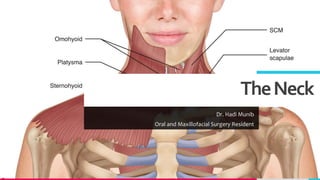
  • 1. TheNeck Dr. Hadi Munib Oral and Maxillofacial Surgery Resident
  • 2. TREYresearch Introduction • The neck is the region of the body that lies between the lower margin of the mandible above and the suprasternal notch and the upper border of the clavicle below. • It is strengthened by the cervical part of the vertebral column, which is convex forward and supports the skull. • Behind the vertebrae is a mass of extensor muscles and in front is a smaller group of flexor muscles. • In the central region of the neck are parts of the respiratory system, namely, the larynx and the trachea, and behind are parts of the alimentary system, the pharynx and the esophagus. • At the sides of these structures are the vertically running carotid arteries, internal jugular veins, the vagus nerve, and the deep cervical lymph nodes Add a footer 2
  • 3. TREYresearch SkinoftheNeck • The natural lines of cleavage of the skin are constant and run almost horizontally around the neck. • This is important clinically because an incision along a cleavage line will heal as a narrow scar. • The one that crosses the lines will heal as a wide or heaped-up scar. • Cutaneous Nerves • The skin overlying the trapezius muscle on the back of the neck and on the back of the scalp as high as the vertex is supplied segmentally by posterior rami of cervical nerves 2 to 5. • The greater occipital nerve is a branch of the posterior ramus of the 2nd cervical nerve. The 1st cervical nerve has no cutaneous branch. • The skin of the front and sides of the neck is supplied by anterior rami of cervical nerves 2 to 4 through branches of the cervical plexus. • The branches emerge from beneath the posterior border of the sternocleidomastoid muscle 3
  • 4. TREYresearch SkinoftheNeck • The lesser occipital nerve (C2) hooks around the accessory nerve and ascends along the posterior border of the sternocleidomastoid muscle to supply the skin over the lateral part of the occipital region and the medial surface of the auricle • The great auricular nerve (C2 and 3) ascends across the sternocleidomastoid muscle and divides into branches that supply the skin over the angle of the mandible, the parotid gland, and on both surfaces of the auricle • The transverse cutaneous nerve (C2 and 3) emerges from behind the middle of the posterior border of the sternocleidomastoid muscle. It passes forward across that muscle and divides into branches that supply the skin on the anterior and lateral surfaces of the neck, from the body of the mandible to the sternum • The supraclavicular nerves (C3 and 4) emerge from beneath the posterior border of the sternocleidomastoid muscle and descend across the side of the neck. They pass onto the chest wall and shoulder region, down to the level of the second rib. Add a footer 4
  • 5. TREYresearch SkinoftheNeck • The medial supraclavicular nerve crosses the medial end of the clavicle and supplies the skin as far as the median plane. • The intermediate supraclavicular nerve crosses the middle of the clavicle and supplies the skin of the chest wall. • The lateral supraclavicular nerve crosses the lateral end of the clavicle and supplies the skin over the shoulder and the upper half of the deltoid muscle; this nerve also supplies the posterior aspect of the shoulder as far down as the spine of the scapula. Add a footer 5
  • 8. TREYresearch SuperficialFascia • The superficial fascia of the neck forms a thin layer that encloses the platysma muscle. • Also embedded in it are the cutaneous nerves, the superficial veins, and the superficial lymph nodes. • Platysma; a thin but clinically important muscular sheet embedded in the superficial fascia. • Superficial Veins; External Jugular Vein • The external jugular vein begins just behind the angle of the mandible by the union of the posterior auricular vein with the posterior division of the retromandibular vein • It descends obliquely across the sternocleidomastoid muscle and, just above the clavicle in the posterior triangle, pierces the deep fascia and drains into the subclavian Vein. • It varies considerably in size, and its course extends from the angle of the mandible to the middle of the clavicle. 8
  • 9. TREYresearch SuperficialFascia • Tributaries • The external jugular vein has the following tributaries: • Posterior auricular vein • Posterior division of the retromandibular vein • Posterior external jugular vein; a small vein that drains the posterior part of the scalp and neck and joins the external jugular vein about halfway along its course • Transverse cervical vein • Suprascapular vein • Anterior jugular vein Add a footer 9
  • 11. TREYresearch AnteriorJugularVein • The anterior jugular vein begins just below the chin, by the union of several small veins. • It runs down the neck close to the midline. • Just above the suprasternal notch, called the jugular arch. • The vein then turns sharply laterally and passes deep to the sternocleidomastoid muscle to drain into the external jugular vein. • Superficial Lymph Nodes • The superficial cervical lymph nodes lie along the external jugular vein superficial to the sternocleidomastoid muscle • They receive lymph vessels from the occipital and mastoid lymph nodes and drain into the deep cervical lymph nodes. Add a footer 11
  • 14. TREYresearch BonesoftheNeck • Cervical Vertebrae • Hyoid Bone • The hyoid bone is a mobile single bone found in the midline of the neck below the mandible and abides the larynx. • It does not articulate with any other bones. • The hyoid bone is U shaped and consists of a body and two greater and two lesser cornua. • It is attached to the skull by the stylohyoid ligament and to the thyroid cartilage by the thyrohyoid membrane. • The hyoid bone forms a base for the tongue and is suspended in position by muscles that connect it to the mandible, to the styloid process of the temporal bone, to the thyroid cartilage, to the sternum, and to the scapula Add a footer 14
  • 18. TREYresearch KeyNeckMuscles • Sternocleidomastoid Muscle • When the sternocleidomastoid muscle contracts, it appears as an oblique band crossing the side of the neck from the sternoclavicular joint to the mastoid process of the skull. • It divides the neck into anterior and posterior triangles. • The anterior border covers the carotid arteries, the internal jugular vein, and the deep cervical lymph nodes; it also overlaps the thyroid gland. • The muscle is covered superficially by skin, fascia, the platysma muscle, and the external jugular vein. The deep surface of the posterior border is related to the cervical plexus of nerves, the phrenic nerve, and the upper part of the brachial plexus Add a footer 18
  • 19. TREYresearch MuscularTrianglesoftheNeck • Anterior Triangle; bounded above by the body of the mandible, posteriorly by the sternocleidomastoid muscle, and anteriorly by the midline; It is further subdivided into the carotid triangle, the digastric triangle, the submental triangle, and the muscular triangle • Posterior Triangle; bounded posteriorly by the trapezius muscle, anteriorly by the sternocleidomastoid muscle, and inferiorly by the clavicle and is further subdivided by the inferior belly of the omohyoid muscle into a large occipital triangle above and a small supraclavicular triangle below Add a footer 19
  • 23. TREYresearch KeyNeckMuscles-ScalenusAnteriorMuscle • A key muscle in understanding the root of the neck. • It is deeply placed and it descends almost vertically from the vertebral column to the 1st rib. • Important Relations • Anteriorly: Related to the carotid arteries, the vagus nerve, the internal jugular vein, and the deep cervical lymph nodes. • The transverse cervical and suprascapular arteries and the prevertebral layer of deep cervical fascia bind the phrenic nerve to the muscle. • Posteriorly: Related to the pleura, the origin of the brachial plexus, and the second part of the subclavian artery. • The scalenus medius muscle lies behind the scalenus anterior muscle. • Medially: Related to the vertebral artery and vein and the sympathetic trunk. • On the left side, the medial border is related to the thoracic duct. 23
  • 24. TREYresearch KeyNeckMuscles–ScalenusAnteriorMuscle • Laterally: Related to the emerging branches of the cervical plexus, the roots of the brachial plexus, and the third part of the subclavian artery Add a footer 24
  • 25. TREYresearch DeepCervicalFascia • The deep cervical fascia supports the muscles, the vessels, and the viscera of the neck. • In certain areas, it is condensed to form well-defined, fibrous sheets called the investing layer, the pretracheal layer, and the prevertebral layer. • It is also condensed to form the carotid sheath. • Investing Layer; a thick layer that encircles the neck; It splits to enclose the trapezius and the sternocleidomastoid muscles. • Pretracheal Layer; a thin layer that is attached above to the laryngeal cartilages; It surrounds the thyroid and the parathyroid glands, forming a sheath for them, and encloses the infrahyoid muscles. • Prevertebral Layer; a thick layer that passes like a septum across the neck behind the pharynx and the esophagus and in front of the prevertebral muscles and the vertebral column; It forms the fascial floor of the posterior triangle, and it extends laterally over the first rib into the axilla to form the important axillary sheath • Carotid Sheath; a local condensation of the prevertebral, the pretracheal, and the investing layers of the deep fascia that surround the common and internal carotid arteries, the internal jugular vein, the vagus nerve, and the deep cervical lymph nodes 25
  • 29. TREYresearch DeepCervicalFascia • Axillary Sheath • All the anterior rami of the cervical nerves that emerge in the interval between the Scalenus anterior and scalenus medius muscles lie at first deep to the prevertebral fascia. • As the subclavian artery and the brachial plexus emerge in the interval between the scalenus anterior and the scalenus medius muscles, they carry with them a sheath of the fascia, which extends into the axilla and is called the axillary sheath. • Cervical Ligaments • Stylohyoid ligament: Connects the styloid process to the lesser cornu of the hyoid bone • Stylomandibular ligament: Connects the styloid process to the angle of the mandible • Sphenomandibular ligament: Connects the spine of the sphenoid bone to the lingula of the mandible • Pterygomandibular ligament: Connects the hamular process of the medial pterygoid plate to the posterior end of the mylohyoid line of the mandible. It gives attachment to the superior constrictor and the buccinator muscles Add a footer 29
  • 30. TREYresearch ArteriesoftheHeadandNeck 30 • Common Carotid Artery • The right common carotid artery arises from the brachiocephalic artery behind the right sternoclavicular joint • The left artery arises from the arch of the aorta in the superior mediastinum • The common carotid artery runs upward through the neck under cover of the anterior border of the sternocleidomastoid muscle, from the sternoclavicular joint to the upper border of the thyroid cartilage. • Here, it divides into the external and internal carotid arteries. • Carotid Sinus; At its point of division, the terminal part of the common carotid artery or the beginning of the internal carotid artery shows a localized dilatation. • The tunica media of the sinus is thinner than elsewhere, but the adventitia is relatively thick and contains numerous nerve endings derived from the glossopharyngeal nerve. • The carotid sinus serves as a reflex presso-receptor mechanism: A rise in blood pressure causes a slowing of the heart rate and vasodilatation of the arterioles.
  • 31. TREYresearch ArteriesoftheHeadandNeck • Carotid Body • The carotid body is a small structure that lies posterior to the point of bifurcation of the common carotid artery • It is innervated by the glossopharyngeal nerve. • The carotid body is a chemoreceptor, being sensitive to excess carbon dioxide and reduced oxygen tension in the blood. • Such a stimulus reflexly produces a rise in blood pressure and heart rate and an increase in respiratory movements. • The common carotid artery is embedded in a connective tissue sheath, called the carotid sheath, throughout its course and is closely related to the internal jugular vein and vagus nerve Add a footer 31
  • 32. TREYresearch ArteriesoftheHeadandNeck • Relations of the Common Carotid Artery • Anterolaterally: The skin, the fascia, the sternocleidomastoid, the sternohyoid, the sternothyroid, and the superior belly of the omohyoid • Posteriorly: The transverse processes of the lower four cervical vertebrae, the prevertebral muscles, and the sympathetic trunk; In the lower part of the neck are the vertebral vessels. • Medially: The larynx and pharynx and, below these, the trachea and esophagus; The lobe of the thyroid gland also lies medially. • Laterally: The internal jugular vein and, posterolaterally, the vagus nerve • Branches of the Common Carotid Artery; Apart from the two terminal branches, the common carotid artery gives off no branches. Add a footer 32
  • 33. TREYresearch ExternalCarotidArtery • One of the terminal branches of the common carotid artery. • It supplies structures in the neck, face, and scalp; it also supplies the tongue and the maxilla. • The artery begins at the level of the upper border of the thyroid cartilage and terminates in the substance of the parotid gland behind the neck of the mandible by dividing into the superficial temporal and maxillary arteries. • Close to its origin, the artery emerges from undercover of the sternocleidomastoid muscle, where its pulsations can be felt. • At first, it lies medial to the internal carotid artery, but as it ascends in the neck, it passes backward and lateral to it. • It is crossed by the posterior belly of the digastric and the stylohyoid. Add a footer 33
  • 34. TREYresearch RelationsoftheExternalCarotidArtery • Anterolaterally: The artery is overlapped at its beginning by the anterior border of the sternocleidomastoid. • Above this level, the artery is comparatively superficial, being covered by skin and fascia. • It is crossed by the hypoglossal nerve, the posterior belly of the digastric muscle, and the stylohyoid muscles. • Within the parotid gland, it is crossed by the facial nerve. • The internal jugular vein first lies lateral to the artery and then posterior to it. • Medially: The wall of the pharynx and the internal carotid artery. • The stylopharyngeus muscle, the glossopharyngeal nerve, and the pharyngeal branch of the vagus pass between the external and internal carotid Arteries. 34
  • 35. TREYresearch BranchesoftheExternalCarotidArtery • Superior thyroid artery • Ascending pharyngeal artery • Lingual artery • Facial artery • Occipital artery • Posterior auricular artery • Superficial temporal artery • Maxillary artery Add a footer 35
  • 36. TREYresearch BranchesofExternalCarotidArtery • Superior Thyroid Artery; Curves downward to the upper pole of the thyroid gland; It is accompanied by the external laryngeal nerve, which supplies the cricothyroid muscle. • Ascending Pharyngeal Artery; Ascends along and supplies the pharyngeal wall. • Lingual Artery; loops upward and forward and supplies the tongue. • Facial Artery; Loops upward close to the outer surface of the pharynx and the tonsil. It lies deep to the submandibular salivary gland and emerges and bends around the lower border of the mandible; It then ascends over the face close to the anterior border of the masseter muscle. The artery then ascends around the lateral margin of the mouth and terminates at the medial angle of the eye. • Branches of the facial artery supply the tonsil, the submandibular salivary gland, and the muscles and the skin of the face. • Occipital Artery; supplies the back of the scalp. • Posterior Auricular Artery; supplies the auricle and the scalp. Add a footer 36
  • 37. TREYresearch BranchesofExternalCarotidArtery • Superficial Temporal Artery; ascends over the zygomatic arch, where it may be palpated just in front of the auricle; It is accompanied by the Auriculotemporal nerve, and it supplies the scalp. • Maxillary Artery; runs forward medial to the neck of the mandible and enters the pterygopalatine fossa of the skull. • Branches of the Maxillary Artery; supply the upper and the lower jaws, the muscles of mastication, the nose, the palate, and the meninges inside the skull. • Middle Meningeal Artery; enters the skull through the foramen spinosum. • It runs laterally within the skull and divides into anterior and posterior branches • The anterior branch is important because it lies close to the motor area of the cerebral cortex of the brain. • Accompanied by its vein, it grooves (or tunnels) through the upper part of the greater wing of the sphenoid bone of the skull and the thin anteroinferior angle of the parietal bone, where it is prone to damage after a blow to the head. Add a footer 37
  • 40. TREYresearch InternalCarotidArtery • The internal carotid artery begins at the bifurcation of the common carotid artery at the level of the upper border of the thyroid cartilage. • It supplies the brain, the eye, the forehead, and part of the nose. • The artery ascends in the neck embedded in the carotid sheath with the internal jugular vein and vagus nerve. • At first it lies superficially; it then passes deep to the parotid salivary Gland. • The internal carotid artery leaves the neck by passing into the cranial cavity through the carotid canal in the petrous part of the temporal bone. It then passes upward and forward in the cavernous venous sinus (without communicating with it). • The artery then leaves the sinus and passes upward again medial to the anterior clinoid process of the sphenoid bone. • The internal carotid artery then inclines backward, lateral to the optic chiasma, and terminates by dividing into the anterior and the middle cerebral arteries. Add a footer 40
  • 41. TREYresearch RelationsoftheInternalCarotidArteryintheNeck • Anterolaterally: Below the digastric lie the skin, the fascia, the anterior border of the sternocleidomastoid, and the hypoglossal nerve. • Above the digastric lie the stylohyoid muscle, the stylopharyngeus muscle, the glossopharyngeal nerve, the pharyngeal branch of the vagus, the parotid gland, and the external carotid artery. • Posteriorly: The sympathetic trunk, the longus capitis muscle, and the transverse processes of the upper three cervical vertebrae • Medially: The pharyngeal wall and the superior laryngeal nerve • Laterally: The internal jugular vein and the vagus nerve. • Branches of the Internal Carotid Artery • There are no branches in the neck. Many important branches, however, are given off in the skull. 41
  • 42. TREYresearch InternalCarotidArtery • Ophthalmic Artery; arises from the internal carotid artery as it emerges from the cavernous sinus. • It passes forward into the orbital cavity through the optic canal, and it gives off the central artery of the retina, which enters the optic nerve and runs forward to enter the eyeball. • The central artery is an end artery and the only blood supply to the retina. • Posterior Communicating Artery; runs backward to join the posterior cerebral artery • Anterior Cerebral Artery; is a terminal branch of the internal carotid artery; It passes forward between the cerebral hemispheres and then winds around the corpus callosum of the brain to supply the medial and the superolateral surfaces of the cerebral hemisphere. It is joined to the artery of the opposite side by the anterior communicating artery. • Middle Cerebral Artery; is the largest terminal branch of the internal carotid artery, and it runs laterally in the lateral cerebral sulcus of the brain. It supplies the entire lateral surface of the cerebral hemisphere except the narrow strip along the superolateral margin (which is supplied by the anterior cerebral artery) and the occipital pole and inferolateral surface of the hemisphere (both of which are supplied by the posterior cerebral artery). • The middle cerebral artery thus supplies all the motor area o the cerebral cortex except the leg area. It also gives off central branches that supply central masses of gray matter and the internal capsule of the brain. 42
  • 43. TREYresearch InternalCarotidArtery • Circle of Willis lies in the subarachnoid space at the base of the brain. It is formed by the anastomosis between the branches of the two internal carotid arteries and the two vertebral arteries. • The anterior communicating, posterior cerebral, and basilar (formed by the junction of the two vertebral arteries) are all arteries that contribute to the circle. Cortical and central branches arise from the circle and supply the brain. • Subclavian Arteries • Right Subclavian Artery; arises from the brachiocephalic artery, behind the right sternoclavicular joint; It arches upward and laterally over the pleura and between the scalenus anterior and medius muscles. At the outer border of the 1st rib, it becomes the axillary artery. • Left Subclavian Artery; arises from the arch of the aorta in the thorax. It ascends to the root of the neck and then arches laterally in a manner similar to that of the right subclavian artery. • The scalenus anterior muscle passes anterior to the artery on each side and divides it into three parts. 43
  • 44. TREYresearch FirstPartoftheSubclavianArtery • Extends from the origin of the subclavian artery to the medial border of the scalenus anterior muscle. • This part gives off the vertebral artery, the thyrocervical trunk, and the internal thoracic artery. • Branches The vertebral artery ascends in the neck through the foramina in the transverse processes of the upper six cervical vertebrae. • It passes medially above the posterior arch of the atlas and then ascends through the foramen magnum into the skull. • On reaching the anterior surface of the medulla oblongata of the brain at the level of the lower border of the pons, it joins the vessel of the opposite side to form the basilar artery. • The basilar artery; ascends in a groove on the anterior surface of the pons. It gives off branches to the pons, the cerebellum, and the internal ear. It finally divides into the two posterior cerebral arteries. • The posterior cerebral artery curves laterally and backward around the midbrain. Cortical branches supply the inferolateral surfaces of the temporal lobe and the visual cortex on the lateral and the medial surfaces of the occipital lobe. 44
  • 45. TREYresearch FirstPartoftheSubclavianArtery • Branches in the neck: Spinal and muscular arteries • Branches in the skull: Meningeal, anterior and posterior spinal, posterior inferior cerebellar, medullary arteries. • The thyrocervical trunk is a short trunk that gives off three terminal branches. • The inferior thyroid artery ascends to the posterior surface of the thyroid gland, where it is closely related to the recurrent laryngeal nerve. It supplies the thyroid and the inferior parathyroid glands. • The superficial cervical artery is a small branch that crosses the brachial plexus. • The suprascapular artery runs laterally over the brachial plexus and follows the suprascapular nerve onto the back of the scapula. • The internal thoracic artery descends into the thorax behind the 1st costal cartilage and in front of the pleura. It descends vertically one fingerbreadth lateral to the sternum; in the 6th intercostal space, it divides into the superior epigastric and the musculophrenic arteries. Add a footer 45
  • 46. TREYresearch SecondPartoftheSubclavianArtery • Lies behind the scalenus anterior muscle. • Branches The costocervical trunk runs backward over the dome of the pleura and divides into the superior intercostal artery, which supplies the 1st and the 2nd intercostal spaces, and the deep cervical artery, which supplies the deep muscles of the neck. • Third Part of the Subclavian Artery • Extends from the lateral border of the scalenus anterior muscle across the posterior triangle of the neck to the lateral border of the 1st rib, where it becomes the axillary artery. Here, in the root of the neck, it is closely related to the nerves of the brachial plexus. • Branches; The third part of the subclavian artery usually has no branches. • Occasionally, however, the superficial cervical arteries, the suprascapular arteries, or both arise from this part. Add a footer 46
  • 47. TREYresearch VeinsoftheHeadandNeck • The veins of the brain, venous sinuses, diploic veins, and emissary veins • The veins of the scalp, face, and neck • Veins of the Brain; thin walled and have no valves. • They consist of the cerebral veins, the cerebellar veins, and the veins of the brainstem, all of which drain into the neighboring venous sinuses. • Venous Sinuses; situated between the periosteal and the meningeal layer of the dura mater. • They have thick, fibrous walls, but they possess no valves. • They receive tributaries from the brain, the skull bones, the orbit, and the internal ear. • The venous sinuses include the superior and inferior sagittal sinuses, the straight sinus, the transverse sinuses, the sigmoid sinuses, the occipital sinus, the cavernous sinuses, and the superior and inferior petrosal sinuses. Add a footer 47
  • 48. TREYresearch VeinsoftheHeadandNeck • Diploic Veins; occupy channels within the bones of the vault of the skull. • Emissary Veins; are valveless veins that pass through the skull bones. • They connect the veins of the scalp to the venous sinuses (and are an important route for the spread of infection). • Veins of the Face and the Neck • Facial Vein; formed at the medial angle of the eye by the union of the supraorbital and supratrochlear veins. • It is connected through the ophthalmic veins with the cavernous sinus. • The facial vein descends down the face with the facial artery and passes around the lateral side of the mouth. • It then crosses the mandible, is joined by the anterior division of the retromandibular vein, and drains into the internal jugular vein. • Superficial Temporal Vein; formed on the side of the Scalp. • It follows the superficial temporal artery and the auriculotemporal nerve and then enters the parotid salivary gland, where it joins the maxillary vein to form the retromandibular vein. 48
  • 49. TREYresearch VeinsoftheHeadandNeck • Maxillary Vein; formed in the infratemporal fossa from the pterygoid venous plexus. • The maxillary vein joins the superficial temporal vein to form the retromandibular vein. • Retromandibular Vein; formed by the union of the superficial temporal and the maxillary veins. • On leaving the parotid salivary gland, it divides into an anterior branch, which joins the facial vein, and a posterior branch, which joins the posterior auricular vein to form the external jugular vein. • External Jugular Vein; formed behind the angle of the jaw by the union of the posterior auricular vein with the posterior division of the retromandibular vein; It descends across the sternocleidomastoid muscle and beneath the platysma muscle, and it drains into the subclavian vein behind the middle of the clavicle. • Tributaries • Posterior external jugular vein from the back of the scalp • Transverse cervical vein from the skin and the fascia over the posterior triangle • Suprascapular vein from the back of the scapula • Anterior jugular vein Add a footer 49
  • 50. TREYresearch VeinsoftheHeadandNeck • Anterior Jugular Vein; descends in the front of the neck close to the midline. • Just above the sternum, it is joined to the opposite vein by the jugular arch. • The anterior jugular vein joins the external jugular vein deep to the sternocleidomastoid muscle. • Internal Jugular Vein; a large vein that receives blood from the brain, face, and neck. • It starts as a continuation of the sigmoid sinus and leaves the skull through the jugular foramen. It then descends through the neck in the carotid sheath lateral to the vagus nerve and the internal and common carotid arteries. • It ends by joining the subclavian vein behind the medial end of the clavicle to form the brachiocephalic vein. • Throughout its course, it is closely related to the deep cervical lymph nodes. • The vein has a dilatation at its upper end called the superior bulb and another near its termination called the inferior bulb. • Directly above the inferior bulb is a bicuspid valve. 50
  • 51. TREYresearch RelationsoftheInternalJugularVein • Anterolaterally: The skin, the fascia, the sternocleidomastoid, and the parotid salivary gland. Its lower part is covered by the sternothyroid, sternohyoid, and omohyoid muscles, which intervene between the vein and the sternocleidomastoid. • Higher up, it is crossed by the stylohyoid, the posterior belly of the digastric, and the spinal part of the accessory nerve. • The chain of deep cervical lymph nodes runs alongside the vein. • Posteriorly: The transverse processes of the cervical vertebrae, the levator scapulae, the scalenus medius, the scalenus anterior, the cervical plexus, the phrenic nerve, the thyrocervical trunk, the vertebral vein, and the first part of the subclavian artery, on the left side, it passes in front of the thoracic duct. • Medially: Above lie the internal carotid artery and the 9th, 10th, 11th, and 12th cranial nerves. Below lie the common carotid artery and the vagus nerve. Add a footer 51
  • 52. TREYresearch TributariesoftheInternalJugularVein • Inferior petrosal sinus • Facial vein • Pharyngeal veins • Lingual vein • Superior thyroid vein • Middle thyroid vein • Subclavian Vein; a continuation of the axillary vein at the outer border of the 1st rib; It joins the internal jugular vein to form the brachiocephalic vein, and it receives the external jugular vein. In addition, it often receives the thoracic duct on the left side and the right lymphatic duct on the right. • Relations • Anteriorly: The clavicle • Posteriorly: The scalenus anterior muscle and the phrenic nerve • Inferiorly: The upper surface of the 1st rib 52
  • 55. TREYresearch LymphDrainageoftheHeadandNeck • Arranged as a regional collar that extends from below the chin to the back of the head and as a deep vertical terminal group that is embedded in the carotid sheath in the neck. • Regional Nodes • Occipital nodes: These are situated over the occipital bone on the back of the skull. They receive lymph from the back of the scalp. • Retroauricular (mastoid) nodes: These lie behind the ear over the mastoid process. They receive lymph from the scalp above the ear, the auricle, and the external auditory meatus. • Parotid nodes: These are situated on or within the parotid salivary gland. They receive lymph from the scalp above the parotid gland, the eyelids, the parotid gland, the auricle, and the external auditory meatus. • Buccal (facial) nodes: One or two nodes lie in the cheek over the buccinator muscle. They drain lymph that ultimately passes into the submandibular nodes. Add a footer 55
  • 56. TREYresearch LymphDrainageoftheHeadandNeck • Submandibular nodes: These lie superficial to the submandibular salivary gland just below the lower margin of the jaw. • They receive lymph from; • Front of the scalp; the nose; the cheek. • The upper lip and the lower lip (except the central part). • The frontal, maxillary, and ethmoid sinuses. • Upper and lower teeth (except the lower incisors) • Anterior two thirds of the tongue (except the tip) • Floor of the mouth and vestibule; and the gums. Add a footer 56
  • 57. TREYresearch LymphDrainageoftheHeadandNeck • Submental nodes: These lie in the submental triangle just below the chin. They drain lymph from the tip of the tongue, the floor of the anterior part of the mouth, the incisor teeth, the center part of the lower lip, and the skin over the chin. • Anterior cervical nodes: These lie along the course of the anterior jugular veins in the front of the neck. They receive lymph from the skin and superficial tissues of the front of the neck. • Superficial cervical nodes: These lie along the course of the external jugular vein on the side of the neck. They drain lymph from the skin over the angle of the jaw, the skin over the lower part of the parotid gland, and the lobe of the ear. • Retropharyngeal nodes: These lie behind the pharynx and in front of the vertebral column. They receive lymph from the nasal pharynx, the auditory tube, and the vertebral column. Add a footer 57
  • 58. TREYresearch LymphDrainageoftheHeadandNeck • Laryngeal nodes: These lie in front of the larynx. They receive lymph from the larynx. • Tracheal (paratracheal) nodes: These lie alongside the trachea. They receive lymph from neighboring structures, including the thyroid gland. • Deep Cervical Nodes; form a vertical chain along the course of the internal jugular vein within the carotid sheath • They receive lymph from all the groups of regional nodes. • The jugulodigastric node, which is located below and behind the angle of the jaw, is mainly concerned with drainage of the tonsil and the tongue. • The juguloomohyoid node, which is situated close to the omohyoid muscle, is mainly associated with drainage of the tongue. • The efferent lymph vessels from the deep cervical lymph nodes join to form the jugular trunk, which drains into the thoracic duct or the right lymphatic duct Add a footer 58
  • 59. TREYresearch References • Chapter 11: The Head and Neck (585 -605) Add a footer 59