
Recommended
More Related Content
Similar to animals , plants , humans cells
Similar to animals , plants , humans cells (20)
More from HUMAMZUHARZUHAR
Recently uploaded
Recently uploaded (20)
animals , plants , humans cells
- 1. CELLS
- 2. Microscopes ● To see cells clearly, you need to use a microscope. ● The kind of microscope used in a school is called a light microscope. The light microscope magnifies up to x 1500.
- 3. An electron microscope magnifies up to x 10 millions.
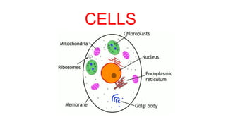
- 6. Cell membrane ● All cells have a cell membrane. ● The cell membrane is a very thin layer of protein and fat.
- 7. ● The cell membrane controls what goes in and out of it. ● It is said to be partially permeable, which means that it will let some substances through but not others.
- 8. Cell wall ● All plant cells surrounded by a cell wall made mainly of cellulose. ● Cell wall protect and support the cell.
- 9. Cytoplasm ● All cells have a cytoplasm. ● It is mainly composed of water in many cells. ● Many different metabolic reactions take place in the cytoplasm.
- 10. Vacuoles ● A vacuole is a space in a cell, surrounded by a membrane, and containing a solution. ● Plant cells have large vacuoles, which contain sugar and other subtances called cell sap. ● Animal cells have much smaller spaces called vesicles which may contain food or water.
- 12. Chloroplast ● They contain the green colouring pigment called chlorophyll. ● Chlorophyll absorbs energy from sunlight and this energy is then used for photosynthesis.
- 13. ● Chloroplasts store starch grains which have been made by photosynthesis. ● Animal cells also have grains similar to starch,called glycogen inside cytoplasm.
- 14. Nucleus ● The nucleus is where the genetic information is stored. ● The information kept on the chromosomes,which are inherited from the organism’s parents. The chromosomes made of DNA.
- 15. Mitochondria ● Almost all cells except prokaryotes have mitochondria.
- 16. ● Mitochondria are the powerhouses of the cell. ● Aerobic respiration occurs in mitochondria ● Cells that use a lot of energy have a lot of mitochondria.(muscle cells, sperm cells, neurons)
- 17. Ribosomes ● Ribosomes are found in all types of cells (bacteria,fungi,animal,plant...) ● Ribosomes are the places where proteins are made. ● They are found in many places around a cell. You might find them floating in the cytoplasm.
- 18. ● Other ribosomes are found on the endoplasmic reticulum. Endoplasmic reticulum with attached ribosomes is called rough ER.
- 19. Micrometres ● Cells, and structures inside them such as mitochondria and ribosomes, are so small that we need a very small unit in which to measure them. The most useful one is the micrometre (µm). ● 1µm = 1 x 10⁻⁶ m ● 1 m = 1 x 106 µm
- 20. Adaptations of Specialised Cells ● Specialised cells are those which have developed certain characteristics in order to perform particular functions. These differences are controlled by genes in the nucleus. ● Cells specialise by undergoing differentiation: this is a process by which cells develop the structure and characteristics needed to be able to carry out their functions. Examples of Specialised Cells in Animals: ★ ciliated cell ★ nerve cell ★ red blood cell ★ sperm cell ★ egg cell (ovum) Examples of Specialised Cells in Plants: ★ root hair cell ★ xylem vessel ★ palisade mesophyll cell
- 21. Ciliated cells
- 22. Nerve cell
- 23. Red blood cells
- 24. Sperm cell
- 25. Egg cell (ovum)
- 26. Root hair cell
- 27. Xylem
- 29. Cells and organisms ● Cell : the smallest unit of life ● Tissue : an aggregate of cells in an organism that have similar structure and function. ● Organ : a group of tissues that perform a specific function or group of functions. ● Organ System : A group of entities or organs that work together to carry out a particular task
- 32. Calculating Magnification & Specimen Size Calculating magnification and specimen size using millimetres as units ● Magnification is calculated using the following equation: Magnification = Drawing size ÷ Actual size A better way to remember the equation is using an equation triangle:
- 33. ● Rearranging the equation to find things other than the magnification becomes easy when you remember the triangle – whatever you are trying to find, place your finger over it and whatever is left is what you do, so: ○ Magnification = image size / actual size ○ Actual size = image size / magnification ○ Image size = magnification x actual size ● Remember magnification does not have any units and is just written as ‘x 10’ or ‘x 5000’
- 34. An image of an animal cell is 30 mm in size and it has been magnified by a factor of x 3000. What is the actual size of the cell? To find the actual size of the cell:
- 35. Calculating Magnification & Specimen Size Using millimetres and micrometres as units ● The table below shows how millimetres are related to two other measures of length
- 37. ● Remember 1mm = 1000µm ● 2000 / 1000 = 2 so the actual thickness of the leaf is 2mm and the drawing thickness is 50mm ● Magnification = image size / actual size = 50 / 2 = 25 ● So the magnification is x 25 (NO UNITS)
- 38. The following diagram may help with unit conversion between mm and µm: