FOETAL SKULL.pptx
•Download as PPTX, PDF•
2 likes•318 views

Foetal Skull, Definition, Parts & Regions of skull, bones of foetal skull, sutures, fontanels, landmark & diameters of foetal skull

Recommended
More Related Content
What's hot
What's hot (20)
Similar to FOETAL SKULL.pptx
Similar to FOETAL SKULL.pptx (20)
Recently uploaded
Recently uploaded (20)
FOETAL SKULL.pptx
- 1. PRESENTED BY: MRS. GURWINDER KAUR
- 2. The foetal skull is the most difficult part of the baby to pass through the mother’s pelvic canal, due to the hard bony nature of the skull. Understanding the anatomy of the foetal skull and its diameter will help you recognise how a labour is progressing, and whether the baby’s head is ‘presenting’ correctly as it comes down the birth canal. INTRODUCTION
- 3. This will give you a better understanding of whether a normal vaginal delivery is likely, or if the mother needs referral because the descent of the baby’s head is not making sufficient progress. The head is the most difficult part to deliver whether it comes first or last. INTRODUCTION
- 4. • Foetal skull is compressible, and made mainly of thin pliable tabular (flat) bones forming the vault. • The skull bone encases and protects the brain. DEFINITION
- 5. VERTEX: It is a quadrangular area bounded anteriorly by the bregma and coronal suture behind by the lambda and lambdoidal sutures and laterally by lines passing through the parietal eminences. BROW: It is an area bounded on one side by the anterior fontanelle and coronal sutures and on the other side by the root of the nose and supra-orbital ridges of either side. PARTS AND REGIONS OF FOETAL SKULL
- 6. PARTS AND REGIONS OF FOETAL SKULL
- 7. FACE: It is the area bounded by the root of the nose and supraorbital ridges and on the other, by the junction of the floor of the mouth with neck. SINCIPUT: It is the area lying in front of the anterior fontanelle and corresponds to the area of brow. OCCIPUT: It is the area limited to the occipital bone. PARTS AND REGIONS OF FOETAL SKULL CONT...
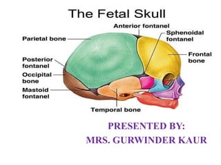
- 8. TWO FRONTAL BONES: Lies in front of the skull. At the centre of each in the frontal eminence. It fuses into single by 8 yrs. of age. These form the forehead or sinciput. TWO PARIETAL BONES: Lies on either side of skull ossification centre of each called the parietal eminence. BONES OF FOETAL SKULL
- 9. ONE OCCIPITAL BONE: Lies at the back of head & at the centre is occipital protuberance. Part of it contributes to the base of skull, as it contains foramen magnum, which protects the spinal cord as it leaves the skull. It forms the region of occiput. In addition to these 5 the upper part of temporal bone is also flat to form small part of vault. BONES OF FOETAL SKULL
- 10. BONES OF FOETAL SKULL
- 11. Flat bones of the vault are united together by the nonosssified membranes attached to the margins of the bones. These are called sutures. These sutures are: The Saggital Suture The Coronal Suture The Lambdoidal Suture The Frontal Suture SUTURES
- 12. SUTURES
- 13. It permits gliding movement of one bone over the other during moulding of the head. Digital palpations of sagittal suture during internal examination in labour gives an idea of the manner of engagement of the head, degree of internal rotation of the head and degree of moulding of the head. IMPORTANCE OF SUTURES
- 14. DEFINITION: Wide gap in the suture line is called fontanelle. There are two types of main fontanelles present in foetal skull and these are: Anterior Fontanelle Posterior Fontanelle FONTANELLES
- 15. ANTERIOR FONTANELLE: Anterior fontanelle is formed by joining of four sutures in midplane. The sutures are Anteriorly frontal, Posteriorly saggital and on either side coronal suture. The shape of the bregma is like a Diamond. Its anterior-posterior and transverse diameters measures approximately 3cm each. The floor is made by a membrane and it is ossified at 18months after birth. FONTANELLES CONT..
- 17. IMPORTANCE OFANTERIOR FONTANELLE: Its palpation through internal examination denotes the degree of flexion of the head. It facilitates moulding of the head. As it remains membranous long after birth, it helps in accommodating the marked brain growth, the brain becoming almost double its size during first year of life. FONTANELLES CONT..
- 18. Palpation of the floor reflects intracranial status depressed in dehydration, elevated in raised intracranial tension. Collection of blood and exchange transfusion, on rare occasion, can be performed through it via the superior longitudinal sinus. Cerebrospinal fluid can be drawn, although rare, through the lateral angle of the anterior fontanelle from the lateral ventricle. FONTANELLES CONT..
- 19. POSTERIOR FONTANELLE: It is also known as lambda and is formed by junction of three line- anteriorly by saggital suture and on the either side by the lamboidal suture. Its triangular in shape and measures about 1.2cm X 1.2cm. The floor is made by a membrane but becomes bony at term. It is ossified at 6-8 weeks after birth. FONTANELLES CONT..
- 21. Occiput- is the occipital bone/external occipital protuberance. Sinciput- is the forehead region of foetal head. Parietal eminences- are the eminences of parietal on either side. Mentum- it is the chin. Vertical point- it is the center of saggital suture. Frontal point- is the root of nose. LANDMARKS OF FOETAL SKULL
- 22. Sub occiput- is the junction foetal neck and occiput. Sub mentum- it is the junction between neck and chin. Bi parietal- is the transverse distance between two parietal eminences. Bi temporal- is the distance between two lower end of coronal suture. LANDMARKS OF FOETAL SKULL
- 23. There are two types of diameters of foetal skull. These are: Anterior-Posterior Diameter Transverse Diameter ANTERIOR-POSTERIOR DIAMETER: Suboccipito-bregmatic: It extends from the nape of the neck to the centre of the bregma. It measures 9.5cm. DIAMETERS OF FOETAL SKULL
- 24. Suboccipito-frontal: It extends from the nape of the neck to the centre of the frontral suture. It measures 10cm. Occipito-frontal: It extends from the occipital eminence to the root of the nose and supra orbital ridges. It measures 11.5cm. Mento-vertical: It extends from the midpoint of the chin to the highest point on the saggital sutre. It measures 14cm. DIAMETERS OF FOETAL SKULL
- 25. Submento-vertical: It extends from the junction of mouth with neck to the highest point on the saggital suture. It measures 11.5cm Submento-bregmatic: It extends from the junction of mouth with neck to centre of bregma. It measures 9.5cm. DIAMETERS OF FOETAL SKULL
- 26. DIAMETERS OF FOETAL SKULL
- 27. TRANSVERSE DIAMETERS: Biparietal: It extends between two parietal eminences. It measures 9.5cm Super-subparietal: It extends from a point placed below one parietal eminence to a point placed above the other parietal eminence of the opposite. It measures 8.5cm. DIAMETERS OF FOETAL SKULL
- 28. Bitemporal: It is the distance between the antero-inferior ends of the coronal suture. It measures 8cm. Bimastoid: It is the distance between the tips of the mastoid processes. It measures 7cm. DIAMETERS OF FOETAL SKULL
- 29. SUMMARIZATION
- 30. RECAPTULIZATION
- 31. Fraser. M. Diane, Cooper. A. Margaret,” Myles Textbook for Midwives”, 14th Edition, Published by Churchill Livingstone Publisher. Pg. No: 157-160. Dutta’s C.D, “Textbook of Obstetrics”, 6th Edition, Published by New central Book Agency (P) Ltd. Pg. No: 86-89. BIBLIOGRAPHY
- 32. Jacob Annamma, “Midwifer casebook A practical record of Maternal Nursing”, 7th Edition, Jaypee Brothers medical publishers (P) Ltd. Pg No: 5-7. BIBLIOGRAPHY