2. research worldwide. It is broadly accepted that novel mechanism
(new-class) agents represent unique and valuable opportunities
to achieve significant advances against bacterial resistance
because they should not be as susceptible to the pre-existing
mechanisms of resistance as are established antibacterial classes,
i.e., they should not exhibit cross-resistance.4,5
This is in contrast
to the more common strategy of introducing multiple new
members within existing antibacterial classes, a strategy that is
typically viewed as an incremental, and thus more temporary and
limited, advance against resistance. The latter situation has
persisted for most of the antibacterial classes used today
including the β-lactams, aminoglycosides, tetracyclines, and
quinolones. The introduction into clinical practice today of an
ATPase inhibitor of gyrase and topo IV would provide a novel
class agent to support our current efforts to combat increasingly
resistant bacterial pathogens, including resistant Gram negative
pathogens such as Pseudomonas aeruginosa, Acinetobacter
baumannii, and Klebsiella pneumoniae.
In this critical analysis, we examine the historical gyrase/topo
IV ATPase inhibitor programs to learn what may have hindered
the development and launch of other such antibacterial agents
during the last five and a half decades. Clearly, the failure to
introduce an improved successor to novobiocin into clinical
practice has not been due to lack of effort. Since the mid-1960s,
major pharmaceutical companies and, more recently, biotechs
have been engaged vigorously in research efforts toward that goal
(Chart 1), yet despite seemingly determined and significant
efforts by many teams of highly experienced drug R&D
professionals over many decades, no antibacterial drug with
novobiocin’s mechanism has progressed even beyond Phase I.
Why?
This narrative-style analysis summarizes individual drug
discovery projects along with relevant key scientific discoveries
beginning with the discovery of novobiocin and the historical
context for which its medical usefulness was established. Over
subsequent decades, the other multiple lines of research into
same-mechanism agents are examined in their own historical
Figure 1. Structures of early coumarin scaffold GyrB/ParE inhibitor antibacterials.
ACS Infectious Diseases Review
DOI: 10.1021/id500013t
ACS Infect. Dis. XXXX, XXX, XXX−XXX
B
3. contexts, and as the history of novobiocin itself continued to
evolve, they are shown to build on and influence one another.
Overall the history can be viewed in three broad phases: (1) a
pre-1990s phase, largely empirically driven with natural products
being the source of drug discovery projects; (2) the decade of the
1990s wherein X-ray crystallography, the discovery of the second
target topo IV, and a growing recognition of the factors
governing resistance development and drug permeation through
bacterial membranes contributed to an expanded scientific
foundation; and (3) drug discovery projects initiated during and
after the late 1990s that employed those new scientific
foundations and shifted lead-finding methods from natural
products to high-throughput screening (HTS) and/or computa-
tional techniques. The numerous drug scaffolds identified during
this latter period are logically grouped together according to
binding mode in the gyrase/topo IV enzymes and more
specifically according to the particular molecular motifs bridging
to a key aspartic acid and structural water unit residing within the
ATP binding pockets of gyrase and topo IV (cf. Figures 7, 10, 12,
16, and 19).
On the basis of this analysis, we argue that the historical lack of
success in developing gyrase/topo IV ATPase inhibiting agents
lies not in any fundamental flaw in the target or mechanism nor in
any of the scientific approaches to the discovery of novel natural
product or synthetic scaffolds to engage that particular
mechanism. On the contrary, the discovery and launch of agents
within this class, especially in light of the considerable insight
gained over the last 10−15 years, should now be one of the most
scientifically achievable strategies for the introduction of a safe
and effective novel mechanism antibacterial agent in the present
era. In reviewing the historical attempts to discover and develop
agents within this class, we discuss various factors that may have
hindered those efforts. With the benefit of accumulated
knowledge and experience, we regard those historical technical
issues either as quite solvable or avoidable. We conclude this
review by providing a broader context for the efforts toward
gyrase/topo IV ATPase inhibitors within the evolving history
and economics of antibacterial drug discovery and offer some
concluding strategic observations that we hope will be useful to
leaders of any new-class antibacterial program. We additionally
hope that this analysis of the research and development of a
specific class of antibacterial drugs might serve as a model for
critical analyses of other lines of antibacterial drug discovery in
the field or even for drug discovery projects in other therapeutic
areas.
1. NOVOBIOCIN, A FIT-FOR-PURPOSE DRUG? HEYDAY
AND CRASH (1955−1960s)
During the late 1940s and early 1950s, not only had the
usefulness of the sulfonamide class of antibacterials declined
drastically because of the emergence of resistant organisms, but
penicillin itself was rapidly losing effectiveness against a range of
medically important pathogens, most notably against Staph-
ylococcus aureus.6,7
During this time, most major pharmaceutical
companies had entered the field of antibacterial R&D and were
making important discoveries of new-class agents, several of
which could potentially be used as clinically effective
antistaphylococcal drugs in place of penicillin. For example
Chart 1. Timeline of GyrB/ParE ATPase Inhibitor Research and Development by Large and Small Biopharmaceutical Companies,
along with Key Relevant Clinical and Scientific Eventsa
a
Compounds or classes that entered human clinical trials are noted. With several exceptions, most project start and termination dates are only
approximate because such estimates are most often inferred from dates of the company’s patent applications and/or published articles. On occasion,
published articles contained specific information on project start and/or termination dates; also a few project leaders were contacted by the authors
for this information.
ACS Infectious Diseases Review
DOI: 10.1021/id500013t
ACS Infect. Dis. XXXX, XXX, XXX−XXX
C
4. erythromycin and vancomycin were discovered during the 1950s
and entered clinical practice as antistaphylococcal agents, among
other indications.
Against this backdrop of intensive antibiotic research and
development, novobiocin was discovered and quickly recognized
as a potentially important antistaphylococcal replacement for
penicillin. Remarkably, this antibiotic was independently
discovered through microbial natural products screening within
the span of 2 years (1955−1956) by four pharmaceutical
companies: Upjohn, Pfizer, Merck, and Lepetit. Each company
initially gave it a different name, but the generic name novobiocin
was eventually agreed upon and Upjohn commercialized it under
the trade name Albamycin.8−11
The in vitro antibacterial spectrum of novobiocin is limited to
Gram positive pathogens and to a few species of Gram negatives
(Table 1, historical data presented).12,13
In vitro, novobiocin was
characterized as being either bactericidal or bacteriostic depend-
ing on the pathogen and test conditions used; against S. aureus it
was described as having a “slow but definite, lethal effect on the
cells”.14,15
Therapeutically, novobiocin was used primarily for
infections due to penicillin-resistant S. aureus or to a lesser extent
for pneumococcal pneumonia, especially when penicillin could
not be used (for example, in individuals with a severe allergy to
penicillin).14,16−19
In an attempt to broaden the microbiological
spectrum, Upjohn also marketed novobiocin as a fixed-dose
combination with tetracycline, called Panalba. Fixed-dose
combinations of antibacterials were commonly marketed during
the 1950s and 1960s, although not without controversy.20
In
particular, one argument against Panalba was that the dose of
each of the two ingredients in the combination was too low and
would encourage resistance development.21
Nevertheless, by
1961 it was estimated that annual production of novobiocin in
the U.S. alone was about 15 000 kg.19
When used as
monotherapy, the oral dosage of novobiocin was typically 1−2
g per day administered in 2 to 4 divided doses.12
It was well
absorbed, with therapeutically useful concentrations readily
achieved in the bloodstream (e.g., average peak serum level of
18.8 μg/mL following 0.5 g oral doses).22
Although highly
protein-bound, its MIC (minimum inhibitory concentration)
values in the presence of serum were still sufficiently potent
against a number of susceptible species to be therapeutically
useful (Table 1, 64-fold increase in MIC for S. aureus 209P).13
The sodium salt of novobiocin is readily soluble in aqueous
solutions, and for intravenous administration, a similar daily dose
(1 to 2 g) was typically employed.23
However, cases of treatment failure were reported because of
spontaneous resistance development during therapy with
novobiocin.7,16,17,24
Rash, sometimes severe, was the most
commonly reported adverse effect associated with novobiocin
use, and occasionally hematological disorders and gastro-
intestinal intolerance were also seen.16,18,25,26
Interestingly,
however, a small clinical study in the early 1990s that employed
novobiocin in the context of methicillin-resistant S. aureus
(MRSA) carriage found a relatively low incidence of rash (1 out
of 45 patients), and the authors proposed that the earlier higher
incidences of rash might have resulted from unspecified
impurities in early batches of novobiocin that had since been
removed.27
This finding of a low incidence of rash compared to
historical rates was further supported by later noninfection
clinical studies wherein novobiocin was administered to patients
in very high oral doses (3−9 g/day, with plasma concentraions of
at least 150 μM sustained for 24 h at a 5.5 g dose) with no serious
associated toxicities observed.28,29
During the 1960s and early 1970s, penicillinase-stable
penicillins (methicillin, oxacillin, etc.) and the first cephalospor-
ins became available, resulting in the decline in use not only of
novobiocin but also of many of the other alternative
antistaphylococcals. In 1969, a combined panel of the National
Academy of Science and National Research Council, which had
been systematically reviewing efficacy claims for over 3000
marketed drug products, stated that oral novobiocin should be
taken off the market because of the “development of safer and
more effective drugs”.21
Although Albamycin remained on the
market (albeit with more restrictive claims and side effect
warnings), the FDA ordered combination antibiotic Panalba off
the U.S. market in 1969.30
By 1978, one commentator stated that
antibacterial therapy with novobiocin “had become more or less
obsolete”.19
2. BRISTOL-MYERS, ROCHE, AND RHONE-POULENC
INVESTIGATE NEW NOVOBIOCIN-MECHANSIM
DRUGS (1965−1970)
Even as antibacterial therapy with novobiocin was beginning to
decline in the 1960s, other companies were eager to explore the
development of structurally similar antibiotics. This was still the
era when profits from antibiotics were key economic drivers for
most pharmaceutical companies.31,32
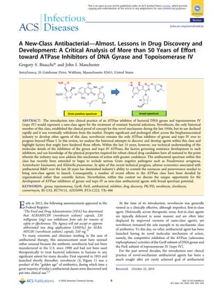
In yet another instance of independent simultaneous antibiotic
discovery, both Roche and Bristol-Myers’ Japanese research unit
reported in 1965 an antibiotic complex called coumermycin, the
most potent member of which was the A1 component (2, Figure
1), which they had both been investigating since about
1960.13,33,34
Of particular interest were the very different
strategies each company followed toward the development of
an antibacterial drug from this common starting point.
Structurally, coumermycin A1 resembles a dimer of
novobiocin; a 2-methylpyrrole ester instead of a primary
carbamate modifies each noviose sugar, and another pyrrole
group asymmetrically links the two coumarin “monomers.”
Both Roche and Bristol-Myers quickly recognized potential
strengths as well as liabilities of coumermycin A1. Coumermycin
A1 demonstrated greater antibacterial potency than novobiocin,
including against S. aureus, and encompassed roughly the same
overall, generally Gram positive spectrum (Table 1). Efficacy
Table 1. MIC Values and s.c. and Oral ED50 Values for
Novobiocin (1) and Coumermycin A1 (2)a
test organism novobiocin (1) coumermycin A1 (2)
MIC (μg/mL)
S. aureus 209P 0.05 0.0025
S. aureus 209P + 50% serum 3.2 0.16
S. aureus Smith 0.039 0.0012
S. aureus 52−34 (multiresistant) 0.78 0.0012
S. pneumoniae Type II 0.78 0.78
S. pyogenes Type 3 0.78 0.78
Neisseria sp. 12.5 12.5
E. coli ATCC 9637 50 6.25
S. flexneri 3.12 6.25
K. pneumoniae Type A 3.13 0.78
P. aeruginosa 100 12.5
ED50 (mg/kg)b
S. aureus s.c. administration 3.0 0.13
S. aureus oral administration 5.6 4.3
a
Data taken from Kawaguchi et al., 1965.13 b
Mouse sepsis model.
ACS Infectious Diseases Review
DOI: 10.1021/id500013t
ACS Infect. Dis. XXXX, XXX, XXX−XXX
D
5. experiments in mice showed coumermycin A1 to be at least as
effective as novobiocin when the compounds were administered
by the s.c. or oral routes.13
Although well tolerated orally in mice, Roche found
coumermycin A1 to be “appreciably toxic” when administered
intravenously.35
Moreover, like novobiocin, coumermycin A1 is
highly protein-bound and shows a significant, though not
therapeutically unacceptable, loss of microbiological potency in
serum (Table 1, 64-fold increase in MIC for S. aureus 209P).
Roche pushed coumermycin A1 forward into human oral studies
but found relatively low oral bioavailability (which could be
somewhat improved by formulation with N-methylglucamine) as
well as gastric irritation.36−38
Preliminary human efficacy studies
using a 1 to 2 g daily dose were reported as satisfactory but
additional testing gave “less encouraging results”, and further
clinical development of coumermycin A1 was terminated by
Roche. Both the low oral bioavailability and i.v. tolerability issues
associated with coumermycin A1 were ascribed to its extremely
poor aqueous solubility.19,34,39
Bristol-Myers, after experiencing similar preclinical and clinical
issues with coumermycin A1,19
decided to embark on a
semisynthetic analog program. At the outset of this program,
they clearly identified four objectives:
(1) improve oral bioavailability,
(2) lower plasma protein binding,
(3) eliminate the dosing irritation liability, and
(4) preserve high activity.39
In particular, improving aqueous solubility was seen by the
Bristol-Myers team as a means both to enhance the oral
bioavailability and to reduce the i.v. dosing irritation liability. The
optimization of aqueous solubility for antibacterial agents, then
as now, is principally motivated by the desire to allow the
formulation of these agents for i.v. administration as well as
(where feasible) for oral adminstration. Dosing requirements for
i.v. administration of many antibiotics typically require
solubilities in the range of ca. 5−20 mg/mL (ca. 10−40 mM),
a high bar that can be challenging to achieve. In terms of the
optimization of free fraction, it was certainly understood during
the 1960s, as it is today, that any significant loss of antibacterial
potency due to the binding to plasma proteins (“serum shifted
MICs”) can be compensated by further optimizing the intrinsic
antibacterial potency of the agents. In practice, however, the co-
optimization of both properties (along with other properties) is
typically undertaken, and a compromise is usually reached.
In their extensive program during the late 1960s, Bristol-Myers
prepared more than 160 analogs of the coumermycin A1
“monomer” with variation principally of the right-hand amide
group attached to the coumarin core. An optimized analog
having an isobutyryl amide group and labeled BL-C43 (3, Figure
1) demonstrated all of the desired improvements, whereas at a
cost of some in vitro potency compared to that of both
coumermycin and novobiocin (Tables 2 and 4). BL-C43 was
shown to be bactericidal against S. aureus.39
Encouragingly,
efficacy from the oral dosing of BL-C43 (3) in several
experimental models of infection in mice demonstrated its
superiority over both novobiocin and coumermycin A1 (Table
5), which was attributed to its greatly improved free fraction
(Table 4) and improved oral absorption. In particular, in
humans, although coumermycin A1 gave blood levels of less than
0.1 μg/mL after a single oral dose of 500 mg, an equivalent dose
of BL-C43 achieved peak blood levels of over 15 μg/mL in 3 h
and the levels were still high after 10 h. The improved
pharmacokinetics of BL-C43 seen in animals and man was
attributed to both improved solubility and to an unexpected
hepatic excretion and reuptake mechanism.40
Safety testing in rats and dogs reported that BL-C43 (3)
caused no gross or microscopic pathological changes at the
maximum oral doses administered (100 mg/kg for 36−39 days),
so a 14 day tolerance study of healthy human volunteers was
initiated. However, after 8−10 days of administration of doses
that were only 10−20% of the highest dose used in dog
toxicology studies, the majority of the 25 volunteers showed
evidence of mild jaundice and/or rash, and one volunteer
Table 2. MIC Values for BL-C43 (3) Compared to Those for
Novobiocin (1)
MIC (μg/mL)
test organism (number of strains) novobiocin (1) BL-C43 (3)
S. aureus (2)a
<0.01 0.5
S. aureus (4)b
0.18 0.9
S. pneumoniae (4) 1 3.2
S. pyogenes (4) 0.5 2
a
Nonpenicillinase producer. b
Penicillinase producer.
Table 3. MIC Values for Clorobiocin (4) Compared to Those
for Novobiocin (1)
MIC (μg/mL)
test organism novobiocin (1) clorobiocin (4)
S. aureus ATCC 29213 <0.06 <0.06
S. aureus 42080 0.25 <0.06
S. aureus 80CR5 >32 4
S. pneumoniae 1/1 serotype 6 8 2
Table 4. Protein Binding and Aqueous Solubility for
Novobiocin (1), Coumermycin A1 (2), Bl-C43 (3), and
Clorobiocin (4)
antibiotic
protein binding; % free
(method) aqueous solubility
novobiocin (1) 0.4% (HSA)a
freely soluble as sodium saltc
coumermycin A
1 (2)
1% (95% serum)b
insoluble or poorly soluble in
any salt formd
BL-C43 (3) 22% (95% serum)b
greatly improved over
coumermycin A1d
clorobiocin (4) 0.01% (HSA)a
very soluble as diethanolamine
saltc
a
Human serum albumin, Coulson et al.233 b
Keil et al.40 c
Berger et al.19
d
Cron et al.39
Table 5. ED50 Values for the Oral Administration of
Novobiocin (1), Coumermycin A1 (2), BL-C43 (3), and
Clorobiocin (4)
ED50 (mg/kg); oral administration; mouse sepsis model
test organism
novobiocin
(1)
coumermycin A
1 (2)c
BL-C
43 (3)
clorobiocin
(4)
S. aureus A9537a
26 8 8 ND
S. aureus A9606b
38 >80 10 ND
S. aureusd
NDe
ND ND 19
S. pneumoniae
A9585
800 >200 196 ND
S. pyogenes A9704 >500 >200 180 ND
a
Nonpenicillinase producer. b
Penicillinase producer. c
Toxic at 400
mg/kg. d
Strain not specified (Berger et al.19
). e
ND = no data.
ACS Infectious Diseases Review
DOI: 10.1021/id500013t
ACS Infect. Dis. XXXX, XXX, XXX−XXX
E
6. developed signs of congestive heart failure. The study was
stopped, and all volunteers who had adverse events made an
uneventful recovery.34
Because of the clear safety liability, further
development of BL-C43 was terminated as was the analog
program. There is no indication from the published records that
Bristol-Myers contemplated developing a backup to BL-C43.
Considering the failure of their animal toxicology panel to predict
human tolerability and without any apparent hypotheses to
explain the underlying cause(s), it is understandable that no
backup program was initiated.
Of interest was an in vitro resistance study for BL-C43, in
comparison to novobiocin, erythromycin, and lincomycin.41
The
development of resistance in S. aureus A9497 to each of the
antibiotics was determined by serially transferring the culture in
subinhibitory concentrations of the agents. The changes in the
MIC for each drug after the transfers are shown (Table 6). S.
aureus developed high-level resistance to BL-C43, novobiocin,
and erythromycin rapidly, and lincomycin showed a much lower
elevation in MIC. These data for novobiocin and erythromycin
were consistent with historical clinical experience where high
rates of resistance development were seen for both drugs during
treatment.17
On the basis of these in vitro data, it might
reasonably be concluded that BL-C43, had its development not
been halted by safety issues, may well have shown the same level
of resistance-development liabilities as novobiocin in clinical
practice.
The investigation of clorobiocin (4, Figure 1), another
coumarin scaffold natural product antibiotic, lagged a few years
behind the coumermycin A1 studies of both Roche and Bristol-
Myers. First reported by Rhone-Poulenc in 1969,42
the structure
of clorobiocin can be viewed as a hybrid, containing the
“monomeric” 2-methylpyrrole-noviose-coumarin functionality
of coumermycin but incorporating the right-hand amide
functionality of novobiocin. Its antibacterial spectrum is very
similar to those of both novobiocin and coumermycin A1 and
was generally regarded as more potent in vitro than novobiocin
(Table 3). Clorobiocin was more highly protein-bound than
novobiocin but had good aqueous solubility as the diethanol-
amine salt. The efficacy (ED50) of clorobiocin in a mouse model
of S. aureus infection was reported to be 5.6 mg/kg s.c. and 19
mg/kg p.o. (Table 5, p.o. value compared to reference agents).19
Little advanced preclinical work was published on clorobiocin,
and neither Rhone-Poulenc nor any other sponsor initiated
clinical work. We speculate that in light of the contemporary
negative experiences from both Roche and Bristol-Myers, as well
as the commercial decline of novobiocin itself, Rhone-Poulenc
may have been reluctant to develop a closely related analog of its
own.
Ironically, starting in 1976, after both the steep decline in
novobiocin clinical use and the abandoned efforts of Bristol-
Myers, Roche, and Rhone-Poulenc in developing their own
coumarin antibiotics, the common mechanism for all these
agents (as well as for the quinolone antibacterial class) began to
be elucidated. These coumarin-scaffold agents inhibited the
ATPase activities of the GyrB and ParE subunits of gyrase and
topo IV, respectively (sections 5 and 6 below).
It is worthwhile to mention that contemporary literature,
without consulting the original source material, frequently lumps
together novobiocin, coumermycin A1, and clorobiocin and
describes the coumarin class as “toxic” and having “poor physical
properties.” In reality, each of those agents had certain individual
drawbacks but also certain strengths. For example, novobiocin
has excellent aqueous solubility and pharmacokinetics, and its
reputation for poor safety seems contradicted by the more recent
clinical experience cited above.
3. RISE OF MRSA: NOVOBIOCIN IS BRIEFLY
RESURRECTED, AND BRISTOL-MYERS STARTS
ANOTHER COUMARIN PROGRAM (1980s)
The emergence of MRSA (methicillin-resistant S. aureus) as a
clinical concern in the early 1980s prompted Bristol-Myers to
reinitiate a program based on semisynthetic analogs of the
coumarin class.43−45
The in vitro susceptibility of clinical MRSA
strains to novobiocin, coumermycin A1, and clorobiocin was
typically quite good, and as expected because of their different
mode of action, there was no cross-resistance to any of the other
classes of existing antibacterials. During this period, there were
few options for the treatment of MRSA especially by the oral or
oral plus i.v. routes. Vancomycin was being used with increasing
frequency against MRSA infections, but was an i.v.-only drug.
Because novobiocin had already proved to be clinically useful
when administered both orally and i.v. against penicillin-
susceptible and -resistant staphylococci, the advantage of a dual
mode of administration undoubtedly added to the attraction of
the coumarin class specifically for the treatment of MRSA.
One aspect of Bristol-Myers’ modest medicinal chemistry
program, reported during the late 1980s to early 1990s,
essentially continued their 1960s strategy by examining addi-
tional amide variations on the right-hand side of the
methylpyrrole-noviose-coumarin core, this time emphasizing
substituents of higher polarity (e.g., charged groups) to ensure
high aqueous solubility (e.g., compound 5, Figure 1). The
Bristol-Myers team additionally examined the replacement of the
2-methylpyrrole with several other substituents and also varied
the point of attachment of the methylpyrrole to the noviose.
Ultimately, however, whereas novel analogs were identified
having sub-μg/mL MIC values against MRSA, the potency was
inferior to that of the parent coumermycin A1. Bristol-Myers
seems to have terminated the program without reporting safety
or in vivo animal efficacy data from any of their new analogs.
Though it would have been both scientifically useful and
technically feasible to generate biochemical data for the
inhibition of gyrase, none was reported.
Concurrent with this Bristol-Myers effort, other groups were
interested in exploring the usefulness of novobiocin itself against
MRSA, for which it was shown to be bactericidal.46
In 1989,
investigators concluded that, on the basis of in vitro susceptibility
data, “novobiocin may have a role in contemporary chemo-
therapy of oxacillin-resistant staphylococcal and other infec-
tions”.47
In 1993, a clinical MRSA colonization eradication study
concluded that novobiocin plus rifampicin was “an effective and
well-tolerated regimen for eradication of MRSA” and,
importantly, that the combination controlled the emergence of
resistance during therapy.27
A second 1993 report, however,
Table 6. Rates of Resistance Development for S. aureus A9497
to BL-C43 (3), Novobiocin (1), and Comparators
number of transfers on antibiotic-containing plates
antibiotic 0 4 8 12
MIC (μg/mL)
BL-C43 (3) 0.63 32 500 1000
novobiocin (1) 0.16 8 250 1000
erythromycin 0.16 16 63 >500
lincomycin 0.32 2 32 63
ACS Infectious Diseases Review
DOI: 10.1021/id500013t
ACS Infect. Dis. XXXX, XXX, XXX−XXX
F
7. suggested that novobiocin and rifampicin combinations may not
be clinically effective in MRSA infections, primarily because of
high serum protein binding of novobiocin.48
Following this
assessment, novobiocin has not been further investigated to any
significant extent clinically for the experimental treatment of
MRSA infections.
4. ROCHE: CYCLOTHIALIDINES (1987−199749
)
During 1992−1993, Roche reported using an in vitro Escherichia
coli DNA supercoiling assay to search for novel natural product
gyrase inhibitors.50−52
Assaying gyrase in this manner would
potentially identify inhibitors working through any mode of
action on gyrase. After screening 20 000 culture broths, a new
lead structure, cyclothialidine (6, Figure 2), was disclosed, and
Roche quickly elucidated its specific GyrB ATP-site inhibitory
mode of action. With parallels to the Bristol-Myers experience,
Roche was also once again investigating GyrB ATPase inhibitors
two decades after the termination of their own coumermycin A1
clinical program. Significantly, this was the first instance of a
noncoumarin inhibitor of the ATPase, an early demonstration of
the tolerance of the ATP binding site of gyrase toward very
different chemical structures.
Roche clearly considered cyclothialidine to be only a “lead
structure” inasmuch as the natural product was extremely weak
antimicrobially, inhibiting the growth of only a few Gram positive
organisms at high MIC (e.g., the MIC vs Streptococcus pyogenes
was 32 μg/mL, Table 7). The high MIC values were presumed to
be due to poor penetration across the bacterial cytoplasmic
membrane, yet intriguingly, the biochemical inhibitory potency
of cyclothialidine against E. coli gyrase was found to be 2-fold
better than that of novobiocin and coumermycin A1 and
significantly superior to that of quinolones, including cipro-
floxacin (29-fold). In addition cyclothialidine appeared to be
“broader spectrum” in its inhibition of gyrases from various
pathogens than reference gyrase inhibitors. Cyclothialidine
furthermore showed a greater selectivity for bacterial gyrase
versus the analogous mammalian Type II topoisomerase
(63 000-fold selective) compared to the coumarin antibiotics
(6700-fold and 1250-fold for novobiocin and coumermycin A1,
respectively) or quinolones such as ciprofloxacin (70-fold).51,52
On the basis of these encouraging findings, Roche must have
certainly felt a powerful incentive to translate the apparent
biochemical superiority and chemical novelty of cyclothialidine
into a family of microbiologically active medicines. After all,
during the previous two decades, quinolone (GyrA/ParC
mechanism) gyrase inhibitors had expanded into a hugely
successful antibacterial class. Over the following years, Roche
optimistically and repeatedly portrayed cyclothialidine as the
Figure 2. Evolution of Roche’s cyclothialidine hit 6, which possessed only weak MIC values, via analogs 7 and 8, to an advanced analog 9 having efficacy
in an animal model of infection.
Table 7. Biochemical Potencies, MIC Values, and ED50 Values for Cyclothialidine (6), Selected Analogs (7−9), and Comparators
enzyme or organism 6 7 8a 8b 9 novobiocin (1) vancomycin
Enzyme Potencies MNEC [IC50] (μg/mL)a
E. coli gyrase (supercoiling assay) 0.05 [0.25] 0.1 [0.4] 0.004 [ND]b
0.005 [ND]b
0.02 [ND]b
0.1 [ND]b
NAc
MIC (μg/mL)
S. aureus 887* or Smith >128* 2* 0.06 0.5 1 0.12 1
S. aureus 25923 NDb
NDb
0.12 0.5 4 0.25 1
S. pyogenes 15 32 8 0.25 0.5 <0.12 1 1
E. faecalis 6 >128 16 0.25 0.25 0.5 4 4
E. coli DC2* or 25922 >128* 64* >64 >64 >64 >64 >64
ED50 (mg/kg), i.v. Mouse Sepsis Model
S. aureus Smith NDb
NDb
>25 12.5 3 3 1.5
a
MNEC = maximum noneffective concentration. b
ND = no data. c
NA = not applicable.
ACS Infectious Diseases Review
DOI: 10.1021/id500013t
ACS Infect. Dis. XXXX, XXX, XXX−XXX
G
8. “basis for a new class of antibacterial agents”a clean break from
the older coumarin class agentsas well as mechanistically
differentiated from the commercially successful quinolone
class.53,54
Roche’s first medicinal chemistry campaign to improve
microbiological potency was described in their original 1992
patent filing, with over 170 analogs of cyclothialidine prepared by
total synthesis, a true synthetic tour de force. During these early
efforts, there was no crystallographic guidance for medicinal
chemists. Nevertheless, critical SAR was quickly discovered,
including the essential 14-position phenolic hydroxyl and and the
nonessentiality of a major portion of the hydroxyl-ProSer motif
on the right-hand side of the molecule. The 12-position hydroxyl
could be methylated, affording for the first time analogs with
modest potency against S. aureus. One such example was Ro46-
9288 (7, Figure 2), which had MIC90 values of 4−8 μg/mL
against S. aureus and S. epidermidus, which were still much less
potent than the corresponding MIC90 values of <0.25−0.5 μg/
mL for novobiocin (Table 7, MIC values). This analog was
described as being “partially” bactericidal (less cidal than
vancomycin, for example).51
No in vivo efficacies were reported
for these early analogs.
In addition to elucidating the SAR for microbiological activity,
Roche monitored the gyrase activity of cyclothialidine analogs
using their in vitro E. coli supercoiling assay. Conventional IC50
values were occasionally reported, but “for practical reasons”,
Roche most often reported the biochemical activity as maximum
non-effective concentration (MNEC) values that were stated to
be “3−5 times lower than the IC50 values” (Table 7).
Parallel to the extensive SAR studies of cyclothialidine lactone,
Roche and Bayer both described acyclic analogs of the
cyclothialidine macrolactone scaffold, which interestingly
retained similar biochemical and microbiological potency
compared to the cyclic analogs, once again demonstrating the
tolerance of the GyrB ATP binding site to changes in inhibitor
structure.55,56
Ro 61-6653 (10, Figure 3) is a representative
example from this acyclic subclass (IC50 and MIC values shown
in Table 8). A protein-binding liability in this class was reported
for the first time: MIC values shifted at least 64-fold in the
presence of 10% horse blood. Therefore, like novobiocin, high
protein binding was also apparently an issue with this new
natural-product-based series.
Mechanistically, even though both the coumarin-class anti-
biotics and the cyclothialidines bind to gyase by partially
overlapping the ATP binding site of GyrB, they do so with
somewhat different sets of interactions. Roche looked at a few
spontaneous mutants resistant to cyclothialidines in S. aureus and
found that most were not cross-resistant to the coumarins.
Conversely coumermycin A1 and novobiocin-resistant mutants
were still sensitive to cyclothialidines.57
In 1996, an X-ray crystal structure of a natural cyclothialidine
variant GR122222X (11, Figure 4, discovered independently by
Glaxo) in complex with the 24 kDa fragment of E. coli GyrB was
solved.58,59
Whereas Roche did briefly discuss the crystal
structure in several of their subsequent SAR publications, it is
unclear how much of their medicinal chemistry effort was truly
influenced by it. At the very least, the role of the key 14-phenolic
hydroxyl was recognized to be involved in a network of hydrogen
bonds with Asp73 and a tightly bound water (Figures 4 and 10).
Reports published during 2003−2004 describe how the
cyclothialidine scaffold had evolved further at Roche during the
1990s, with several analogs specifically highlighted, among them
compound 8a (Figure 2), which displayed outstanding gyrase
inhibitory as well as antibacterial activity against Gram positive
pathogens (Table 7).54,60
However, it was also appreciated that
the hard-won improvements in microbiological potency were not
translating into in vivo efficacy because of a combination of
factors, i.e., moderate to high protein binding (88−96% bound)
as well as glucuronidation of the key 14-hydroxy phenol leading
to high clearance. Although aqueous solubility data were not
reported for this series, a solubility issue was implied in the
published report. As a consequence, a hydroxymethyl group was
incorporated into 8b and related analogs not only to lower the
lipophilicity of the scaffold (presumably to lower both the
protein binding and mitigate the glucuronidation clearance
mechanism) but also to “increase their water solubility”. When
dosed i.v., analog 8b demonstrated an ED50 of 12.5 mg/kg
compared to 3 and 1.5 mg/kg for novobiocin and vancomycin,
respectively, in a mouse sepsis model of S. aureus infection (Table
7).
Further optimization of the cyclothialidine lactone scaffold
provided analogs with excellent antibacterial activities, expressed
as MIC90 values against panels of contemporary multiresistant
Gram positive (S. aureus, Enterococcus faecalis, Enterococcus
faecium, and Streptococcus pneumoniae) and respiratory Gram
negative pathogens (Haemophilus influenzae and Moraxella
catarrhalis).61
Roche had clearly recognized by this time that
optimizing physical properties, e.g., fine tuning lipophilicity, was
critical to achieving optimal in vivo efficacy. Its earliest analogs
(including cyclothialidine itself) displayed poor MIC values
because of high polarity and the consequent inability to penetrate
the lipophilic cytoplasmic membrane efficiently, yet the most
microbiologically potent compounds were highly lipophilic,
leading to high protein binding and high metabolic clearance due
to glucuronidation. Hence, Roche tried to optimize within a
narrow physical property window. Similar to the Bristol-Myers
experience leading to BL-C43 30 years earlier, Roche ultimately
Figure 3. Structure of cyclothialidine acyclic analog 10, which both
Roche and Bayer had investigated.
Table 8. IC50 and MIC Values for Acyclic Analog 10 and Effect
of Protein Binding by the Addition of 10% Horse Blood
enzyme or organism 10 10 + 10% horse blood
IC50 (μg/mL)
E. coli gyrase (supercoiling assay) 0.1 NAa
human DNA Type II topoisomerase 60 NAa
MIC (μg/mL)
S. aureus 133 0.25 16 (64-fold)
S. pyogenes 4851 0.25 16 (64-fold)
E. faecium L4001 0.25 32 (128-fold)
E. coli Neumann >64 >64
a
NA = not applicable.
ACS Infectious Diseases Review
DOI: 10.1021/id500013t
ACS Infect. Dis. XXXX, XXX, XXX−XXX
H
9. found that the optimization of physicochemical properties and
exposure came at the expense of some microbiological potency
against S. aureus. Thus, the somewhat more polar diamide analog
9 (Figure 2), wherein the phenol 12-methyl was additionally
replaced by a bromo group, was 16- to 32-fold and 2- to 8-fold
less potent microbiologically against S. aureus compared to more
lipophilic thioamide analogs 8a and 8b, respectively, yet it gained
a considerable free fraction (resulting in a negligible serum-
shifted MIC) along with mitigation of the glucuronidation
liability. The net result was that analog 9 achieved a superior
ED50 of 3 mg/kg when dosed i.v. in an S. aureus mouse sepsis
model, now comparable to the efficacies of novobiocin and
vancomycin (Table 7).
Little safety data was reported by Roche for their optimized
series. Although a few optimized analogs were monitored for
cytotoxicity in HeLa cell culture, showing variable margins, no
safety investigations in animals have been reported.
For historical context, it is worth mentioning that Roche’s
original gyrase screening campaign 25 years ago was one of the
earliest examples of a biochemical, rather than a “whole cell”
(phenotypic), screen for antibiotics. It is a testament to the
innovation and perseverance of the Roche scientists to have
evolved such a microbiologically weak and structurally complex
natural product screening hit into simpler molecules with potent
MIC values against Gram positive pathogens and with a
demonstration of excellent efficacy in S. aureus animal models
of infection.
5. DISCOVERY OF TOPOISOMERASE IV AND THE
CONCEPT OF POTENT DUAL INHIBITION: A KEY
CRITERION FOR CONTROLLING RESISTANCE?
(1990s ONWARD)
First characterized from E. coli, DNA gyrase has been recognized
since 1976 as a bacterial target of both the coumarin and
quinolone classes.62−64
Only much later did evidence for the
existence of a physiologically relevant “twin” to gyrase, namely,
topoisomerase IV (topo IV), begin to accumulate, and by 1990−
1992, E. coli topo IV had been characterized biochemically.65,66
Additionally, during the 1990s, gyrase and topo IV from S. aureus
Figure 4. Illustrations of key ligand−enzyme binding interactions of novobiocin, GR12222X, clorobiocin, and ADPNP revealed by X-ray crystal
structures using the 24 kDa N-terminal fragment of E. coli GyrB. Interactions of the critical Asp73−water motif with residues in each compound are
shown in red. The adenine ring of ADPMP engages the Asp73−water motif as illustrated, but the triphosphate analog moiety largely binds in a pocket
distinct from the regions that the inhibitor ligands occupy; see also Figure 5B.
ACS Infectious Diseases Review
DOI: 10.1021/id500013t
ACS Infect. Dis. XXXX, XXX, XXX−XXX
I
10. and S. pneumoniae were isolated and characterized.67−71
Investigators also worked out details of the mechanism for
these Type II topoisomerases.72
Briefly, gyrase and topo IV are
both ATP-fueled heterotetramers operating by analogous
mechanisms. Both enzymes transiently break double-stranded
DNA, pass an intact DNA strand through the opening, and then
reseal the double-strand nicks. Gyrase is primarily involved in the
negative supercoiling of DNA during replication, and topo IV is
involved primarily in DNA decatenation. The gyrase tetramer is
composed of two subunits of GyrB and two of GyrA
[(GyrB)2(GyrA)2], and the topo IV tetramer is composed of
two subunits of ParE and two of ParC [(ParE)2(ParC)2] (Figure
5A). The GyrB and ParE subunits contain the ATP binding site
(Figure 5B) and are thus the site of action for the coumarin
antibiotics, cyclothialidines, and other more recent GyrB-
directed synthetic inhibitors, discussed later. Members of the
quinolone class, by contrast, interact with the GyrA and ParC
subunits, binding to each in a ternary manner together with the
covalently bound (and cleaved) DNA strand.73,74
During studies with quinolones in the 1990s, the extent of
inhibition of each enzyme (gyrase and topo IV) by individual
agents was found to be variable, depending on both the
quinolone structure and the pathogen tested. In general, in Gram
negative pathogens, gyrase tended to be inhibited more strongly
by quinolones, and topo IV tended to be more sensitive in Gram
positive pathogens.75
Eukaryotic Type II topoisomerases exist,
but their structures are sufficiently different from the bacterial
Type II enzymes that achieving a safe margin of selectivity at the
enzyme level, can typically be accomplished.
During the last 15 years, it has been suggested that for any
bacterial Type II topoisomerase inhibitor the simultaneous and
potent inhibition of both gyrase and topo IV is needed in order to
mitigate the development of target-based resistant mutants as
measured in vitro or as observed during drug treatment.70,76−81
This view first grew out of quinolone studies (GyrA/ParC
inhibitors) but was then extended to the ATP competitive
antibacterial agents (GyrB/ParE inhibitors). For ATP com-
petitive agents, according to this view, the goal is to effect potent
inhibition of ATP binding at both gyrase and topo IV. The
desirability of inhibiting multiple bacterial targets simultaneously
to mitigate spontaneous target-based resistance development is
an old concept, with the strategy of combination therapy for
treating tuberculosis (TB) being a prime example. More
generally, numerous thought leaders in the antibacterial field
have stated the benefits of antibacterial “multitargetting” as a
means to mitigate target-based resistance, either by the use of a
single agent inhibiting two or more critical targets or by the use of
a combination of drugs, with each inhibiting a single separate
target. In the context of quinolones, David Hooper has remarked
that
“...for a quinolone congener with equal activity against both
topo IV and DNA gyrase, two mutations, one in each target
would need to be present simultaneously for the first step in
resistance due to an altered target to occur, and thus
resistance would be substantially less frequent. This
association of a low frequency of resistant mutants
(<10−10
) and similar drug activities against both gyrase
and topo IV has been seen with clinafloxacin in S.
pneumoniae and with garenoxacin and gemifloxacin in S.
aureus.”77
The initial characterization of mutants resistant to coumarin
and quinolone antibacterials began in 1976, when bacterial
gyrase was first reported. Gellert et al. isolated spontaneous
resistant mutants of E. coli grown in the presence of novobiocin,
although it was reported (without data or further explanation)
that the resistance mechanism was due to reduced permeability,
as opposed to target mutations. This assessment of the resistance
mechanism conflicted with later work (1993) that claimed no
evidence of permeability changes in novobiocin-resistant
mutants of E. coli.82
Nevertheless, in 1982 Hooper, in an
important early study, measured the frequencies of resistance of
E. coli exposed to a panel of coumarin class inhibitors that
included novobiocin, clorobiocin, and coumermycin A1 as well
as Bristol-Myers clinical agent BL-C43.83
In particular, the
Figure 5. (A) Models for bacterial topoisomerase II tetramers: DNA
gyrase (left) and topoisomerase IV (right). The arrows show the relative
locations of the ATP binding sites in GyrB and ParE and the DNA
cleavage/ligation catalytic sites in GyrA and ParC that are shown to bind
the so-called “gateway” segment of DNA. Each monomer within the two
tetramers is defined by a different color or shade of color. The models
were constructed with S. pneumoniae ParC (PDB code 4I3H),230
E. coli
ParE (1S16),162
C. psychrerythraea 34H GyrA (3LPX), and E. coli GyrB
(1EI1)231
using the X-ray crystal structure of the complete tetramer
from S. cerevisiae (4GFH)232
as a template. (B) ATP binding site of E.
coli GyrB (PDB code 1EI1) in complex with the nonhydrolyzable ATP
analog, ADPNP (adenosine 5′-(β,γ-imido)triphosphate). Most of the
binding affinity for the nucleotide is derived from interactions between
the enzyme and the phosphates. However, the adenine is positioned by a
hydrogen-bond network with Asp73 and a bound water (left panel), and
there are additional interactions between the base and ribose and the P-
loop (or “ATP lid”, shown in blue-gray). In the dimer, the N-terminus of
the other GyrB subunit (green) packs against the P-loop and interacts
with the Arg136, a key residue for binding affinity and selectivity among
all known inhibitors of the ATPase. In addition to preventing the
binding of ATP, SAR suggests that inhibitor binding disrupts dimer
formation by displacing the N-terminus so that the binding pocket
relevant to inhibitor design consists of a small hydrophobic pocket
(apparent as a gray surface in the right panel, which is colored by atom
type) distal to Asp73, an H-bond network with Asp 73 itself, and a
largely hydrophobic surface that leads proximally to the basic arginine
binding site.
ACS Infectious Diseases Review
DOI: 10.1021/id500013t
ACS Infect. Dis. XXXX, XXX, XXX−XXX
J
11. frequencies of resistance in E. coli induced by both novobiocin
and BL-C43 ((2−4) × 10−6
and 10−6
, respectively) were at a
level that, by today’s standards, would strongly suggest a risk for
clinical resistance development, at least for E. coli. The data for E.
coli are qualitatively consistent with the earlier comparative
resistance development studies done with novobiocin and BL-
C43 using the serial passage of S. aureus (Table 6), both of which
showed relatively high rates of spontaneous resistance develop-
ment.
In 1993, the biochemical inhibition of gyrase and topo IV by
novobiocin was measured for the first time: novobiocin inhibited
E. coli gyrase at nanomolar levels (IC50 6−160 nM), whereas it
inhibited topo IV at a much higher concentration (IC50 2.7
μM).84
Subsequent investigators repeated and confirmed the
relatively poor inhibition of E. coli topo IV by novobiocin and
moreover found that clorobiocin and coumermycin A1 gave
similar trends with the E. coli enzymes. Starting in the mid-2000s,
the extents of inhibition of S. aureus gyrase and topo IV by
novobiocin, clorobiocin, and coumermycin were measured, with
topo IV again showing less sensistivity to inhibition by all three
agents.85,86
In particular, as measured by several investigators for
the S. aureus enzymes, novobiocin demonstrated gyrase IC50 (or
Ki) values of between <0.004 and 0.19 μM and topo IV inhibition
of between 0.9 and 35 μM. (The factors influencing the large
spread of inhibition values for each enzyme are discussed further
below.)
Moreover, the frequencies of spontaneous resistance of S.
aureus grown in the presence of novobiocin at 4-fold the MIC
have been determined by several investigators over the past
decade and are in the range of 10−7
to 10−8
.85,87,88
Although not
as high as the frequencies for E. coli determined by Hooper in the
early 1980s ((2−4) × 10−6
), this in vitro level of resistance
frequency normally suggests a risk for clinical resistance
development. Furthermore, when one considers the exceedingly
Table 9. Comparative Biochemical Potencies for Novobiocin (1), Clorobiocin (4), and Coumermycin A1 (2) for the Inhibition of
E. coli and S. aureus Gyrase and Topo IV Using Different Assay Formats in Different Laboratories, with Associated Selected MIC
Values
enzyme or
bacterium
enzyme assay method or
bacteria ID unit
novobiocin
(1)
clorobiocin
(4)
coumermycin A1
(2) lab (year of publication)
E. coli gyrase ATPase IC50 (μg/mL) 0.045 Biota (2013)91
E. coli topo IV ATPase 0.18
S. aureus ATCC 29213 MIC (μg/mL) 0.12
E. coli gyrase supercoiling IC50 (μg/mL) 0.098 Institute of Microbial Chemistry, Tokyo
(2012)209
E. coli topo IV decatenation >12.5
S. aureus Smith MIC (μg/mL) 0.5 0.008
FDA209P 0.025
MS16526 MRSA 1
E. coli gyrase supercoiling (700 mM K-Glu) IC50 (μM) 0.08 0.03 0.03 Heide (2011)234
E. coli topo IV decatenation (100 mM K-Glu) 10 3 5
S. aureus gyrase supercoiling (700 mM K-Glu) 0.01 0.006 0.006
S. aureus topo IV decatentation (100 mM K-
Glu)
20 10 100
S. aureus ATCC 29213 MIC (μg/mL) 0.25 <0.06 <0.06
E. coli gyrase supercoiling IC50 (μM) 0.5 Merck (2011)89
E. coli topo IV decatenation 10
E. coli gyrase ATPase 0.023
E. coli topo IV ATPase 0.45
S. aureus gyrase supercoiling <0.004
S. aureus topo IV decatenation 35
S. aureus MB 5957 MIC (μg/mL) 0.5 0.008
MB2865 MSSA 0.25
MB5393 MRSA-COL 0.06
E. coli gyrase ATPase Ki (μM) 0.013 Vertex (2006, 2008)85,163
E. coli topo IV ATPase 0.160
S. aureus gyrase ATPase 0.019
S. aureus topo IV ATPase 0.900
S. aureus 54 strains MIC90 (μg/
mL)
0.5
E. coli gyrase supercoiling IC50 (μg/mL) 0.25 0.15 Hoechst Marion Roussel (2000)124,125
S. aureus gyrase supercoiling 0.5
S. aureus 011HT3 MIC (μg/mL) ≤0.04 ≤0.04
011GO64 OfloOxaEry-R ≤0.04
E. coli gyrase supercoiling IC50 (μM) 0.49 0.21 0.082−0.14 Hooper (1982)83
ACS Infectious Diseases Review
DOI: 10.1021/id500013t
ACS Infect. Dis. XXXX, XXX, XXX−XXX
K
12. low plasma levels of free (plasma-unbound) novobiocin
following the historically recommended dosing protocol in
man, it seems apparent that the free concentrations required to
inhibit the S. aureus targets biochemically, especially topo IV
(IC50 between 0.9 and 35 μM), cannot be adequately achieved or
maintained clinically. In particular, with novobiocin’s average
peak (total) serum level of 18.8 μg/mL (ca. 30 μM) following
administration of the historically recommended 0.5 g oral doses,
the peak free (plasma unbound) levels would be 0.075 μg/mL,
below the levels for inhibition of topo IV and possibly not at
sufficient multiples of the IC50 of gyrase to ensure sustained
coverage during trough periods. In retrospect, the historical
clinical observation of rapid resistance development to
novobiocin when used at those doses is consistent with the
moderately high in vitro frequency of the spontaneous resistance
of S. aureus (resulting presumably from potent inhibition at only
one target, gyrase) as well as with the likely inability to sustain
free drug levels during treatment above the inhibition
concentration constants of both biochemical targets for a
sufficient length of time.
This analysis suggests that modern GyrB/ParE inhibitors
should be designed not only to be highly potent against both
targets but also to be sufficiently unbound in plasma to fully
manifest the biochemical potency in vivo in order to limit
resistance development in addition to driving efficacy. In the
authors’ experience, agents having greater than 5−10% free
plasma levels typically show relatively small serum MIC shifts
and a higher propensity to demonstrate in vivo efficacy.
It should be pointed out that the measured values of
biochemical inhibition of gyrase and topo IV for any given
pathogen are greatly dependent on the assay techniques
employed, leading to a high degree of lab-to-lab variation in
reported data as illustrated above by the wide ranges reported for
the inhibition of S. aureus gyrase and topo IV by novobiocin.
Moreover some investigators report inhibition data in terms of Ki
values whereas others report IC50 values (not to mention Roche’s
custom MNEC inhibition units, see Section 4). The assay format
(e.g., gel supercoiling/decatenation versus ATPase format) or
differences in concentration of certain assay reagents (e.g.,
potassium glutamate) can affect the absolute values of the
inhibition concentrations by several orders of magnitude. For
example, Vertex (2006) using an ATPase assay format reported
S. aureus gyrase and topo IV Ki values for novobiocin of 0.019 and
0.90 μM, respectively, (a 45-fold difference between the two
enzymes), and Merck (2011) reported IC50 values of <0.004 and
35 μM in supercoiling (gyrase) and decatenation (topo IV)
assays, respectively, a >2000-fold difference (Table 9).85,89
In the absence of a single, standard enzyme format or even the
rigorous use of a common reference compound such as
novobiocin, it is not possible to assess quantitatively what
“potent dual inhibition” precisely means. Fortunately, the
majority of the published data agree qualitatively at least that
novobiocin is a more potent inhibitor of gyrase compared to topo
IV (Table 9), yet large differences in the absolute values for
inhibition of any given target enzyme, as well as the variations in
spread (fold difference) between the two target enzymes, leads to
confusion in our mutual understanding of what potent inhibition
actually means in practical, quantifiable terms. This uncertainty is
potentially problematic because investigators use enzyme
inhibition values to guide their medicinal chemistry programs,
employing this data as temporary surrogates to forecast in vitro
frequencies of spontaneous resistance, which in turn serve as a
surrogates to estimate the likelihood and extent of clinical
resistance development. Although to date several investigators
have associated potent dual target inhibition values with low
frequencies of spontaneous resistance in bacteria, the variable
data resulting from different enzyme assay methods employed
from lab to lab suggests that it is not yet possible to provide a
generalizable, quantitative recommendation of the actual extent
of potency required at each enzyme in order to achieve
adequately low frequencies of resistance (Table 9). Furthermore,
there is at the present time no clinical experience beyond
novobiocin to set expectations regarding the extent of develop-
ment of clinical resistance based on laboratory frequencies of
resistance for other GyrB/ParE inhibitors. Additional drugs will
need to be brought to the clinic to allow the further assessment of
any meaningful correlations between in vitro measurements of
potent dual inhibition, in vitro frequencies of spontaneous
resistance, and the extent of clinical resistance for this class of
antibacterials. To complicate matters further, any such
correlations based purely on target mutation considerations
will likely hold even less well for Gram negative pathogens (E.
coli, K. pneumoniae, P. aeruginosa, etc.), which, compared to Gram
positives, can more readily develop resistance via nontarget-
based mechanisms, especially mechanisms governing drug
accumulation (lower permeation and greater efflux) as alluded
to earlier in this section.
Finally, it is worth mentioning that the different mechanism of
inhibition of gyrase and topo IV displayed by fluoroquinolones
compared to ATP site inhibitors leads to some differences in the
mode of bacterial cell killing. Whereas fluoroquinolones and
GyrB/ParE ATP site inhibitors are both generally regarded as
bactericidal, the cidal action of the fluoroquinolones is partially
mediated by the toxic action of double strand breaks resulting
from the enzyme-DNA-quinolone complexes.90
Perhaps because
of these differences the rate of cell killing by fluoroquinolones has
been generally found to be more rapid than that achieved by ATP
site inhibitors.85,91
However, the clinical relevance of bactericidal
versus bacteriostatic mechanisms in the treatment of infections is
currently controversial;92,93
therefore, varying degrees of cidality
may or may not make a significant difference in the clinical use of
these agents.
6. X-RAY CRYSTALLOGRAPHY: A STIMULUS FOR NEW
DRUG DISCOVERY EFFORTS (MID-1990s ONWARD)
The X-ray crystal structure of the 43 kDa N-terminal fragment of
E. coli gyrase B subunit in complex with ADPNP (adenosine 5′-
(β,γ-imido)triphosphate), a nonhydrolyzable ATP analog, at 2.5
Å resolution was reported in 1991.94
This was the first high-
resolution structure of the bacterial ATPase domain and revealed
that adenine forms a hydrogen bond network with conserved
residue Asp73 and a bound water (Figure 4). In 1996, crystal
structures were published for novobiocin and GR12222X (11), a
member of the cyclothialidine (section 4, above) natural product
family.59
In these cases, the complex was with a 24 kDa N-
terminal fragment derived from the 43 kDa fragment. Finally in
1997, both the Wigley lab and the Zeneca lab independently
solved the structure of clorobiocin with the 24 kDa fragment of
GyrB.95
The availability of these structures energized GyrB-targeted
drug discovery efforts starting in the mid-1990s. As will be seen in
the following sections, this new structural information
immediately provided confidence to a number of biopharma-
ceutical companies for the rational redesign of known scaffolds or
(more significantly) the pursuit of de novo approaches to lead
discovery with the benefit of structure-informed lead optimiza-
ACS Infectious Diseases Review
DOI: 10.1021/id500013t
ACS Infect. Dis. XXXX, XXX, XXX−XXX
L
13. tion. In terms of therapeutic motivation, during the mid to late
1990s there was still the pressing need for new, effective agents
against MRSA as well as against increasingly resistant enter-
ococcus pathogens. At that time vancomycin, a parenteral-only
drug, was essentially the only reliable agent for MRSA infections,
and its effectiveness was waning against some enterococci,
especially E. faecium. ATP-site GyrB/ParE inhibitors in theory
could address those medical needs. The biopharmaceutical
companies that entered the GyrB/ParE inhibitor field rapidly set
up their own in-house crystallography efforts to facilitate iterative
drug design.
The cocrystal structures of ADPNP, novobiocin, clorobiocin,
and GR12222X with GyrB (and later ParE) ATPase domain
fragments delineated several critical drug−protein interaction
themes that were frequently utilized in subsequent drug design
efforts over the years. Several of these observed interactions
coincided with information derived from prior resistant mutant
sequencing studies, thus validating the practical relevance of
these drug−enzyme molecular interactions.
The most critical, and seemingly indispensable, interaction is a
set of hydrogen bonds provided by Asp73 (E. coli gyrase
numbering) and an associated water molecule. The adenine of
ATP, as assessed through its analog ADPNP, forms a donor/
acceptor motif as shown in Figures 4 and 5B. As it happens, the
binding sites of ATP and those of all of the reported bacterial
GyrB/ParE inhibitors overlap essentially only at this one key site
of interaction. Asp73 is so important for anchoring ATP
substrate turnover that no resistant mutants have been reported
with a change at this amino acid. The remainder of the
interactions for GyrB and ParE inhibitors are in a region
extending away from the triphosphate binding site of ATP and
make use of Arg136, a “hydrophobic floor” consisting of Pro79,
Ile78 and Ile94, and a π-stacking “ceiling” formed by Arg76
within a Glu50-Arg76 salt bridge. An important additional
hydrophobic pocket revealed by the clorobiocin structure is
composed of Val 71, Val43, Val 167, and Ala 47 and is the binding
site for the clorobiocin methylpyrrole moiety (Figure 4). The
higher enzymatic potency of clorobiocin (and coumermycin A1)
has been rationalized by the presence of this additional
interaction. The greater microbiological potency of both
clorobiocin and coumermycin A1 has been explained by a
combination of this greater enzymatic potency along with better
membrane permeability. Finally, there are two additional
conserved residues (Asn46 and Asp49) that several recent series
of inhibitors have targeted (the azaindole scaffold from
AstraZeneca and the pyrrolopyrimidine and tricyclic scaffolds
from Trius; sections 14 and 16).
From a target drugability standpoint, the binding pocket
presents an attractive opportunity for binding small molecules
having a balance of polarity and hydrophobicity targeting
multiple residues. The ATPase GyrB and ParE domains are
highly homologous across species, and in particular, the residues
labeled in Figure 4 are largely conserved across pathogenic
species and between GyrB and ParE. Thus, the target offers an
excellent opportunity for the design and discovery of broad-
spectrum antibacterial agents. At the same time, there is low
conservation among residues that do not interact directly with
the nucleotide in homologous ATPases, particularly in the
vicinity of Arg136.
Humans express two Type II topoisomerases, topo IIα and IIβ,
which are highly homologous. Despite low sequence homology
with bacterial gyrase and topo IV, the human homologues adopt
a similar fold.96
However, the binding pocket is significantly more
occluded in human topo II, and the critical Arg136 maps to a Glu
(residue 185), making the binding of bacterial topoisomerase
inhibitors less favorable. Indeed, in programs at AstraZeneca we
routinely saw >3 orders of magnitude selectivity for the bacterial
isozymes versus human.
Regarding selectivity versus other ATPases, gyrase is a
member of the GHL (gyrase, HSP90, and MutL) subfamily of
ATPases that exhibit striking differences from other ATPases97
and adopt a similar nucleotide binding fold. Here again,
important differences in the binding pockets proximal to the
adenine binding region make interactions with these related
enzymes unlikely. In HSP90, there is an occlusion of the binding
pocket, loss of the π-stacking interaction via Arg76, and the
mutation of Arg136 to histidine.98
Similarly, in MutL, among
other differences, Arg76 is replaced with a cysteine and Arg136 is
replaced with a threonine.99
Finally, in PMS2, a more recently
identified GHL ATPase, there are again large differences,
including significant occlusion of the “arginine binding region”
by the shift of a helix and the loss of Arg76 to cysteine.100
Whereas X-ray crystal structures are useful in guiding design
decisions for the optimization of enzyme potency, improvements
in antibacterial activity require the co-optimzation of enzyme
potency with factors governing cell permeation (see section
below), the avoidance of strong plasma protein binding, and
other factors governing free plasma concentrations. We have also
seen with the Roche cyclothialidine project that the antibacterial
potency can be successfully optimized in the absence of enzyme
structural information. Nevertheless, with a bounty of newly
revealed structural information coupled with the motivation to
develop novel antibacterial classes effective against MRSA, it was
with renewed energy that the industry approached gyrase drug
discovery in the mid-1990s.
7. PROBLEM OF DRUG PENETRATION THROUGH
BACTERIAL MEMBRANES: A GROWING
AWARENESS YET LARGELY EMPIRICAL SOLUTIONS
(1990s ONWARD)
One challenging feature of gyrase and topo IV as drug targets is
their location in the cytoplasm, requiring the inhibitors of those
enzymes to traverse the hydrophobic cytoplasmic membrane
efficiently. For efficacy against Gram negative pathogens,
compounds must traverse both the cytoplasmic membrane and
the outer membrane, with its opposite physical property
requirements and general preference for small, polar com-
pounds.101,102
As early as the 1960s, Bristol-Myers observed the effect of
compound polarity on S. aureus antibacterial activity during their
coumarin analog SAR efforts. In particular, increasing the carbon
chain length of the right-hand acylamino group gradually
improved the antibacterial potency against S. aureus up to a
maximum of 10 carbons, but further increases in chain length led
to a precipitous loss in MIC potency.103
Bristol-Myers did not
specifically link the concept of the degree of polarity (or
lipophilicity) to the extent of bacterial cell penetration, however.
By contrast, in 1986 when gyrase assays were available, Upjohn
reported that both novobiocin and its truncated free amino
analog (lacking the lipophilic isopentenyl hydroxybenzoate
moiety) inhibited E. coli gyrase equally, yet the polar novobiocin
fragment was completely inactive microbiologically against S.
aureus. Upjohn attributed that lack of bacterial activity to a
membrane permeation deficiency associated with the more polar
novobiocin fragment.104
By the 1990s, both the Roussel-Uclaf
ACS Infectious Diseases Review
DOI: 10.1021/id500013t
ACS Infect. Dis. XXXX, XXX, XXX−XXX
M
14. coumarin program (described below) and the Roche cyclo-
thialidine program employed the modulation of analog lip-
ophilicity as a specific design strategy to optimize antibacterial
potency by facilitating cytoplasmic membrane permeability. Both
Roche and AstraZeneca used log D measurements to help guide
their programs, and both companies concluded, on the basis of
simple ratios of enzyme to antibacterial (MIC) potencies in
comparison to corresponding log D values that for Gram positive
bacteria a specific range of low to moderate lipophilicity (log D ≈
0−3) was typically optimal for passive cytoplasmic drug
permeation.61,105,106
Complicating any simplistic lipophilicity−
MIC correlations, however, are the additional effects that
lipophilicity may have on protein binding, aqueous solubility,
and enzyme potency itself because of potential interactions of the
drug with any of several hydrophobic patches within the GyrB
and ParE binding pockets (e.g., the hydrophobic floor).95
Recently, substantial progress toward achieving antibacterial
activity against important Gram negative pathogens such as P.
aeurginosa, A. baumannii, and K. pneumoniae from the GyrB/ParE
inhibitor series has been reported, most thoroughly documented
by Trius (now Cubist) and described in detail below.107−109
AstraZeneca and Biota have also recently seen potent MIC values
against Gram negative pathogens.110,111
The scaffolds from these
programs are thus able to penetrate both the outer membrane
and the cytoplasmic membrane of these pathogens. Expansion of
the antibacterial spectrum of GyrB/ParE inhibitors to include
many problematic Gram negative pathogens is a highly
significant scientific accomplishment in the field. New-class
drugs to treat infections caused by such pathogens are critically
needed because of the rapidly diminishing therapeutic options
for treating life-threatening infections caused by increasingly
resistant organisms.
At present, however, beyond imperfect correlations of log D
and ionic charge with antibacterial potency in Gram positive
bacteria, there is disappointingly little further quantifiable and
generalizable understanding of those specific factors that
facilitate (or hinder) intracellular drug accumulation, especially
those special sets of physical property factors that favor
simultaneous permeation though both cytoplasmic and outer
membranes in Gram negative bacteria and/or the avoidance of
bacterial efflux pumps. Nevertheless, it has been suggested, for
example, that the zwitterionic fluoroquinolones are able to
permeate both membranes of Gram negative bacteria efficiently
because of a combination of unique structural features, most
important among them being a small size (for facile outer
membrane porin transit) and an ability to adjust the polarity by
altering ionic charge states within the zwitterionic mani-
fold.112,113
According to this view, fluoroquinolones may achieve
the needed polarity to transit outer membrane porins as well as
the needed lipophilicy to transit the cytoplasmic membrane via
adjustment of the charged states, although this model may be an
oversimplification.114
The utility of the traditional and widely
used informal measure of susceptibility of antibacterial drugs to
efflux, i.e., the comparison of the wild-type pathogen MIC to the
corresponding efflux-null mutant MIC, has been called into
question.115,116
It is argued that such measures can be misleading
in the absence of direct measurements of permeation; for
example, a slow rate of permeation could be misinterpreted as a
high rate of efflux. Unfortunately, there are still no reliable
general assays for the direct, routine measurement of drug
permeation through bacterial membranes or for the intracellular
accumulation of drug in bacteria, although some recent progress
has been made toward such assays.117,118
Encouragingly, there is
ongoing interest and active research to elucidate the molecular
influences of drug accumulation (both permeation and efflux) in
ways that can be practically applied to new inhibitor
design.113,119,120
8. ROUSSEL-UCLAF (HOECHST MARION ROUSSEL)
RATIONALLY REDESIGNS NOVOBIOCIN: RU79115
(LATE 1990s)
Roussel-Uclaf (later as Hoechst Marion Roussel, HMR), at the
Roumainville site near Paris, mounted a determined effort
starting in the mid-1990s to develop an antibacterial clinical
candidate based on the structure of the original coumarin
class.121−129
It appeared that their effort was partially motivated
by the increasing problem of MRSA and partially by the new
crystallographic information that could now, for the first time,
influence such a medicinal chemistry program. Like Bristol-
Myers’ earlier efforts, the Roussel team clearly recognized the
opportunities for improvement on the coumarin scaffold and
announced their vision as the “rational drug design of structurally
diverse analogs with improved pharmacological profile and
physico-chemical properties”. Moreover, differentiated from the
two Bristol-Myers coumarin analog efforts, the Roussel group
devised a completely synthetic (as opposed to semisynthetic)
route to new analogs that allowed maximum design flexibility in
ways not previously possible.
After several years of systematic and structure-guided
exploration of SAR at many positions around the coumarin
scaffold, optimized analog RU79115 (12, Figure 6) was reported
during 1999−2000.125,130
The advantage of RU79115 was its
potency both in vitro and in vivo against a range of susceptible
Figure 6. Comparison of 12, RU79115, Roussel’s 1999 structure-guided
optimized analog of novobiocin with 3, BL-C43, and Bristol-Myers
optimized analog of coumermycin A1 from 30 years earlier. Several
ligand−enzyme interactions are indicated, with Asp73−water inter-
actions shown in red. The binding mode of 3 is inferred on the basis of
other inhibitor crystal structures.
ACS Infectious Diseases Review
DOI: 10.1021/id500013t
ACS Infect. Dis. XXXX, XXX, XXX−XXX
N
15. and multiply resistant S. aureus strains (Tables 10 and 11). In
vitro MIC values against S. aureus were comparable to
novobiocin and clorobiocin and, not surprisingly, on the basis
of structural similarity, there was some cross-resistance with
novobiocin as seen with S. aureus strain 011HT1. MIC values for
RU79115 were somewhat higher than for novobiocin against
other Gram positive pathogens such as S. pyogenes and E. faecium.
RU79115 was reported to be bactericidal against both S. aureus
and E. faecium. Favorable pharmacokinetics were shown for
RU79115 in mice, with a half-life of 2.5 h and oral bioavailability
of 62%; the Cmax was 5.4 μg/mL following a 10 mg/kg oral dose.
Although SAR for the inhibition of S. aureus gyrase was
developed within the series, neither topo IV inhibition values
nor frequencies of spontaneous resistance were reported for
RU79115 or its analogs, so it is unclear whether rapid resistance
development might be a liability. What is also missing from the
published descriptions of RU79115 (as well the majority of other
coumarin analogs from the Roussel team) is quantitative
assessments of solubility, protein binding (or, alternatively,
serum shift MIC measurements), and in vitro or in vivo safety
assessments. The absence of reported physicochemical data
seems at odds with the original vision statement that set
improvements in physical properties as a clear goal. Nevertheless,
the low ED50 values of orally dosed RU79115 (mouse sepsis
model) in comparison to the values for oxazolidinones and
vancomycin, and the fact that RU79115 seemed to have been
tolerated at the moderate doses used for the efficacy studies,
suggest that any physicochemical or safety issues were not severe
(Table 11). It is interesting to compare the structure of RU7115
to Bristol-Myers’ clinical analog BL-C43 (3) of 30 years earlier
(Figure 6). Compared to novobiocin/clorobiocin, both of these
advanced analogs have similar highly truncated right-hand sides,
and both seem to be able to achieve similar binding interactions
within the enzyme. Although comparisons of data separated by
30 years comes with caveats, both compounds demonstrated
rather comparable ED50 values in S. aureus mouse sepsis models
(1−7 mg/kg for RU7115 versus 8−10 mg/kg for BL-C43). After
the early 2000s, no further information was disclosed on this
program, and it appeared to have been terminated.
9. ZENECA’S AMINOTRIAZINE STRUCTURE-BASED
EFFORT (LATE 1990s)
Concurrent with the publication of the novobiocin-GyrB crystal
structure in 1996, Zeneca was conducting a medicinal chemistry
program focused on fully synthetic novobiocin and clorobiocin
analogs constrained by a spiroketal moiety.131
None of these
analogs, however, were biochemically or microbiologically active,
which the authors attributed to several nonoptimal enzyme
interactions, including poor stacking with Arg76, based on
modeling. That series was abandoned. Biochemical and micro-
biological activity was, however, achieved by Zeneca in a follow-
on series of di- and trisubstituted triazines disclosed initially in a
1999 patent application.132−134
This Zeneca triazine project was
noteworthy because it represented the first reported GyrB/ParE
inhibitor medicinal chemistry effort not based on natural
products. The scaffold was identified from high-throughput
screening of the Zeneca corporate compound collection. Both X-
ray crystal structure information (for 13, Figure 8) and protein
NMR structure work (for 14) confirmed the binding mode
within this series (also depicted more generally in Figure 7).
Analog 15 inhibited gyrase in a supercoiling assay with an IC50 of
0.55 μg/mL, and analog 16 had an MIC range of 2−4 μg/mL
against a small panel of susceptible and resistant Gram positive
pathogens (Table 12). The Zeneca team formulated a hypothesis
correlating cytoplasmic membrane penetration to lipophilicity
values (expressed as log P or C log P) for these analogs. Major
challenges centered on issues of poor solubility and a potentially
undesirable second off-target mechanism.133
Although this project was not pursued further at Zeneca, these
triazine structures did have some influence on later GyrB/ParE
inhibitor projects at other companies, for example, the Roche
hybrid effort discussed directly below and, we speculate, aspects
of the Trius effort (compare triazine 15 with Trius pyrrolopyr-
Table 10. IC50 and MIC Values for RU79115 (12) Compared
to Those for Novobiocin (1) and Clorobiocin (4)
enzyme or organism
RU
79115 (12)
novobiocin
(1)
clorobiocin
(4)
IC50 (μg/mL)
S. aureus gyrase (supercoiling
assay)
0.2 0.5 0.3
MIC (μg/mL)
S. aureus 011HT3a
<0.04 <0.04 <0.04
S. aureus 011GO64b
<0.04 <0.04 NDd
S. aureus 011HT1c
1.2 10 0.6
S. pyogenes 02A1UC1 0.15 <0.04 <0.04
E. faecium 02D31P2 0.15 0.6 <0.04
a
Susceptible strain. b
Ofloxacin-, oxacillin-, erythromycin-resistant.
c
Novobiocin-resistant. d
ND = no data.
Table 11. ED50 and MIC Values for RU79115 (12) Compared
to Those for Eperezolid (or Linezolid) and Vancomycin
ED50 oral (mg/kg)
[MIC μg/mL]a
ED50 s.c. (mg/kg) [MIC μg/mL]a
test organism 12, RU79115
eperezolid (e) or
linezolid (l) vancomycin
S. aureus
HT18
5.6 [<0.04] 20 [0.6] (e) 7.8 [1.2]
S. aureus
GR56b
1 [0.08] 5 [1.2] (e) 4 [1.2]
S. aureus
HT17
2.8 [0.15] 5.8 [1.2] (l) ND [ND]c
S. aureus GO3 7.1 [0.3] 8.7 [2.5] (l) ND [ND]c
S. pyogenes
A1UC1
37.6 [0.15] 22.8 [<0.15] (e) <1.5 [0.6]
E. faecium
D3AP9
44 [0.6] 8.7 [1.2] (e) >50 [>40]
a
Mouse sepsis model. b
Oxacillin-resistant. c
ND = no data.
Figure 7. General binding motif for Zeneca’s triazines, Roche’s hybrid
series, AstraZeneca’s arylaminopyrimidine, and Trius’s pyrrolopyrimi-
dines. R = H, ring carbon, or aryl; n = 0, 1; Z = C, CH or N. Binding motif
for ATP but only for Asp73-H20 and Asn46 interactions. The key
Asp73−water interaction is shown in red.
ACS Infectious Diseases Review
DOI: 10.1021/id500013t
ACS Infect. Dis. XXXX, XXX, XXX−XXX
O
16. imidine 33, Figure 15). More broadly, this series was the first
example of a group of related scaffolds, discussed below,
incorporating a 2-amino nitrogen-containing heterocycle motif
that hydrogen-bonded with the critical Asp73−water and that
allowed suitably designed functionality to project toward other
key sites of interaction on the protein (Figure 7).
10. ROCHE’S HYBRID SCAFFOLD: A BLEND OF ATP,
NOVOBIOCIN, AND THE ZENECA AMINOTRIAZINE
(LATE 1990s)
In parallel with their continuing cyclothialidine efforts during the
1990s, Roche disclosed three additional lines of research
exploring ATP competitive GyrB inhibition: a hybrid scaffold
program, discussed in this section, and indazole and phenol
“needle” (fragment-based) programs, described in sections 11
and 12 below.
In 2000, Roche published a series of hybrid GyrB inhibitors
that blended features of ATP, novobiocin, and the Zeneca
triazine scaffold (Figure 9).135
One analog from this series was
cocrystallized with a 23 kDa fragment of S. aureus DNA GyrB,
and the structure was solved by X-ray crystallography. As
anticipated, the binding mode demonstrated that the NH2 group
of the core heterocycle formed critical hydrogen bonds to Asp73
and the associated water molecule (Figure 7). The best
compound from this limited survey was analog 17 (Figure 9),
which showed, at best, comparable MIC values compared to
those of novobiocin (Table 13). An important design element in
this series discovered by Roche was the recognition that a small
lipophilic ethyl group contributed to enzymatic potency. This
ethyl group occuped the same lipophilic pocket in GyrB as the
propargyl group of RU79115 (12) and the pyrrole methyl group
of clorobiocin (4). Roche provided no physicochemical, efficacy,
or safety data for this hybrid series.
11. ROCHE: A PHENOL FRAGMENT-BASED EFFORT
(LATE 1990s)
In the same year (2000) that Roche published their hybrid
scaffold (Figure 9), they also published an effort called “needle
screening”, essentially an in-silico structure- and fragment-based
approach to identify novel leads for GyrB inhibition.136
This
strategy was different from Roche’s other GyrB projects that had
focused on natural product starting points (coumarins and
cyclothialidine).
One Roche de novo lead discovery effort was based on phenol
18 (Figure 11), a “needle” screening hit.137
The Roche scientists
stated that because of their extensive medicinal chemistry
experience with the phenolic cyclothialidine series including
knowledge of X-ray structural information, they could reasonably
evolve 18 in some productive directions. One optimized analog,
19, was certainly an improvement over 18 on the basis of both E.
coli gyrase activity and MIC values for susceptible and resistant S.
aureus (Table 14). No X-ray data verifying that 19 bound in the
expected orientation in GyrB was disclosed, but it is reasonable to
assume that these analogs were competitive ATP GyrB inhibitors
that bound according to the general motif shown in Figure 10.
No physicochemical or other biological data were reported for
the series. Because of the phenol moiety, this scaffold might suffer
from the same glucuronidation liability and resultant rapid
clearance as the early cyclothialidines.
Figure 8. Arylaminotriazine motif efforts at Zeneca. The substructure shown in red interacts with the critical Asp73−water motif in gyrase and topo IV
(E. coli gyrase numbering) as illustrated in the general binding mode of Figure 7. The aryl groups depicted to the left of the triazine interact with the
lipophilic pocket, and the right-hand aryl group interacts with the Arg136/Arg76 motifs. The additional substituted aryl of 14 interacts with Ile94.
Table 12. MIC Values for Zeneca’s Triazine 16
test organism 16 MIC (μg/mL)
S. aureus Oxford 2
S. aureus novobiocin-resistant 2
S. aureus MRQSa
4
S. aureus MRQRb
2
S. pyogenes C203 4
E. faecalis 4
S. aureus Oxford 2
a
Methicillin-resistant, quinolone-sensistive. b
Methicillin-resistant, qui-
nolone-resistant.
ACS Infectious Diseases Review
DOI: 10.1021/id500013t
ACS Infect. Dis. XXXX, XXX, XXX−XXX
P
17. 12. ROCHE AND DAINIPPON: FRAGMENT-BASED
INDAZOLE AND PYRAZOLE SCAFFOLDS
(1990S−EARLY 2000S)
Roche also identified simple indazole needle 20 (Figure 13) that
was evolved, using a structure-guided approach, to analogs 21
and 22. The binding modes of 20 and 22 were determined by X-
ray from a cocrystal with the S. aureus GyrB 24 kDa fragment.
Indazole ring NH and N of both analogs are shown to interact
with Asp73 and a bound water, and for 22, the benzoic acid
substituent interacted with Arg136 and the phenyl interacted
with Ile94 on the “hydrophobic floor” of GyrB (general binding
mode depicted in Figure 12). Gyrase activity (expressed as
MNEC) for analog 21 was 8-fold better than that for novobiocin
(Table 15). No physicochemical data or any microbiological data
were reported by Roche for this series.
Figure 9. Roche hybrid scaffold 17. Substructures shown in red interact with the critical Asp73−water motif in gyrase and topo IV (E. coli gyrase
numbering), as illustrated in Figure 4 and general Figure 7. The ethyl group of 17 interacts with the left-hand hydrophobic pocket.
Table 13. Biochemical Potencies and MIC Values for Roche
Hybrid Analog 17 Compared to Those for Novobiocin (1)
enzyme or organism 17 novobiocin (1)
MNEC (μg/mL)a
E. coli gyrase (supercoiling) 0.5 0.25
MIC (μg/mL)
S. aureus ATCC 25923 4 0.25
S. aureus Smith 4 0.25
S. pyogenes b15 2 2
E. faecium van A E23−8 16 8
a
MNEC = maximum noneffective concentration.
Figure 10. General binding motif for cyclothialidines and acyclic analogs
from Roche, Glaxo, and Bayer and for the phenol “needle” optimization
program from Roche; X = H, OH, OMe; Y = Me, Br. The key Asp73−
water interaction is shown in red.
Figure 11. Roche phenol needle optimization effort. The phenol
hydroxy shown in red interacts with the critical Asp73−water motif in
gyrase and topo IV (E. coli gyrase numbering) as illustrated in the
general binding mode diagram of Figure 10
Table 14. Biochemical Potencies and MIC Values for Roche
Phenol Lead 18 and Optimized Analog 19 Compared to
Those for Novobiocin (1)
enzyme or organism 18 19 novobiocin (1)
MNEC (μg/mL)a
E. coli gyrase (supercoiling assay) 5 0.13 0.25
MIC (μg/mL)
S. aureus Smith 16 1 0.25
S. aureus QR-54 16 0.5 0.12
E. faecium van A E23-8 16 >32 8
a
MNEC = maximum noneffective concentration.
ACS Infectious Diseases Review
DOI: 10.1021/id500013t
ACS Infect. Dis. XXXX, XXX, XXX−XXX
Q
18. Dainippon investigated pyrazole 24 (Figure 13), a somewhat
related gyrase hit of their own, and studied the Roche indazole
series in depth in an attempt to inform and expand their own
series.138,139
Accordingly, Dainippon resynthesized Roche’s
more advanced analog 21 and prepared several related novel
analogs of their own, including indazole 23, which was
specifically highlighted in their paper. As reported by Dainippon,
Roche’s indazole 21 was equipotent to novobiocin in a gyrase
assay and was selective over human topoisomerase II but showed
no antibacterial activity, likely because of poor cellular
penetration (Table 15). Their own indazole analog 23 lost 16-
fold gyrase activity compared to 21 yet showed moderate MIC
values of 4−8 μg/mL against susceptible and resistant S. aureus
and E. faecium. We speculate that better physical properties
(overall polarity) of Dainippon indazole 23 compared to those of
Roche indazole 21 contributed to improvements in micro-
biological activity despite the decrease in target potency, leading
to better penetration through the bacterial cytoplasmic
membrane. However, in 10% horse blood, the antibacterial
activity of 23 was reported as weak as a result of high protein
binding.
Dainippon employed what they learned from the Roche
indazole series to evolve their own weak pyrazole hit 24 to the
more optimized analog 25 (Figure 13), which bears some
structural resemblance to indazole 23. Even though the gyrase
activity for 25 was comparable to that of novobiocin, the
antibacterial activity was modest to weak (Table 15). Dainippon
did not report the extent of protein binding for their own series.
No further work has since been reported by Roche or Dainippon
for these related indazole and pyarazole series.
13. VERTEX, QUOREX, AND CUBIST: ADDITIONAL
INDAZOLE- AND PYRAZOLE-BASED SCAFFOLDS
(2000s)
Three other companies investigated pyrazole- or indazole-based
scaffolds, and in each case, those heterocycles were attached to a
Table 15. Biochemical Potencies and MIC Values for Roche
and Dainippon Indazole (21 and 23) and Pyrazole (24 and
25) Analogs
enzyme or organism 21 23 24 25
novobiocin
(1)
MNEC (μg/mL)c
E. coli gyrase (supercoiling
assay)
0.03 NDd
NDd
NDd
0.25
IC50 (μg/mL)e
E. coli gyrase (supercoiling
assay)
0.25 4 128 0.5 0.25
E. coli topo IV
(decatenation assay)
>128 NDd
128 NDd
25
human Type II
topoisomerase
>400 NDd
200 NDd
>400
MIC (μg/mL)e
S. aureus FDA 209P >128 4 64 8 0.25
S. aureus KMP9a
>128 4 64 16 0.25
E. faecium ATCC 29212 >128 8 64 64 2
E. faecium KU 1777b
>128 4 64 32 2
a
Sparfloxacin-, clarithromycin-, and ampicillin-resistant. b
Sparfloxacin-
and vancomycin-resistant. c
Maximum noneffective concentration;
Roche data. d
ND = no data. e
Dainippon data.
Figure 12. General binding motif for the pyrazole and indazole scaffolds
from Roche, Dainippon, Quorex, and Vertex and for the pyrazolopyr-
idone scaffold from Cubist. The key Asp73−water interaction is shown
in red.
Figure 13. Pyrazole- and indazole-based scaffolds from Roche and Dainippon. Substructures shown in red interact with the critical Asp73−water motif
in gyrase and topo IV (E. coli gyrase numbering) as illustrated in the general binding mode of Figure 12.
ACS Infectious Diseases Review
DOI: 10.1021/id500013t
ACS Infect. Dis. XXXX, XXX, XXX−XXX
R
19. central thiazole ring. About the same time that Roche was
exploring its indazole series, Vertex was working on a pyrazole
scaffold (Figure 14).140−142
Vertex appended to the pyrazole ring
small lipophilic groups to interact with the left-hand lipophilic
pocket of the enzyme targets. In this regard, Vertex’s use of a
propargyl group in compound 26 was analogous to Roussel’s use
of propargyl for their optimized coumarin candidate RU79115,
compound 12 (Figure 6). A number of examples from this Vertex
pyrazole series had potent GyrB Ki values, although ParE topo IV
Ki were not disclosed. Only modest MIC values were achieved
for some analogs against S. aureus (8−32 μg/mL) and S.
pneumoniae (1.5−4 μg/mL), and many compounds reportedly
suffered from poor solubility and/or chemical instability. Other
analogs, although highly potent biochemically (as low as 0.140
μM vs S. aureus gyrase and <0.004 μM vs E. coli gyrase), were
devoid of antibacterial activity against wild-type pathogens for
reasons that were not discussed. Vertex apparently deprioritized
this series, focusing thereafter on an ethylurea benzimidazole
scaffold (section 16).
Quorex Pharmaceuticals developed a series of indazole
inhibitors that were disclosed during 2004−2005.117−223
The
detailed data for this series were to be included in posters at the
Interscience Conference on Antimicrobial Agents and Chemo-
therapy (ICAAC) in 2004, but the posters were unexpectedly
withdrawn and the only disclosed data were contained in the
conference abstracts.143−148
In those abstracts, the data were
associated with compound numbers, but no structures were
shown, whereas specific structures, but without associated
biological data, were contained in a 2005 patent application. A
representative example from the patent is indazole 27 (Figure
14).149
Quorex’s ICAAC abstracts provided data for two
compounds, QX-5-2987 and QX-5-3073, that seemed to be
similar in their biological profiles. According to the abstracts,
crystallographic information was obtained using the N-terminal
domain of E. faecalis ParE. These crystal structures have not been
disclosed, but the binding mode is assumed to be similar to that
of the other indazoles and pyrazoles by analogy (Figure 12).
Enzyme Ki values and MIC90 values for a panel of Gram positive
pathogens are shown in Table 16 for QX-5-2987, which was
described as bactericidal at concentrations a few fold above the
MIC. Compared to the prior pyrazole and indazole efforts of
Roche, Daiichi, or Vertex, this series demonstrated higher
biochemical and microbiological potency. A “low incidence of
resistance” was reported, ascribed to dual gyrase/topo IV
inhibition. Oral and i.v. pharmacokinetics were determined in
mice, rats, and dogs, and it was stated that the exposure was
sufficient to support efficacy. Efficacy in mice was reportedly run
in both sepsis and neutropenic thigh models, and ED50 values
“better than 50 mg/kg” against several pathogens were claimed.
The only safety data were in vitro cytotoxicity values in six human
cell lines (TC50 values reported as >40 μM). Physicochemical
characterization data (solubility, protein binding) were not
reported. Quorex was acquired by Pfizer in 2005, and further
characterization of the series was carried out there. The
compounds fared poorly in animal models of infection at Pfizer
because of high clearance caused by rapid glucuronidation of the
indazole ring nitrogen. Attempts to solve this issue by modifying
the electronics or sterics of the indazole ring with a variety of
different substituents were not successful, and the series was
terminated.150
In a 2013 ICAAC poster, Cubist described a S. aureus GyrB
NMR-based fragment screening effort that led to a series of
pyrazolopyridones, a representative example being compound
28 (Figure 14).151
An X-ray cocrystal structure was obtained with
the 24 kDa fragment of S. aureus GyrB that demonstrated the
binding mode illustrated in Figure 12. IC50 values for S. aureus
ParE inhibition were 30-fold less potent than for GyrB (Table
17); nevertheless, the spontaneous resistance frequency for 28
was reported to be low (3 × 10⁻⁹
) in S. aureus. MIC values against
a Gram positive panel were low (0.06 to 0.25 μg/mL). The
program is in its early stages, and no further characterization data
Figure 14. Pyrazole, indazole, and pyrazolopyridone scaffolds from Vertex, Quorex, and Cubist. Substructures shown in red interact with the critical
Asp73−water motif in gyrase and topo IV (E. coli gyrase numbering) as illustrated in the general binding mode of Figure 12
Table 16. Ki and MIC90 Values and an ED50 Value for Quorex
Analog QX-5-2987 (Structure Not Disclosed)
enzyme or species (number of strains) QX-5-2987
Ki (nM)
E. faecalis GyrB (ATPase) 1.8
E. faecalis ParE (ATPase) 10
MIC90 (μg/mL)
S. aureus (26) 0.08
S. pyogenes (15) 0.16
S. pneumoniae (18) 0.316
E. faecalis VREa
(23) 0.63
ED50
b
“for one or more S. aureus, S pneumoniae, and E. faecalis” <50 mg/kg
a
Vancomycin-resistant enterococcus. b
Mouse sepsis model; dosing tid,
1 day; route of administration not specified.
Table 17. Ki and MIC Values for Cubist Pyrazolopyridone
Analog 28
enzyme or organism 28
Ki (nM)
S. aureus GyrB (ATPase assay) 6
S. aureus ParE (ATPase assay) 183
MIC (μg/mL)
S. aureus 1118 (MRSA) 0.06
S. aureus 1721 (MRSA, ciproR)a
0.06
S. pneumoniae ATCC 6303 0.25
E. faecalis ATCC 6569 0.125
a
Ciprofloxacin-resistant.
ACS Infectious Diseases Review
DOI: 10.1021/id500013t
ACS Infect. Dis. XXXX, XXX, XXX−XXX
S