

Recommended
More Related Content
What's hot
What's hot (20)
Similar to Foetal assessment11
Similar to Foetal assessment11 (20)
More from P V GREESHMA
More from P V GREESHMA (18)
Recently uploaded
Recently uploaded (20)
Foetal assessment11
- 3. Definition: •"Assessment" means is to "evaluate" .i.e. here we gather the information of client starts and it identifies the specific needs of a client by which better care can be given to the client and her developing fetus.
- 4. Aims of antenatal fetal monitoring: • The primary objective for antenatal assessment is to avoid fetal death through out pregnancy. • To ensure satisfactory growth and well being of the fetus. • To screen out the high risk factors that affect the growth of the fetus. • Prevent fetal injury and death. • Improve long-term neurologic outcome through optimal timing of delivery • Avoiding unnecessary intervention, such as cesarean delivery or preterm delivery.
- 5. Common indications for antepartum fetal monitoring : • Pregnancy with obstetric complications: • IUGR, Multiple pregnancy, Polyhydramnios or Oligohydramnios, Rhesus alloimmunization. • Pregnancy with medical complications: • Diabetes mellitus, Hypertension, Epilepsy, Renal or Cardiac disease, Infection (Tuberculosis), SLE. • Others: • maternal age (> 35 years), previous still birth or recurrent abortion, previous birth of a baby with structural (anencephaly, spina bifida) or chromosomal (autosomal trisomy) abnormalities. • Routine antenatal testing.
- 6. Procedures of antenatal examination First visit: History collection, physical examination, recording of the obstetrical examination for the mother with following test. Hb estimation Urine culture Serological test for syphilis ABO and Rh typing PPBS & GTT Other test in case of unexplained recurrent abortion/still birth
- 7. At subsequent visit: following clinical parameters are taken in to account of the satisfactory progress of gestation: 1. Maternal weight gain 2. Blood pressure 3. Assessment of size of uterus & height of fundus 4. Clinical assessment of liquor 5. Oedema of feet 6. Abdominal girth in last trimester
- 9. biochemical Maternal serum alpha foeto protein It is the onco foeto protein. Produced by yolk sac and foetal liver. The highest level of the AFP in foetal serum and amniotic fluid reached around 13 weeks and thereafter it decreases. Maternal serum level peak around 32 weeks. Maternal screening access the quantity of the maternal serum protein level.can be perform any time between 15 7 21 weeks of gestation (17 weeks is ideal).
- 12. Elevated at Wrong gestational age Open neural tube defect of the foetus Multiple pregnancy IUFD Anterior abdominal wall defects Renal anomalies
- 13. Low at • Down syndrome • Gestational trophoblastic diseases.
- 15. Triple test It is combined test in which includes MSFP, hCG and UE3 (unconjugated oestriol). It is used for detection of down syndrome. Performed at 15-18 weeks.
- 16. Condition MSAFP UE3 hCG Neural tube defect Increased Normal Normal Trisomy 21 Low Low Increased Trisomy 18 Low Low Increased Molar pregnancy Low Low Very high Multiple gestation Increased Normal Increased Fetal death Increased Low Low
- 17. Coomb’s test The Coomb’s test (also known as Antiglobulin Test or AGT) refers to two clinical blood tests used in immunohematology which are done to find certain antibodies that cause autoimmune haemolysis of red blood cells (erythrocytes). Used to detect presence or absence of the maternal antibodies on fetal red cells. If there is no antibodies, the blood is retested at 28 and 34 weeks of pregnancy.
- 18. Types Direct Coombs Test The direct Coombs test (also known as the direct antiglobulin test or DAT) is used to detect if antibodies or complement system factors have bound to RBC surface antigens in vivo. A blood sample is taken and the RBCs are washed and then incubated with antihuman globulin. If this produces agglutination of RBCs, the direct Coombs test is positive, a visual indication that antibodies are bound to the surface of red blood cells.
- 20. Indirect Coombs Test The indirect Coombs test (also known as the indirect antiglobulin test or IAT) is used to detect in-vitro antibody-antigen reactions. It is used to detect very low concentrations of antibodies present in a patient’s plasma/serum prior to a blood transfusion. In antenatal care, this test is used to screen pregnant women for antibodies that may cause hemolytic disease of the newborn. The IAT can also be used for compatibility testing, antibody identification, RBC phenotyping, and titration studies.
- 22. Interpretation If the blood coagulates, it can be concluded that the patient’s red blood cells have been bound by (his/her own) immunoglobulins. Of course, this isn’t the normal state of affairs, and implies that the patient is experiencing an autoimmune haemolysis of his/her red cells.
- 23. Biophysical It is a screening test for uteroplacental insufficiency. The fetus biophysical activities are initiated, modulated and regulated through fetal nervous system. The fetus CNS is very much sensitive to diminished oxygenation. Hypoxia → metabolic acidosis → CNS depression → changes in fetal biophysical activity.
- 24. It is performed at two stages : • Antepartum • Intrapartum A. Antepartum tests: 1. Fetal movement monitoring 2. The non stress test (NST) 3. The contraction stress test (CST) 4. The biophysical profile 5. Doppler usg of fetal umbilical artery blood flow B. Intrapartum assessment of fetal well-being: 1. Continuous electronic fetal monitoring: a. The fetal heart rate (FHR) b. Uterine activity 2. Fetal scalp blood gas
- 25. Antepartum tests 1. Fetal movement monitoring Cardif ‘count 10’ formula: Daily fetal movement count (DFMC):
- 27. Factors affecting movement • Maternal obesity • Excessive liquor • Placental site (?) • Fetal malformations
- 28. The non stress test (NST): A test monitors the fetal heart in response to fetal movements in order to assess the integrity of fetal central nervous system and cardio vascular system. NST involves application of the fetal monitor to record the fetal heart rate.
- 29. Purposes To assess the fetal ability to cope with continuation of a high risk pregnancy. To determine the projected ability of a fetus to withstand the stress of labour. To assess the fetal status in women for whom contraction stress is contraindicated such as previous CS, placenta previa or preterm.
- 30. Indication (maternal) Post dated pregnancy Rh sensitization Maternal age 35 or more Chronic renal disease Hypertension Collagen disease Sickle cell disease Diabetes Premature rupture of membrane History of still birth Trauma Vaginal bleeding in 2nd an d3rd trimester
- 31. Indications (fetal) • Decreased fetal movement • Intrauterine growth retardation (IUGR) • Fetal evaluation after amniocentesis • Oligohydramnios/polyhydramnios
- 34. Vibroacoustic stimulation • Introduced by Zimmer et all, 1993. • Stimulation via an artificial larynx, over the fetal head along with NST attached & producing vibratory acoustic stimulus of approx 80Hz and 82dB. • A healthy fetus responds with a sudden movement (Startle Response) followed by FHR acceleration.
- 35. The contraction stress test (CST) Performed during pregnancy to verify whether or not the unborn baby’s heart is strong enough to withstand labor.
- 36. Types of CST Nipple stimulation test: Oxytocin challenge test: The contraction stress test (CST)
- 37. Results Negative CST(normal): is represented by late or variable decelerations of the fetal heart rate. Positive CST is late or variable decelerations of the fetal heart with 50% or more of the contractions in the absence of hyperstimulation of the uterus. Equivocal: it contains decelerations but with less than 50% of the contractions, or the uterine activity shows the hyperstimulation of the uterus.
- 38. Fetal cardiotocography (ctg) Is a technical means of recording (-graphy) the fetal heartbeat (cardio-) and the uterine contractions (-toco-) during pregnancy, typically in the third trimester.
- 39. Recordings are performed by two separate transducers, one for the measurement of the fetal heart rate and a second one for the uterine contractions. Each of them may be either external or internal.
- 42. Interpretation of a CTG tracing requires both qualitative abd quantitative description: Uterine activity (contractions) Baseline fetal heart rate Baseline FHR variability Presence of accelerations Periods or episodic decelerations Changes or trends of FHR patterns over time.
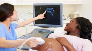
- 46. Doppler ultrasonography of fetal umbilical artery blood flow:
- 47. cytogenic
- 48. AMNIOCENTESIS
- 49. Indications • Neural tube defects (such as spina bifida or anencephaly) • Blood type of the fetus (which can be important if the mother's blood contains antibodies that can react with the fetus's red blood cells) • Genetic disorders in the fetus, such as sickle cell anemia • Infection in the fetus • Readiness of the fetus's lungs to live outside the uterus (if done late in pregnancy)
- 50. Complications • Leakage of amniotic fluid • Injury to the fetus • Infection • Miscarriage
- 51. cordocentesis
- 52. INDICATIONS • Rapid Karyotype In Fetuses Detected With Anomalies On USG. • Fetal Hemolytic Disease • Suspected Fetal Viral Infection • Non Immunologic Hydrops Fetalis • Suspected Fetal Thrombocytopenia • Twin To Twin Transfusion • Fetal Heamoglobinopathies
- 53. RISKS • BLEEDING FROM PUNCTURE SITE • VASO VAGAL REFLEX • FETAL BRADYCARDIA
- 55. Indications: •Family history of a genetic disorder •Wife & husband are carriers for genetic disorders, such as fragile X, Tay Sachs disease, or cystic fibrosis. • Prenatal screening tests (blood tests or ultrasound) show that fetus is at increased risk of having a genetic disorder.
- 56. Types of procedure • Transcervical CVS — In the transcervical CVS technique, the physician inserts a small tube through the cervix into the placenta. This is done while ultrasound guides the physician.
- 57. Transabdominal CVS — In the transabdominal CVS technique, the physician inserts a needle through the abdomen into the placenta. This is also done with ultrasound to guide the physician
- 59. FLUORESCENCE IN SITU HYBRIDIZATION
- 60. • Fluorescence in situ hybridization (FISH) is a cytogenetic technique that uses fluorescent probes that bind to only those parts of the chromosome with a high degree of sequence complementarity • It is used to reveal the location of specific nucleic acid sequences on chromosomes or in tissues, a crucial step for understanding the organization, regulation, and function of genes.