Conceição et al, 2009. characterization of a new bioactive peptide from potam...
poster grace print
1. Cytotoxic
Effects
of
Electronic
Cigare3es
on
16HBE
Human
Bronchial
Epithelial
Cells
In
Vitro
INTRODUCTION
Promoted
as
a
means
of
reducing
smoking,
Electronic
cigare5es
(ECs)
have
been
the
subject
of
much
interest
contribu=ng
to
its
open
considera=on
as
a
safer
smoking
alterna=ve.
Recently
approved
by
the
Britain’s
medicine
regulator
for
this
purpose,
sales
are
expected
to
grow
significantly
in
the
next
few
years,
where
prescrip=on
through
the
NHS
could
become
readily
available1.
However,
healthcare
prac==oners,
remain
uncertain
of
the
safety
and
efficacy
of
electronic
cigare5es
as
a
consequence
of
limited
evidence,
inconsistencies
in
results,
methodologies
and
absence
of
long-‐term
con=nuous
studies.
Besides
chemical
evalua=ons2,
limited
studies
have
performed
in
vitro
on
the
airway
epithelial;
therefore
no
definite
conclusions
can
be
drawn
on
the
poten=al
cytotoxic
effects
and
safety
of
ECs.
Thus,
in
order
to
compare
cellular
reac=ons
induced
of
E.liquid
and
it’s
aerosol,
the
current
project
aimed
to
implement
a
realis=c
simula=on
of
E.C
use.
We
developed
an
in
vitro
cytotoxicity
model,
analyzing
a
high
nico=nic
content
(18mg/ml)
ice
mint
flavor,
Bri=sh
e-‐liquid,
in
order
to
evaluate
the
cytotoxic
poten=al,
with
and
without
pH
adjustments,
in
addi=on
to
cellular
levels
of
poten=al
pro-‐inflammatory
cytokine
release
IL-‐6
and
TER
of
airway
epithelial
cells
16HBE.
RESULTS
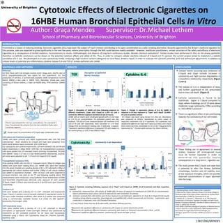
Figure
1:
DisrupGon
of
16HBE
cell
lines
following
exposure
to
different
treatments,
(Vape
and
E.liquid
at
1.25%
v/v
and
a
control)
for
different
exposure
duraGons
(4
and
26
hours).
16HBE
cells
on
inserts
were
challenged
apically
with
E.C
and
Vape
at
1.25
%
v/v
concentra?ons.
An
untreated
control
was
also
analyzed.
TER
(Ω
cm2)
was
measured
before
cell
treatment
(t=0)
and
at
4h
and
26h
respec?vely.
Data
calculated
as
a
%
mean
change
from
pre-‐treatment
reading
±SD,
4
replicates,
3
repeats.
*represents
significant
difference
in
measured
TER
with
respect
to
the
control
group;
p<
0.05;
2
way
ANOVA
Tukey.
Figure
2:
Change
in
expression
release
of
IL-‐6
by
16HBE
in
response
to
24
hour
exposure
to
“Vape”
and
E.liquid
(0.306%-‐
5%
v/v)
or
posiGve
control.
Il-‐6
release
was
assed
using
Human
Il-‐6
Elisa
set.
Absorbance
was
measured
at
450nm,
represented
as
mean
values
to
respec?ve
treatments
±SD
of
4
replicates.
IL-‐6
expression
was
significantly
different
for
vape
and
E.liquid
(p<0.001);
and
E.liquid
from
control
(p<0.05),
remarkably
at
5%
v/v
for
E.liquid
(p<0.05)
represented
by
*;
Kruskal-‐wallis
and
post
hoc
Mann-‐Whitney.
Figure
3:
Cytotoxic
screening
following
exposure
of
a)
“Vape”
and
E.liquid
on
16HBE,
b)
pH
treatment
and
their
respecGve
controls
a) Cytotoxicity,
measured
from
LDH
ac?vity
of
16HBE
aber
24
hours
of
exposure
to
treatments
at
0.306-‐5%
v/v
concentra?ons.
Data
is
presented
as
mean
values
±SD
of
10
replicates
for
each
treatment,
18
controls.
b)
Cytotoxic
assessed
from
LDH
ac?vity
post
24
hour
exposure
to
pH
treatment
adjusted
to
7.3
from
8.23
(Vape)
8.53
(E.liquid)
revealing
strong
alkaline
proper=es,
physiologically
incompa=ble
with
cellular
environment
and
func=on.
.
Experiments
were
conducted
in
4
replicates;
error
±SD
(Standard
Devia?on).
13815280/MENDES
Disrup.on
of
Epithelial
Barrier
Func.on
a)
b)
CONCLUSIONS
à
Cellular
events
occurring
post
treatment
of
E.liquid
and
Vape
include
increase
in
cytotoxicity
and
=ght
junc=on
degrada=on
in
a
dose/=me
rela=onship
respec=vely
(Figure
1
and
3)
à The
release
of
IL-‐6
is
independent
of
dose,
and
further
suppressed
at
5%,
presumably
due
to
mass
cell
death.
(Figure
2)
à Cellular
cytotoxicity
is
found
to
be
sta=s=cally
higher
in
E.liquid
compared
to
Vape,
where
4
readings
out
of
10
were
above
moderate
range
cytotoxicity
(70%)
according
to
-‐ISO
10993-‐5
protocol.3
à There
is
a
significant
effect
of
pH
contribu=ng
towards
the
cytotoxicity
of
our
cell
model.
à These
finding
are
in
agreement
to
several
studies,
however
pH
unrecognized
issue
must
be
further
exploited
in
order
to
d e t e r m i n e
t h e
p o t e n = a l
h e a l t h
consequences
in
a
long-‐term
E.
cigare5e
use.
à The
study
proves
that
E.liquid
and
vape
have
a
poten=al
to
alter
the
Airway
Epithelial
morphology,
func=on
and
cell
viability,
even
at
low
exposure
strengths,
which
are
possibly
observed
concentra=ons
of
vapor
absorbed
into
the
lungs.
MATERIALS
AND
METHODS
Materials:
Ice
Mint
flavor
with
full
strength
nico=ne
levels
18mg,
and
a
VG/PG
ra=o
of
65:35
(Liqualites,Bolton,UK)
was
opted
for
this
experiment.
For
the
produc=on
of
extracts,
a
commercially
available
160W
temperature
control
device
(SMOK
x
box
cube
II,
SMOK
Tech,
Shenzhen,
China)
was
used,
consis=ng
of
lithium
ba5ery,
a
triple
coil
Ni200
alloy,
TFV4
atomizer.
(SMOK
Tech)
Known
exact
%
concentra=on
of
E.liquid
vape
condensate
used.
Cell
culture
and
Treatment
preparaGon:
16HBE
cells
were
cultured
with
MEM
supplemented
with
10%
FBS
(PAA
Laboratories)
.
Stock
solu=ons
for
E.liquid
and
Vape
were
prepared,
from
which
serial
dilu=ons
were
conducted.
(5%-‐0.036
%v/v).
For
cytotoxicity
and
cytokine
experiments,
cell
were
seeded
in
96
and
48
well
plates
respec=vely
in
100μL
MEM
+10%
FBS.
For
transepithelium
resistance,
cells
were
seeded
into
12
transwell
inserts
(Corning
Incorporated,
NY,USA)
with
DMEM
,
Hams
F-‐12
mix
(1:1)
(GE
HealthcarePAA
Laboratories,
Austria).
pH
stocks
were
adjusted
to
pH
7.3
from
8.23
(Vape)
8.53
(E.liquid).
Transepithelial
resistance
(TER):
Prior
seeding
16HBE
cells
into
the
12
transwell
inserts,
200μl
of
collagen
was
added
onto
each
insert
coa=ng
(Pure
col).
16HBE
cells,
were
then
seeded
into
the
apical
chamber
at
a
seeding
density
of
4.3
x
105
cells/well
in
500μl
of
appropriate
cell
culture
medium
and
further
1500μl
of
cell
culture
medium
was
added
to
basolateral
chamber.
Aner
24
hours
cells
were
subjected
to
air-‐liquid
interface,
and
used
on
the
7th
day
following
seeding
where
TER
measurements
were
conducted
using
Epithelial
Tissue
Voltohmeter
(EVOM)
and
hand-‐held
chops=ck-‐type
electrode
prior
exposure
of
each
variable
(E.liquid,
Vaped
1.25%v/v
and
control)
and
at
4
and
26
hours
post
treatment
respec=vely.
Cytokine
IL-‐6:
Cells
were
seeded
with
a
density
of
1.5
x
104
cells/well
and
treated
with
Vape,
E.lqiuid
or
posi=ve
control
(vanadyl
sulphate)
in
appropriate
media
for
24
hours
under
standard
condi=ons,
aner
which
IL-‐6
release
was
measured
using
a
commercially
available
Human
IL-‐6
ELISA
kit
(BD OptEIA™,
Biosciences
Pharmingen
,USA).
LDH
Cytotoxicity
assay:
Cells
were
seeded
with
a
density
of
1.0
x
104
cells/well
in
96-‐well
microplates
,in
appropriate
media
overnight.
Medium
was
subs=tuted
by
treatments
or
len
untreated
(control)
for
24
hours
and
successively,
evaluated
using
a
Pierce
LDH
Cytotoxicity
Assay
Kit.
(Thermo
Scien=fic,
Rockford,
USA)
The
E.C
was
ac=vated
for
2-‐2.5
sec.
every
30
secs.
for
a
period
of
1
hour.
Successively
the
extracts
from
the
two
collec=ons
flasks
were
combined
together.
Cytotoxicity
is
pH
dependent
a) 2
way
Anova;
Kukey
Post
Hoc
b)
Kruskal-‐wallis,
and
Mann-‐Whitney
post
hoc.
*Represents
the
significant
difference
in
cytotoxicity
with
respect
to
control
treatments
p<0.05.
nRepresents
the
significant
difference
between
cytotoxicity
with
respect
to
Treatment
concentra?on
5
%v/v.
p<0.05
✚Represents
significant
difference
between
cytotoxicity
of
E.liquid
and
Vape
p<0.05.
✓
References
1-‐
Nico=ne
without
smoke
Tobacco
harm
reduc=on
A
report
by
the
Tobacco
Advisory
Group
of
the
Royal
College
of
Physicians
(April
2016)
h5ps://www.rcplondon.ac.uk/file/3563/
download?token=uV0R0Twz
(accessed
05.05.16)
2-‐
Famele,
M.,
C.
Ferran=,
C.
Abenavoli,
et
al.
'The
Chemical
Components
of
Electronic
Cigare5e
Cartridges
and
Refill
Fluids:
Review
of
Analy=cal
Methods',
Nico?ne
&
Tobacco
Research,
vol.
17/no.
3,
(2015),
pp.
271-‐279.
3-‐ISO
10993:5
Standard.
Biological
Evalua=on
of
Medical
Devices
—Part
5:
Tests
for
in
vitro
Cytotoxicity,
2009.
Available
online:
h5p://www.iso.org/iso/home/store/catalogue_tc/
catalogue_detail.htm?csnumber=36406
(accessed
on
14
March
2016).
*