2. 22 CHAPTER 2 Microbial growth kinetics
On integration Eq. (2.1) gives:
= µ
x x e
t
t
0
(2.2)
where x0 is the original biomass concentration, xt is the biomass concentration after
the time interval, t hours, e is the base of the natural logarithm.
On taking natural logarithms, Eq. (2.2) becomes:
µ
= +
x x t
ln ln
t 0
(2.3)
Thus, a plot of the natural logarithm of biomass concentration against time should
yield a straight line, the slope of which would equal to µ. During the exponential
phase nutrients are in excess and the organism is growing at its maximum specific
growth rate, µmax. It is important to appreciate that the µmax value is the maximum
growth rate under the prevailing conditions of the experiment, thus the value of µmax
will be affected by, for example, the medium composition, pH, and temperature.
Typical values of µmax for a range of microorganisms are given in Table 2.1.
It is easy to visualize the exponential growth of single celled organisms that rep-
licate by binary fission. Indeed, animal and plant cells in suspension culture will
behave very similarly to unicellular microorganisms (Griffiths, 1986; Petersen &
Alfermann, 1993). However, it is more difficult to appreciate that mycelial organisms,
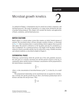
FIGURE 2.1 Growth of a Typical Microbial Culture in Batch Conditions
3. 23
Batch culture
which grow only at the apices of the hyphae, also grow exponentially. The filamentous
fungi and the filamentous bacteria (particularly the genus Streptomyces) are signifi-
cant fermentation organisms and thus an understanding of their growth is important.
Plomley (1959) was the first to suggest that filamentous fungi have a “growth unit”
that is replicated at a constant rate and is composed of the hyphal apex (tip) and a short
length of supporting hypha. Trinci (1974) demonstrated that the total hyphal length
of a mycelium and the number of tips increased exponentially at approximately the
same rate indicating that a branch is initiated when a certain hyphal length is reached.
Robinson and Smith (1979) demonstrated that it is the volume of a fungal hypha rath-
er than simply the length, that is, the branch initiation factor and Riesenberger and
Bergter (1979) confirmed the same observation for Streptomyces hygroscopicus. Thus,
branching in both fungi and streptomycetes is initiated when the biomass of the hyphal
growth unit exceeds a critical level. This is equivalent to the division of a single celled
organism when the cell reaches a critical mass. Hence, the rate of increase in hyphal
mass, total length, and number of tips is dictated by the specific growth rate and:
µ
=
dx
dt
x,
µ
=
dH
dt
H,
µ
=
dA
dt
A
where H is total hyphal length and A is the number of growing tips. Although the
growth of both filamentous fungi and streptomycetes are described by identical
kinetics, the mechanisms associated with apical growth differ. The movement of
materials to the fungal growing tip is dependent on a microtubule-based transport
system (Egan, McClintock, & Reck-Peterson, 2012), whereas that in Streptomyces
is facilitated by the coiled coil protein DivIVA that recruits other proteins to the
growing site forming multiprotein assemblies termed polarisomes (Flardh, Richards,
Hempel, Howard, & Butner, 2012).
Table 2.1 Some Representative Values of µmax (Obtained Under the
Conditions Specified in the Original Reference) for a Range of Organisms
Organism µmax (h–1
) References
Vibrio natriegens 4.24 Eagon (1961)
Methylomonas methanolytica 0.53 Dostalek et al. (1972)
Aspergillus nidulans 0.36 Trinci (1969)
Penicillium chrysogenum 0.12 Trinci (1969)
Fusarium graminearum Schwabe 0.28 Trinci (1992)
Plant cells in suspension culture 0.01–0.046 Petersen and Alfermann (1993)
Animal cells 0.01–0.05 Lavery (1990)
4. 24 CHAPTER 2 Microbial growth kinetics
In submerged liquid culture (shake flask or fermenter), a mycelial organism may
grow as dispersed hyphal fragments or as pellets (as shown in Fig. 2.2) and whether
the culture is filamentous or pelleted can have a significant influence on the products
produced by a mycelial organism (Krull et al., 2013). As discussed in more detail in
Chapter 6, the key factors influencing hyphal morphology in submerged culture are
the concentration of spores in the inoculum, medium design, and shear conditions.
The influence of morphology on culture rheology and oxygen supply is discussed in
Chapter 9. The growth of pellets will be exponential until the density of the pellet
results in diffusion limitation. Under such limitation, the central biomass of the pellet
will not receive a supply of nutrients, nor will potentially toxic products diffuse out.
Thus, the growth of the pellet proceeds from the outer shell of biomass that is the
actively growing zone and was described by Pirt (1975) as:
= +
M kt M
1/3
0
1/3
where M0 and M are the mycelium mass at time 0 and t, respectively. Thus, a plot
of the cube root of mycelial mass against time will give a straight line, the slope of
which equals k.
FIGURE 2.2 Morphological Forms of Aspergillus sp
(a) profile view of conidiophores (diameter 200 µm) on solid agar medium, (b) single
spore, (c) spore package (spore diameter 5 µm), (d) germinated tube (length approx.
250 µm), (e) coagulated type of mycel, in which single ungerminated spores adhere
to germinated hyphal tubes (length approx. 100 µm), (f) dispersed mycel, (g) exposed
hyphae of a pellet (pellet hair) (length approx. 100 µm), (h) pellet slice (diameter approx.
1000 µm), (i) hairy biopellet (pellet diameter approx. 1000 µm), and (j) submerged
biopellets. (Krull et al., 2013)
5. 25
Batch culture
It is possible for new pellets to be generated by the fragmentation of old pellets
and, thus, the behavior of a pelleted culture may be intermediate between exponential
and cube root growth.
DECELERATION AND STATIONARY PHASES
Whether the organism is unicellular or mycelial, the foregoing equations predict that
growth will continue indefinitely. However, growth results in the consumption of nu-
trients and the excretion of microbial products; events which influence the growth of
the organism. Thus, after a certain time the growth rate of the culture decreases until
growth ceases. The cessation of growth may be due to the depletion of some essential
nutrient in the medium (substrate limitation), the accumulation of some autotoxic
product of the organism in the medium (toxin limitation) or a combination of the two.
The nature of the limitation of growth may be explored by growing the organ-
ism in the presence of a range of substrate concentrations and plotting the biomass
concentration at stationary phase against the initial substrate concentration, as shown
in Fig. 2.3. From Fig. 2.3 it may be seen that over the zone A to B an increase in
initial substrate concentration gives a proportional increase in the biomass produced
at stationary phase, indicating that the substrate is limiting. The situation may be
described by the equation:
= −
x Y S s
( )
R
(2.4)
where x is the concentration of biomass produced, Y is the yield factor (g biomass
produced g–1
substrate consumed), SR is the initial substrate concentration, and s is
the residual substrate concentration.
Over the zone A to B in Fig. 2.3, s equals zero at the point of cessation of growth.
Thus, Eq. (2.4) may be used to predict the biomass that may be produced from a
certain amount of substrate. Over the zone C to D an increase in the initial substrate
concentration does not give a proportional increase in biomass. This may be due to
either the exhaustion of another substrate or the accumulation of toxic products. Over
FIGURE 2.3 The Effect of Initial Substrate Concentration on the Biomass Concentration at the
Onset of Stationary Phase, in Batch Culture
6. 26 CHAPTER 2 Microbial growth kinetics
the zone B to C the utilization of the substrate is deleteriously affected by either the
accumulating toxins or the availability of another substrate.
The yield factor (Y) is a measure of the efficiency of conversion of any one sub-
strate into biomass and it can be used to predict the substrate concentration required
to produce a certain biomass concentration. However, it is important to appreciate
that Y is not a constant—it will vary according to growth rate, pH, temperature, the
limiting substrate, and the concentration of the substrates in excess.
The decrease in growth rate and the cessation of growth, due to the depletion of
substrate, may be described by the relationship between µ and the residual growth-
limiting substrate, represented in Eq. (2.5) and in Fig. 2.4 (Monod, 1942):
µ µ
= +
s K s
/( )
s
max
(2.5)
Where, s is the substrate concentration in the presence of the organism, Ks is the
substrate utilization constant, numerically equal to substrate concentration, when µ is
half µmax and is a measure of the affinity of the organism for its substrate.
The zone A to B in Fig. 2.4 is equivalent to the exponential phase in batch cul-
ture where substrate concentration is in excess and growth is at µmax. The zone C
to A in Fig. 2.4 is equivalent to the deceleration phase of batch culture where the
growth of the organism has resulted in the depletion of substrate to a growth-limiting
concentration which will not support µmax. If the organism has a very high affinity
for the limiting substrate (a low Ks value), the growth rate will not be affected until
the substrate concentration has declined to a very low level. Thus, the deceleration
phase for such a culture would be short. However, if the organism has a low affin-
ity for the substrate (a high Ks value) the growth rate will be deleteriously affected
at a relatively high substrate concentration. Thus, the deceleration phase for such a
culture would be relatively long. Typical values of Ks for a range of organisms and
FIGURE 2.4 The Effect of Residual Limiting Substrate Concentration on the Specific Growth
Rate of a Hypothetical Bacterium
7. 27
Batch culture
substrates are shown in Table 2.2, from which it may be seen that such values are
usually very small and the affinity for substrate is high. It will be appreciated that
the biomass concentration at the end of the exponential phase is at its highest and,
thus, the decline in substrate concentration will be very rapid so that the time period
during which the substrate concentration is close to Ks is very short. While the con-
cept of Ks facilitates the quantitative description of the relationship between specific
growth rate and substrate concentration it should not be regarded as a true constant.
There are many cases in the literature of microorganisms expressing different en-
zyme systems, achieving the same metabolic end point, depending on the concentra-
tion of substrate. Harder and Dijkhuizen’s review (1983) and that of Ferenci (1999)
cite many such examples for carbon and nitrogen metabolism in which high affinity
(low Ks) systems are expressed under limitation and low affinity systems (high Ks)
expressed under nutrient excess conditions, thus enabling organisms to “scavenge”
for substrate under conditions of nutrient stress.
The stationary phase in batch culture is that point where the growth rate has
declined to zero. However, it is important to appreciate that the cessation of growth
is not the microbiological equivalent of a car running out of fuel. Although the two
situations may be the result of fuel limitation, microorganisms have evolved strate-
gies that avoid the consequences of coming to a halt in the fast lane. The kinetic de-
scriptions discussed so far ignore the physiological adaptations that microorganisms
undergo during a period of declining growth rate—adaptations that equip them to
survive periods of nutrient starvation. Stationary phase cells are not simply exponen-
tial phase cells that have stopped growing—they are physiologically different.
Sigma factors are bacterial protein transcription factors that facilitate promoter
recognition by RNA polymerase, thus enabling gene transcription and, ultimately,
gene expression. Each RNA polymerase molecule consists of one sigma factor and a
core enzyme (consisting of several units)—the nature of the sigma factor dictates the
promoters that may be recognized. All bacteria have one sigma factor that recognizes
the promoters of “housekeeping” genes enabling growth. However, they also have
a range of sigma factors that recognize the promoters of other genes that may be
switched on under specific circumstances. Thus, the deployment of particular sigma
factors under specific prevailing circumstances enables the organism to adapt to its
environment and change its gene expression profile and hence its phenotype. E. coli
Table 2.2 Some Representative Values of Ks for a Range of Microorganisms
and Substrates
Organism Substrate Ks (mg dm–3
) References
Escherichia coli Glucose 6.8 × 10⁻²
Shehata and Marr (1971)
Saccharomyces
cerevisiae
Glucose 25.0 Pirt and Kurowski (1970)
Pseudomonas sp. Methanol 0.7 Harrison (1973)
8. 28 CHAPTER 2 Microbial growth kinetics
has seven sigma factors (see Table 2.3) one of which, σ38
or σS
, recognizes genes tran-
scribed uniquely during the stationary phase (Landini, Egli, Wolf, & Lacour, 2014).
Bacteria have been shown to modify their physiology in response to both growth
rate and biomass concentration. The response to biomass concentration is referred
to as “quorum sensing”—a phenomenon in which the expression of certain genes
only occurs when the culture reaches a threshold biomass. In this system, each cell
produces a signal molecule, the concentration of which in the environment is then
dependent on the number of bacteria producing it. Thus, as biomass concentration
increases so does that of the signal molecule, until it reaches the threshold level and
specific genes are induced. The nature of the signal molecules and some of the pro-
cesses controlled by quorum sensing are shown in Table 2.4. An example of quorum
sensing in the induction of secondary metabolism is discussed in detail in Chapter 6.
However, in an elegant continuous culture experiment (see in later sections), Ihssen
and Egli (2004) demonstrated that the level of σS
in E. coli is controlled by growth
Table 2.4 Quorum Sensing Systems
Signal Molecule Controlled Property Taxonomic Group
Gamma-butyrolactones Initiation of secondary metabolism
and morphological differentiation
Streptomyces spp.
Acyl homoserine
lactones
Bacterial bioluminescence
Virulence
Antibiotic synthesis
Gram negative bacteria
Oligopeptides Biofilm formation
Competence
Sporulation
Virulence
Gram positive bacteria
Table 2.3 The Sigma Factors of Escherichia coli
Sigma Factor Function
σ70
or σD
(RpoD) Housekeeping sigma factor—recognizes genes required for
growth
σ19
or σI
(FecI) The ferric citrate sigma factor, recognizes the fec gene for iron
transport
σ24
or σE
(RpoE) Regulates and responds to extracytoplasmic functions
σ28
or σF
(RpoF) Control of flagella and pilli synthesis
σ32
or σH
(RpoH) Controls the production of heat shock proteins
σ38
or σs
(RpoS) Controls the general stress response of cells entering the
stationary phase
σ54
or σN
(RpoN) Controls the response to nitrogen limitation
9. 29
Batch culture
rate and not by biomass concentration with σS
levels being enhanced at low growth
rates—that is, under conditions of nutrient depletion or toxin accumulation akin to
the deceleration and stationary phases. The expression of the genes recognized by σS
results in the expression of a raft of phenotypes, protecting the cells from a range of
stresses that may be experienced in the stationary phase. The range of σS
influenced
characteristics include:
• cell size—stationary phase cells are smaller than those from the exponential
phase, thus increasing the surface area to volume ratio and facilitating the
enhanced uptake of limiting nutrients;
• production of detoxifying enzymes such as catalase and superoxide dismutase;
• repair and protection systems including DNA repair and protein protection by
chaperonins;
• resistance to osmotic stress;
• resistance to high temperatures;
• resistance to adverse pH.
The σS
governed responses involve approximately 500 genes, accounting for 10%
of the genome and the overall process has been termed the “general stress response”
(Hengge-Aronis, 1996). However, only about 140 genes are expressed simply as a re-
sult of enhanced σS
levels—the control of the remainder is mediated by both σS
and
specific environmental stresses. Such an orchestrated wide-reaching process would
have a significant energy demand—a requirement that is at odds with the energy
status of stationary phase organisms. Landini et al. (2014) discusses the “general
stress response” as an immediate reaction to nutrient deprivation by cells which still
have the metabolic activity to take the necessary action to protect themselves from
impending stress—that is, cells which have not yet entered the stationary phase but
are experiencing growth rates less than the maximum. The ubiquitous nature of the
response means that the organism is then protected against a range of adverse condi-
tions that may develop. The control of σS
synthesis and activity is a complex inter-
action of initiation of transcription, modulation of the mRNA transcripts and their
translation and the regulation of the degradation of σS
and its affinity for promoters.
Landini et al. (2014) summarize these control systems in their excellent review.
While E. coli responds to nutrient limitation by modulating its physiology, other
bacteria respond more dramatically by undergoing complex differentiation process-
es that enable the production of cell types capable of surviving adverse conditions.
Bacillus subtilis produces a range of cell types including endospores (dormant cells),
cannibal cells that prey on vegetative cells (of the same species), and thus over-
come nutrient limitation, matrix producing cells that form biofilms and motile cells
bearing flagella. The streptomycetes (filamentous bacteria) produce aerial hyphae
bearing exospores. As in E.coli, sigma factors also play key controlling roles in the
transition from exponential growth to stationary phase in these differentiating organ-
isms. In Bacillus subtilis, there are at least 17 alternative sigma factors with sigma-H
being paramount in a transcription cascade controlling the development of the endo-
spore. Sigma-H has been shown to control the expression of 87 genes in B. subtilis
10. 30 CHAPTER 2 Microbial growth kinetics
(Britton et al., 2002). While the “stationary phase response” in E.coli has been attrib-
uted to the organism’s titration of its decreasing growth rate (due to nutrient limita-
tion), in B. subtilis the transition to sporulation and other morphological types is a
response to the complex interaction of the detection of both biomass level (quorum
sensing) and nutrient limitation (Lazazzera, 2000; Britton et al., 2002). The degree
of nutrient limitation modulates the quorum sensing response, again enabling the
organism to undergo a series of energy-dependent transformations to adapt to im-
minent starvation conditions before the source of that energy is completely depleted.
The production of aerial hyphae and sporulation by the streptomycetes under
nutrient limitation is a highly complex process that is responding to environmental
conditions and accompanied by other stress responses such as protection against free
radicals. Streptomyces coelicolor has 63 different sigma factors (Hopwood, 2007),
49 of which belong to the ECF family (extracytoplasmic function) and detect envi-
ronmental change, including nutrient limitation and oxidative stress. It is interesting
to note that morphological differentiation in Streptomyces griseus is governed by
quorum sensing whereas that in S. coelicolor is not. Thus, closely related organisms
have evolved different mechanisms to accomplish the same end point. The filamen-
tous fungi also produce a range of taxonomically dependent spore types, again re-
sponding to environmental signals. However, it is important to appreciate that many
fungi and streptomycetes will not undergo complete differentiation in submerged
liquid culture, as this is not their natural habitat. Sporulation of mycelial organisms
in submerged culture is considered in Chapter 6 (inoculum development) from which
it can be appreciated that it has been observed far more frequently for fungal systems
than for streptomycete ones. The differentiation of Streptomyces spp. has been stud-
ied extensively in the last 10 years and has been shown to involve a programmed cell
death (PCD) process. As the name suggests, PCD is a carefully controlled process
resulting in cell death that is actually beneficial to the development and survival of
the colony as a whole. Although PCD is more associated with eukaryotic organisms
(see later) it has been observed in a number of prokaryotes, particularly when grown
on solidified medium enabling the development of defined colonies (Tanouchi, Pai,
Buchler, & You, 2012). The development of Streptomyces antibioticus on solidified
medium involves a number of distinct stages (Manteca, Fernandez, & Sanchez, 2005;
Yague, Lopez-Garcia, Rioseras, Sanchez, & Manteca, 2012, 2013):
• Compartmentalized young mycelium (termed MI) develops from germinated
spores.
• Selected compartments of the MI mycelium die, controlled by a highly ordered
process; the remaining viable segments then develop into a multinucleated second
(MII) mycelium, presumably utilizing substrate from the “sacrificed” cells.
• The MII mycelium develops in the agar medium until it undergoes a second
PCD event and the surviving MII express the synthesis of an outer hydrophobic
layer and grow into the air. Again, aerial mycelium develops at the expense of
the dead cells.
• The aerial mycelium produces aerial spores.