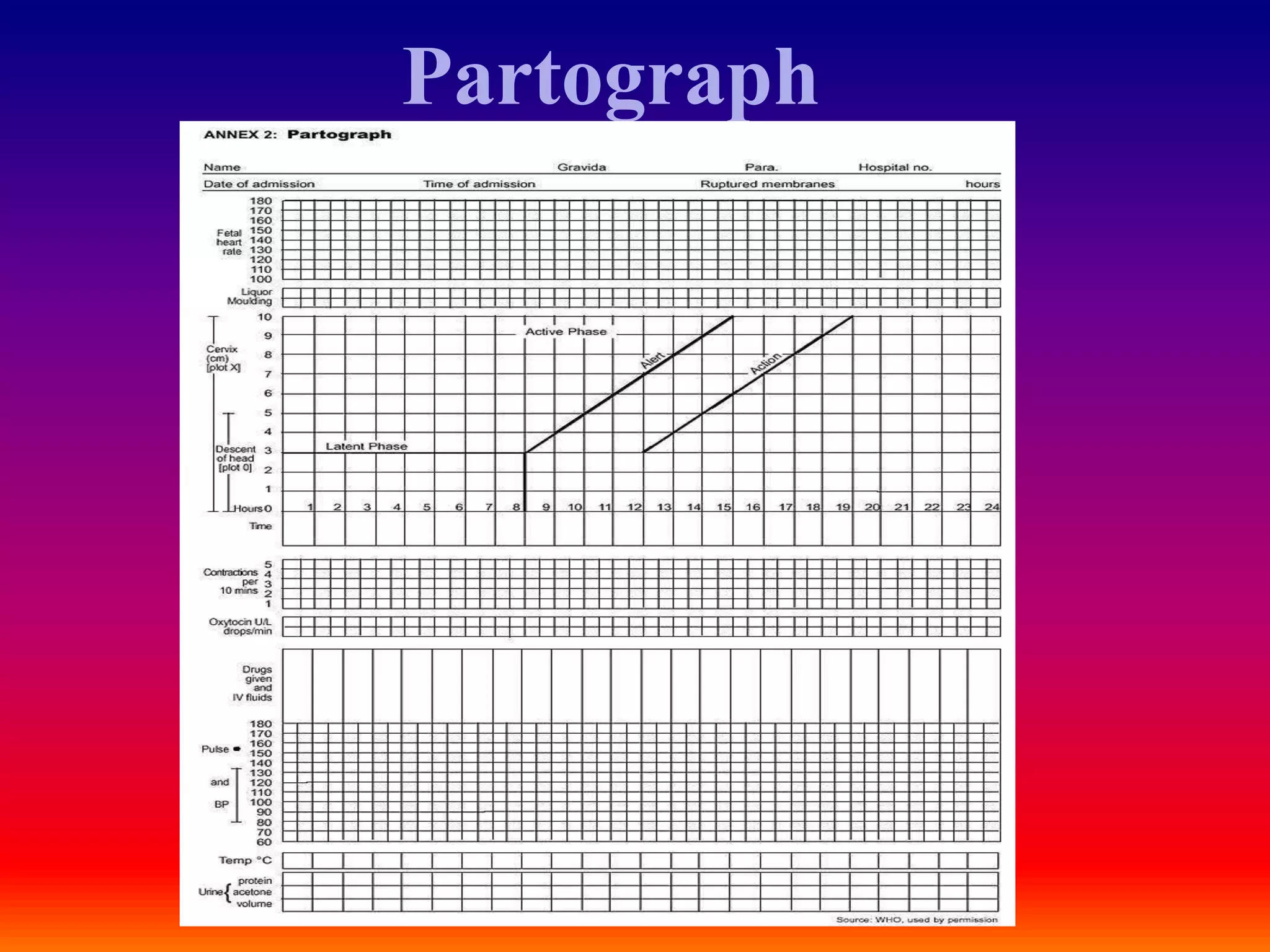
MANAGEMENT OF LABOUR
USING PARTOGRAPH
ALICE SHALUA
Partograph
Partograph
• A partograph is a graphical
record of the observations made
of a women in labor
• For progress of labor and
conditions of the mother and
the fetus
• It was developed and
extensively tested by the world
health organization WHO
History Of Partogram
• Friedman's partogram devised in
1954 was based on observations of
cervical dilatation and fetal station
against time elapsed in hours from
onset of labour. The time onset of
labour was based on the patient's
subjective perception of her
contractility. Plotting cervical
dilatation against time yielded the
typical sigmoid or 'S' shaped curve
and station against time gave rise
to the hyperbolic curve. Limits of
normal were defined
WHO partograph
Objectives
• early detection of abnormal progress of a labour
• prevention of prolonged labour
• recognize cephalo pelvic disproportion long before obstructed labour
• assist in early decision on transfer , augmentation , or termination of
labour
• increase the quality and regularity of all observations of mother and
fetus
• early recognition of maternal or fetal problems
• the partograph can be highly effective in reducing complications from
prolonged labor for the mother (postpartum hemorrhage, sepsis,
uterine rupture and its sequel) and for the newborn (death, anoxia,
infections, etc.).
Partograph function
• The partograph is designed for use in all maternity settings ,
but has a different level of function at different levels of
health care
• in health center, the partograph critical function is
to give early warning if labour is likely to be prolonged and
to indicate that the woman should be transferred to hospital
(alert line function )
• in hospital settings, moving to the right of alert line serves
as a warning for extra vigilance , but the action line is the
critical point at which specific management decisions must
be made
• other observations on the progress of labour are also
recorded on the partograph and are essential features in
management of labour
Components of the partograph
• Part 1 : fetal condition
( at top )
• Part 2 : progress of
labour ( at middle )
• Part 3 : maternal
condition ( at bottom )
Part 1 : Fetal condition
• this part of the graph is used to monitor and assess fetal
condition
• 1 - Fetal heart rate
• 2 - membranes and liquor
• 3 - molding the fetal skull bones. Caput
Fetal heart rate
< 160 beats/min =tachycardia
> 120 beats/min = bradycardia
>100beats/min=severe
bradycardia
Decelerations? yes/no
Relation to contractions?
 Early
 Variable
 Late
membranes and liquor
• intact membranes ……………………………………….I
• ruptured membranes + clear liquor …………………….C
• ruptured membranes + meconium- stained liquor ……..M
• ruptured membranes + blood – stained liquor …………B
• ruptured membranes + absent liquor…………………....A
Molding the fetal skull bones
• Molding is an important indication of how adequately the pelvis can
accommodate the fetal head. Increasing molding with the head high
in the pelvis is an ominous sign of Cephalopelvic disproportion.
• separated bones . sutures felt easily……….O
• bones just touching each other……………..+
• overlapping bones …………… …………...++
• severely overlapping bones ( notable ) ……..+++
Part 2 – progress of labour
. Cervical dilatation
• Descent of the fetal head
• Uterine contractions
• this section of the paragraph has as its central feature a graph
of cervical dilation against time
• it is divided into a latent phase and an active phase
latent phase :
• it starts from onset of labour until the cervix
reaches 3 cm dilatation
• once 3 cm dilatation is reached , labour enters the
active phase
• lasts 8 hours or less
• each lasting < 20 seconds
• at least 2/10 min contractions
Active phase :
• Contractions at least 3 / 10 min
• each lasting < 40 seconds
• The cervix should dilate at a rate
of 1 cm / hour or faster
Alert line ( health facility line )
• The alert line drawn from 3 cm dilatation
represents the rate of dilatation of 1 cm /
hour
• Moving to the right or the alert line means
referral to hospital for extra vigilance
Action line ( hospital line )
• The action line is drawn 4 hour to the right
of the alert line and parallel to it
• This is the critical line at which specific
management decisions must be made at the
hospital
Cervical dilatation
• It is the most important information and the surest way to
assess progress of labour , even though other findings discovered
on vaginal examination are also important
• when progress of labour is normal and satisfactory , plotting of
cervical dilatation remains on the alert line or to left of it
• if a woman arrives in the active phase of labour , recording of
cervical dilatation starts on the alert line
• when the active phase of labor begins , all recordings are
transferred and start by platting cervical dilatation on the alert line
• When labor goes from latent to active phase , plotting of
the dilatation is immediately transferred from the latent
phase area to the alert line
Descent of the fetal head
• It should be assessed by abdominal
examination immediately before doing
a vaginal examination, using the rule of
fifth to assess engagement
• The rule of fifth means the palpable
fifth of the fetal head are felt by
abdominal examination to be above the
level of symphysis pubis
• When 2/5 or less of fetal head is felt
above the level of symphysis pubis ,
this means that the head is engage , and
by vaginal examination , the lowest
part of vertex has passed or is at the
level of ischial spines
Assessing descent of the fetal head by vaginal
examination;
0 station is at the level of the ischial spine
(Sp).
Uterine contractions
• Observations of the contractions are made every hour in the
latent phase and every half-hour in the active phase
• frequency how often are they felt ?
• Assessed by number of contractions in a 10 minutes period
• duration how long do they last ?
Measured in seconds from the time the contraction is first felt
abdominally , to the time the contraction phases off
• Each square represents one contraction
Palpate number of contraction in ten
minutes and duration of each contraction in
seconds
• Less than 20 seconds:
• Between 20 and 40 seconds:
• More than 40 seconds:
Part 3: maternal condition
Name / Age /Gestation
Medical / Obstetrical issues
Assess maternal condition regularly by monitoring :
• drugs , IV fluids , and oxytocin , if labour is augmented
• pulse , blood pressure, Temperature, Urine volume , analysis for
protein and acetone
Management of labour using
the partograph
Diagnosis of labour
Regular painful contractions resulting
in progressive change of the
Cervix
+/- show
+/- rupture of membranes
Components of normal labour
Patient
pain , bladder empty , dehydration , exhaustion
Powers
Uterine contractions
Maternal effort
Passages
Maternal pelvis ( Inlet - Outlet )
Maternal soft tissue
Passenger
Fetal ( size - presentation - position – Moulding)
cord
placenta
membranes
- latent phase is less than 8 hours
- progress in active phase
remains on or left of the alert
line
• Do not augment with Oxytocin if
latent and active phases go
normally
• Do not intervene unless
complications develop
• Artificial rupture of membranes
( ARM )
• No ARM in latent phase
• ARM at any time in active phase
Between alert and action lines
• In health center , the women must be transferred to
a hospital with facilities for cesarean section ,
unless the cervix is almost fully dilated
• Observe labor progress for short period before
transfer
• Continue routine observations
• ARM may be performed if membranes are still
intact
!
At or beyond action line
• Conduct full medical assessment
• Consider intravenous infusion / bladder catheterization /
analgesia
• Options
- Deliver by cesarean section if there is fetal distress or
obstructed labour
- Augment with Oxytocin by intravenous infusion if there are
no contraindications
ABNORMAL PROGRESS OF
LBOR
• One of the main functions of the partograph
is to detect early deviation from normal
progress of labor
Moving to the right of alert line
• This means warning
• Transfer the woman from health center to
hospital
• reaching the action line
• This means possible danger
• Decision needed on future management
(usually by obstetrician or resident )
Prolonged latent phase
• If a woman is admitted in labor
in the latent phase ( less than 3
cm dilatation ) and remains in the
latent phase for next 8 hours
• Progress is abnormal and she
must be transferred to a hospital
for a decision about further
action
• This is why there is a heavy line
drawn on the partograph at the
end of 8 hours of the latent phase
Prolonged Active phase
• In the active phase of labor , plotting
of cervical dilatation will normally
remain on or to the left of the alert
line .But some cases will move to the
right of the alert line and this warns
that labor may be prolonged
• This will happen if the rate of
cervical dilatation in the active
phase of labor is not 1 cm / hour or
faster
• A woman whose cervical dilatation
moves to the right of the alert line
must be transferred and managed in a
hospital with adequate facilities for
obstetric intervention unless delivery
is near
• at the action line , the woman must
be carefully reassessed for why labor
is not progressing and a decision
made on further management
Secondary arrest of
cervical dilatation
• Abnormal progress of labor
may occur in cases with
normal progress of cervical
dilatation then followed by
secondary arrest of dilatation
Secondary arrest of head descant
• Abnormal progress of labor may occur with normal progress of
descent of the fetal head then followed by secondary arrest of
descent of fetal head
• It is important to realize that the partograph is a tool for
managing labor progress only
• The partograph does not help to identify other risk
factors that may have been present before labor started
POINTS TO REMEMBER
• Only start a partograph when you have checked
that there are no complications of pregnancy that
require immediate action
• A partograph chart must only be started when a
woman is in labor, Be sure that she is contracting
enough to start a partograph
• If progress of labor is satisfactory , the plotting of
cervical dilatation will remain or to the left of the
alert line
• When labor progress well , the dilatation should not move to
the right of the alert line
• The latent phase . 0 – 3 cm dilatation , is accompanied by
gradual shortening of cervix . normally , the latent phase
should not last more than 8 hours
• The active phase , 3 – 10 cm dilatation , should progress at
rate of at least 1 cm/hour
• When admission takes place in the active phase , the
admission dilatation, is immediately plotted on the alert line
• Dilatation of the cervix is plotted ( recorded with an X ,
descent of the fetal head is plotted with an O , and uterine
contractions are plotted with differential shading
• Descent of the head should always be assessed by abdominal
examination ( by the rule of fifths felt above the pelvic brim
) immediately before doing a vaginal examination
• Assessing descent of the head assists in detecting progress of
labor
• Increased molding with a high head is a sign of
Cephalopelvic disproportion
• Vaginal examination should be performed infrequently
as this is compatible with safe practice ( once every 4
hours is recommended )
• When the woman arrives in the latent phase , time of
admission is 0 time
• A woman whose cervical dilatation moves to the right
of the alert line must be transferred and managed in an
institution with adequate facilities for obstetric
intervention , unless delivery is near
• When a woman‘s partograph reaches the action line , she must be
carefully reassessed to determine why there is lack of progress , and
a decision must be made on further management ( usually by an
obstetrician or resident )
• When a woman in labor passes the latent phase in less than 8 hours
i.e., transfers from latent to active phase , the most important feature
is to transfer plotting of cervical dilatation to the alert line using the
letters TR,
• Leaving the area between the transferred recording blank. The
broken transfer line is not part of the process of labor
• DO not forget to transfer all other findings vertically
IMPORTANT
COSIDERATIONS
OXYTOCIN
• Oxytocics must be preserved in a cool ,
dark place, A local regime may be used.
• Oxytocin should be titrates against
uterine contractions and increased every
half- hour until contractions are 3 or 4
in10 minutes , each lasting 40 – 50
seconds
• It may be maintained at the rate
throughout the second stage of labor
• Stop Oxytocin infusion if there is
evidence of uterine hyperactivity and /
or fetal distress
• Oxytocin must be used with caution in
multiparous women and rarely , if at all ,
in women of Para 4 or more
• Augment with Oxytocin only after
artificial rupture of membranes and
provided that the liquor is clear
MEMBRANES
• if membranes have been ruptured for 12 hours
or more , antibiotics should be given
• As a first defense against serious infections, give a combination of
antibiotics:
- ampicillin 2 g IV every 6 hours;
- PLUS gentamicin 5 mg/kg body weight IV every 24 hours;
- PLUS metronidazole 500 mg IV every 8 hours.
Note:
If the infection is not severe, amoxicillin 500 mg by mouth every 8
hours can be used instead of ampicillin. Metronidazole can be given
by mouth instead of IV.
FETAL DISTRESS
• If a woman is laboring in a health center . transfer her to a
hospital with facilities for operative delivery
• In a hospital , immediately : Conduct a vaginal
examination to exclude cord prolapse and observe amniotic
fluid
- Provide adequate hydration
- Administer oxygen , if available stop Oxytocin
-Turn the woman or her left side
PARTOGRAPH IN MANAGING LABOUR 2021.ppt

PARTOGRAPH IN MANAGING LABOUR 2021.ppt

  • 1.
    MANAGEMENT OF LABOUR USINGPARTOGRAPH ALICE SHALUA
  • 2.
  • 3.
    Partograph • A partographis a graphical record of the observations made of a women in labor • For progress of labor and conditions of the mother and the fetus • It was developed and extensively tested by the world health organization WHO
  • 4.
    History Of Partogram •Friedman's partogram devised in 1954 was based on observations of cervical dilatation and fetal station against time elapsed in hours from onset of labour. The time onset of labour was based on the patient's subjective perception of her contractility. Plotting cervical dilatation against time yielded the typical sigmoid or 'S' shaped curve and station against time gave rise to the hyperbolic curve. Limits of normal were defined
  • 5.
  • 6.
    Objectives • early detectionof abnormal progress of a labour • prevention of prolonged labour • recognize cephalo pelvic disproportion long before obstructed labour • assist in early decision on transfer , augmentation , or termination of labour • increase the quality and regularity of all observations of mother and fetus • early recognition of maternal or fetal problems • the partograph can be highly effective in reducing complications from prolonged labor for the mother (postpartum hemorrhage, sepsis, uterine rupture and its sequel) and for the newborn (death, anoxia, infections, etc.).
  • 7.
    Partograph function • Thepartograph is designed for use in all maternity settings , but has a different level of function at different levels of health care • in health center, the partograph critical function is to give early warning if labour is likely to be prolonged and to indicate that the woman should be transferred to hospital (alert line function ) • in hospital settings, moving to the right of alert line serves as a warning for extra vigilance , but the action line is the critical point at which specific management decisions must be made • other observations on the progress of labour are also recorded on the partograph and are essential features in management of labour
  • 8.
    Components of thepartograph • Part 1 : fetal condition ( at top ) • Part 2 : progress of labour ( at middle ) • Part 3 : maternal condition ( at bottom )
  • 10.
    Part 1 :Fetal condition • this part of the graph is used to monitor and assess fetal condition • 1 - Fetal heart rate • 2 - membranes and liquor • 3 - molding the fetal skull bones. Caput
  • 11.
    Fetal heart rate <160 beats/min =tachycardia > 120 beats/min = bradycardia >100beats/min=severe bradycardia Decelerations? yes/no Relation to contractions?  Early  Variable  Late
  • 12.
    membranes and liquor •intact membranes ……………………………………….I • ruptured membranes + clear liquor …………………….C • ruptured membranes + meconium- stained liquor ……..M • ruptured membranes + blood – stained liquor …………B • ruptured membranes + absent liquor…………………....A
  • 13.
    Molding the fetalskull bones • Molding is an important indication of how adequately the pelvis can accommodate the fetal head. Increasing molding with the head high in the pelvis is an ominous sign of Cephalopelvic disproportion. • separated bones . sutures felt easily……….O • bones just touching each other……………..+ • overlapping bones …………… …………...++ • severely overlapping bones ( notable ) ……..+++
  • 14.
    Part 2 –progress of labour . Cervical dilatation • Descent of the fetal head • Uterine contractions • this section of the paragraph has as its central feature a graph of cervical dilation against time • it is divided into a latent phase and an active phase
  • 15.
    latent phase : •it starts from onset of labour until the cervix reaches 3 cm dilatation • once 3 cm dilatation is reached , labour enters the active phase • lasts 8 hours or less • each lasting < 20 seconds • at least 2/10 min contractions
  • 16.
    Active phase : •Contractions at least 3 / 10 min • each lasting < 40 seconds • The cervix should dilate at a rate of 1 cm / hour or faster
  • 17.
    Alert line (health facility line ) • The alert line drawn from 3 cm dilatation represents the rate of dilatation of 1 cm / hour • Moving to the right or the alert line means referral to hospital for extra vigilance
  • 18.
    Action line (hospital line ) • The action line is drawn 4 hour to the right of the alert line and parallel to it • This is the critical line at which specific management decisions must be made at the hospital
  • 19.
    Cervical dilatation • Itis the most important information and the surest way to assess progress of labour , even though other findings discovered on vaginal examination are also important • when progress of labour is normal and satisfactory , plotting of cervical dilatation remains on the alert line or to left of it • if a woman arrives in the active phase of labour , recording of cervical dilatation starts on the alert line • when the active phase of labor begins , all recordings are transferred and start by platting cervical dilatation on the alert line
  • 20.
    • When laborgoes from latent to active phase , plotting of the dilatation is immediately transferred from the latent phase area to the alert line
  • 21.
    Descent of thefetal head • It should be assessed by abdominal examination immediately before doing a vaginal examination, using the rule of fifth to assess engagement • The rule of fifth means the palpable fifth of the fetal head are felt by abdominal examination to be above the level of symphysis pubis • When 2/5 or less of fetal head is felt above the level of symphysis pubis , this means that the head is engage , and by vaginal examination , the lowest part of vertex has passed or is at the level of ischial spines
  • 22.
    Assessing descent ofthe fetal head by vaginal examination; 0 station is at the level of the ischial spine (Sp).
  • 23.
    Uterine contractions • Observationsof the contractions are made every hour in the latent phase and every half-hour in the active phase • frequency how often are they felt ? • Assessed by number of contractions in a 10 minutes period • duration how long do they last ? Measured in seconds from the time the contraction is first felt abdominally , to the time the contraction phases off • Each square represents one contraction
  • 24.
    Palpate number ofcontraction in ten minutes and duration of each contraction in seconds • Less than 20 seconds: • Between 20 and 40 seconds: • More than 40 seconds:
  • 25.
    Part 3: maternalcondition Name / Age /Gestation Medical / Obstetrical issues Assess maternal condition regularly by monitoring : • drugs , IV fluids , and oxytocin , if labour is augmented • pulse , blood pressure, Temperature, Urine volume , analysis for protein and acetone
  • 26.
    Management of labourusing the partograph
  • 27.
    Diagnosis of labour Regularpainful contractions resulting in progressive change of the Cervix +/- show +/- rupture of membranes
  • 28.
    Components of normallabour Patient pain , bladder empty , dehydration , exhaustion Powers Uterine contractions Maternal effort Passages Maternal pelvis ( Inlet - Outlet ) Maternal soft tissue Passenger Fetal ( size - presentation - position – Moulding) cord placenta membranes
  • 29.
    - latent phaseis less than 8 hours - progress in active phase remains on or left of the alert line • Do not augment with Oxytocin if latent and active phases go normally • Do not intervene unless complications develop • Artificial rupture of membranes ( ARM ) • No ARM in latent phase • ARM at any time in active phase
  • 30.
    Between alert andaction lines • In health center , the women must be transferred to a hospital with facilities for cesarean section , unless the cervix is almost fully dilated • Observe labor progress for short period before transfer • Continue routine observations • ARM may be performed if membranes are still intact
  • 31.
    ! At or beyondaction line • Conduct full medical assessment • Consider intravenous infusion / bladder catheterization / analgesia • Options - Deliver by cesarean section if there is fetal distress or obstructed labour - Augment with Oxytocin by intravenous infusion if there are no contraindications
  • 32.
  • 33.
    • One ofthe main functions of the partograph is to detect early deviation from normal progress of labor
  • 34.
    Moving to theright of alert line • This means warning • Transfer the woman from health center to hospital • reaching the action line • This means possible danger • Decision needed on future management (usually by obstetrician or resident )
  • 35.
    Prolonged latent phase •If a woman is admitted in labor in the latent phase ( less than 3 cm dilatation ) and remains in the latent phase for next 8 hours • Progress is abnormal and she must be transferred to a hospital for a decision about further action • This is why there is a heavy line drawn on the partograph at the end of 8 hours of the latent phase
  • 36.
    Prolonged Active phase •In the active phase of labor , plotting of cervical dilatation will normally remain on or to the left of the alert line .But some cases will move to the right of the alert line and this warns that labor may be prolonged • This will happen if the rate of cervical dilatation in the active phase of labor is not 1 cm / hour or faster • A woman whose cervical dilatation moves to the right of the alert line must be transferred and managed in a hospital with adequate facilities for obstetric intervention unless delivery is near • at the action line , the woman must be carefully reassessed for why labor is not progressing and a decision made on further management
  • 37.
    Secondary arrest of cervicaldilatation • Abnormal progress of labor may occur in cases with normal progress of cervical dilatation then followed by secondary arrest of dilatation
  • 38.
    Secondary arrest ofhead descant • Abnormal progress of labor may occur with normal progress of descent of the fetal head then followed by secondary arrest of descent of fetal head
  • 39.
    • It isimportant to realize that the partograph is a tool for managing labor progress only • The partograph does not help to identify other risk factors that may have been present before labor started POINTS TO REMEMBER
  • 40.
    • Only starta partograph when you have checked that there are no complications of pregnancy that require immediate action • A partograph chart must only be started when a woman is in labor, Be sure that she is contracting enough to start a partograph • If progress of labor is satisfactory , the plotting of cervical dilatation will remain or to the left of the alert line
  • 41.
    • When laborprogress well , the dilatation should not move to the right of the alert line • The latent phase . 0 – 3 cm dilatation , is accompanied by gradual shortening of cervix . normally , the latent phase should not last more than 8 hours • The active phase , 3 – 10 cm dilatation , should progress at rate of at least 1 cm/hour • When admission takes place in the active phase , the admission dilatation, is immediately plotted on the alert line
  • 42.
    • Dilatation ofthe cervix is plotted ( recorded with an X , descent of the fetal head is plotted with an O , and uterine contractions are plotted with differential shading • Descent of the head should always be assessed by abdominal examination ( by the rule of fifths felt above the pelvic brim ) immediately before doing a vaginal examination • Assessing descent of the head assists in detecting progress of labor • Increased molding with a high head is a sign of Cephalopelvic disproportion
  • 43.
    • Vaginal examinationshould be performed infrequently as this is compatible with safe practice ( once every 4 hours is recommended ) • When the woman arrives in the latent phase , time of admission is 0 time • A woman whose cervical dilatation moves to the right of the alert line must be transferred and managed in an institution with adequate facilities for obstetric intervention , unless delivery is near
  • 44.
    • When awoman‘s partograph reaches the action line , she must be carefully reassessed to determine why there is lack of progress , and a decision must be made on further management ( usually by an obstetrician or resident ) • When a woman in labor passes the latent phase in less than 8 hours i.e., transfers from latent to active phase , the most important feature is to transfer plotting of cervical dilatation to the alert line using the letters TR, • Leaving the area between the transferred recording blank. The broken transfer line is not part of the process of labor • DO not forget to transfer all other findings vertically
  • 45.
  • 46.
    OXYTOCIN • Oxytocics mustbe preserved in a cool , dark place, A local regime may be used. • Oxytocin should be titrates against uterine contractions and increased every half- hour until contractions are 3 or 4 in10 minutes , each lasting 40 – 50 seconds • It may be maintained at the rate throughout the second stage of labor • Stop Oxytocin infusion if there is evidence of uterine hyperactivity and / or fetal distress • Oxytocin must be used with caution in multiparous women and rarely , if at all , in women of Para 4 or more • Augment with Oxytocin only after artificial rupture of membranes and provided that the liquor is clear
  • 47.
    MEMBRANES • if membraneshave been ruptured for 12 hours or more , antibiotics should be given • As a first defense against serious infections, give a combination of antibiotics: - ampicillin 2 g IV every 6 hours; - PLUS gentamicin 5 mg/kg body weight IV every 24 hours; - PLUS metronidazole 500 mg IV every 8 hours. Note: If the infection is not severe, amoxicillin 500 mg by mouth every 8 hours can be used instead of ampicillin. Metronidazole can be given by mouth instead of IV.
  • 48.
    FETAL DISTRESS • Ifa woman is laboring in a health center . transfer her to a hospital with facilities for operative delivery • In a hospital , immediately : Conduct a vaginal examination to exclude cord prolapse and observe amniotic fluid - Provide adequate hydration - Administer oxygen , if available stop Oxytocin -Turn the woman or her left side