2. Avance´es (Orle´ans, France). Marilyn mice, of the B6 RAG-2Ϫ/Ϫ
genetic back-
ground, expressing the TCR␣ (V␣1.1-J␣35) and TCR (V6-J2.3) chains
from Marilyn, a CD4ϩ
T cell clone specific for the complex of a male Ag
(H-Y) peptide with I-Ab
, have been described previously (23). These mice
were crossed with CD45.1ϩ
B6 mice to obtain CD45.1ϩ
Marilyn mice. The
H-Y peptide (NAGFNSNRANSSRSS) was synthesized by EPYTOP (Nimes,
France), purified by reversed-phase HPLC (Ͼ99%), and purity was verified by
mass spectroscopy.
Cells
D1 is a DC line of B6 splenic origin that in the presence of growth factors
is continuously maintained in the immature state (24). Primary culture of
bone marrow(BM)-derived DCs from B6 mice and their I-AϪ/Ϫ
counter-
parts were obtained as described elsewhere (25). For both D1 and BM-
DCs, maturation was induced by 20-h treatment with 10 g/ml LPS. CD4ϩ
T lymphocytes from female Marilyn mice were obtained from lymph nodes
of female mice ages 6–8 wk. To obtain activated CD4ϩ
Marylin T cells,
1 ϫ 106
naive cells were injected into female B6 RAG-2Ϫ/Ϫ
mice subse-
quently immunized with 3 ϫ 106
mitomycin-treated CD3Ϫ/Ϫ
male spleno-
cytes. Seven days later, activated CD4ϩ
Marylin T cells were recovered
from spleen by negative selection (Spin Sep Murine CD4ϩ
T Cell Enrich-
men kit StemCell Technologies, Vancouver, BC, Canada). For the com-
parison of activated/naive T cells (see Fig. 5), we used as naive T cells
splenocytes from female Marilyn mice purified as described above. Purity
and phenotype of activated and naive CD4ϩ
T cells were verified by FACS
analysis. Syngeneic CD4ϩ
T cells were obtained as previously described (22).
FACS analysis
Phenotypic analysis of D1 cells and BM-DCs was performed using the
following Abs: FITC-conjugated anti-mouse CD11c, I-Ab
, CD40, CD86,
and the corresponding FITC-conjugated isotype controls (BD PharMingen,
Le Pont de Claix, France). Loading of the E␣ peptide was assessed by
staining peptide-loaded D1 cells with 10 g/ml of biotinylated Y-Ae Ab
followed by 5-(4,6-dichlorotriazinyl)aminofluorescein-conjugated strepta-
vidin (Immunotech, Marseille, France). To measure up-regulation of
CD69, immature or mature D1 cells were preincubated with dilutions
of H-Y peptide for 3 h at 37°C. Free peptide was removed by three rounds
of washing in complete medium. DCs were incubated with Marilyn T cells
at 1:5 ratio in 96-well plates in complete IMDM (Sigma-Aldrich, St. Louis,
MO). After 12 h, cells were stained for FACS analysis using FITC-
conjugated anti-mouse CD4, Tricolor-conjugated anti-mouse V6, and
biotin-conjugated anti-mouse CD69 followed by PE-conjugated streptavi-
din. To follow proliferation of CD4ϩ
T cells, plates were prepared as
described for CD69 using CFSE-loaded T cells (1 M; Molecular Probes,
Eugene, OR). At days 2–5 of the coculture, cells were analyzed by FACS
using PE-conjugated anti-mouse CD44 and Tricolor-conjugated anti-
mouse V6. All Abs were purchased from BD PharMingen.
Adhesion assay and FACS analysis of conjugate formation
DCs (immature or activated by overnight treatment with 10 g/ml LPS)
pulsed or not with different doses of H-Y peptide (3 h at 37°C), were
collected, washed twice with PBS, and immobilized on poly-L-lysine-
coated coverslips for 20 min at room temperature (1 ϫ 105
cells/coverslip).
PBS was then removed and replaced with complete medium and the cov-
erslips were incubated for 1 h at 37°C. The number of DCs that remained
attached to the coverslips under these conditions was 1 ϫ 104
. Marilyn T
cells (at 1 ϫ 106
/ml) in complete medium were added as a drop of 100 l
on each coverslip (ratio T:DCs ϭ 10:1) and incubated for 1 h. After in-
cubation, the coverslips were washed with 200 l of PBS several times (as
indicated in the figure legends), taking extreme care to ensure homogenous
washing. Coverslips were then mounted onto glass slides using a Mowiol
solution (Calbiochem). To quantify adhesion, each coverslip was divided
into four quadrants and differential interference contrast (DIC) images of
two random fields from each quadrant were acquired using a ϫ63 objec-
tive. For each field, we counted the total numbers of DCs, which are readily
distinguishable by size and shape (around 30 cells/field and 240 cells/
coverslip). On the same fields T cells forming clear contacts with DCs were
quantified blindly (Ͻ1% of the T cells were not conjugated to DCs after the
washes). Values are expressed as T cell:DC ratios. SD are referred to du-
plicates of coverslips or experiments performed on different days.
To quantify conjugate formation by FACS analysis, we prestained T
cells with 0.1 M CFSE and DCs with 1 M (5-(and-6)-(((4-chloromethyl)
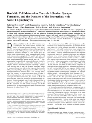
FIGURE 1. Peptide loading on immature and mature DCs. A, Phenotypic analysis of lineage-specific (CD11c) and maturation markers (I-Ab
, CD40,
CD86) on D1 cells. Cells were either untreated (immature, upper panels) or stimulated for 20 h with 10 g/ml LPS (mature, lower panels) and stained with
fluorescent Abs as indicated. B, Binding of E␣ to immature and LPS-activated DCs. DCs were incubated for 3 h with increasing concentrations of peptide
(pE␣) as indicated. The binding was revealed using a biotin-conjugated mAb specific for I-Ab
/pE␣ (Y-Ae). Fluorescence intensity (mean fluorescence
intensity (MFI)) has been corrected for background binding in the absence of pE␣. C, Loading of H-Y peptide on immature and mature DCs. DCs were
loaded with 30 M (immature) or 15 M (mature) of pE␣ to obtain comparable mean fluorescence intensities. YAe binding was competed with increasing
concentrations of H-Y peptide, corresponding to 2-, 4-, and 8-fold the initial pE␣ concentrations (30, 60, and 120 M and 60, 120, and 240 M for mature
and immature DCs, respectively). As a control, we added 120 or 240 M of an irrelevant peptide (HEL103–117) to mature and immature DCs, respectively
(cont immature, cont mature).
293The Journal of Immunology
3. benzoil)amino)tetramethylrodamine) (Molecular Probes). T cells and DCs
(prepulsed or not with the H-Y peptide) were mixed at 1:1 ratio, spun for
3 min at 300 rpm (4°C), and incubated at 37°C for 20 min. Tubes were
transferred on ice and promptly analyzed by FACS. The results are ex-
pressed as percentage of T cells that form conjugates with DCs as calcu-
lated by the ratio of two-color events to total T cells events.
Time lapse videomicroscopy and kinetic of contacts
For the dynamic analysis of conjugate formation in living cells, coverslips
coated with 3 ϫ 105
D1 cells were placed into a chamber on the micro-
scope at 37°C in a 5% CO2 atmosphere. DIC images were acquired using
63ϫ 1.32 aperture objective and a cooled charge-coupled device camera 5
Micromax Princeton Instruments, Trenton, NJ). One minute after the ad-
dition of 3 ϫ 105
T cells (t ϭ 0), we started to collect images every 10 s
for 20 min. To create quick-time files, the DIC images were accelerated
ϫ60. To quantify the duration of contacts established by individual T cells,
we analyzed the fate of single T cells along the length of the movie by
scrolling images one by one. Repetitive contacts were scored without tak-
ing into accounts whether they are formed with the same or with
different D1.
Immunolabeling of DC-T conjugates, quantification of
clustering, three-dimensional reconstitution
Conjugates between D1 and CD4ϩ
T cells were formed as described for the
adhesion assay. Incubation was stopped after 30 min and coverslips were
washed five times with PBS. Cells were fixed for 10 min with 3% para-
formaldehyde and permeabilized with PBS, 2% BSA (Sigma-Aldrich), and
0.05% saponin (ICN Biomedicals, Costa Mesa, CA). For the “not washed”
condition in the experiment shown in Fig. 6D (not washed), T cells were
removed and coverslips were fixed after a gentle wash with 200 l of PBS.
Primary and secondary fluorescent Abs were diluted in PBS, 2% BSA, and
0.05% saponin and incubated for 1 h or 30 min, respectively. Abs used for
single labeling were as follows: biotin-conjugated hamster anti-mouse
CD3⑀ (CD3-⑀ 145-2C11; BD PharMingen) followed by Alexa 488-conju-
gated streptavidin (Molecular Probes); anti-LAT (rabbit polyclonal IgG;
Upstate Biotechnology, Lake Placid, NY) followed by Texas Red-conju-
gated anti-rabbit IgG (Jackson ImmunoResearch Laboratories, West
Grove, PA); monoclonal rat anti-mouse LFA-1 (ATCC TIB-127) followed
by Cy3-conjugated anti-rat IgG (Jackson ImmunoResearch Laboratories);
mouse anti-tubulin (clone Ab-1; Oncogene Research Products, San Diego,
CA) followed by anti-mouse Alexa 488 (Molecular Probes); and rat anti-
mouse CD43 (clone S7; BD PharMingen) followed by anti-rat Cy3.
To acquire images of conjugates, we used a Leica TCS SP2 confocal
scanning microscope (Leica, Deerfield, IL) equipped with a 100ϫ 1.4 ap-
erture HCX PL APO oil immersion objective. To quantify redistribution of
molecules at the site of contact, T-DC doublets were chosen by DIC images
and then scored as negative or positive by evaluating the corresponding
fluorescent images along for sections on the z plane. “En face” view of the
T-DC contact zone (xz) was reconstructed from series of xy sections spaced
by 0.3 m (Metamorph software; Universal Imaging).
Results
Mature, but not immature, DCs activate naive CD4ϩ
T
lymphocytes
We have analyzed the interaction of immature and mature DCs
with naive CD4ϩ
T lymphocytes using either a growth factor-
dependent DC line, called D1 (24), or fresh BM-DCs. Immature
D1 cells grow continuously in the presence of a GM-CSF-contain-
ing conditioned medium. As shown in Fig. 1A, immature D1 cells
express CD11c, intermediate levels of I-Ab
and CD86, but no
CD40. These cells also express ICAM-1 and LFA-1 (23). After
20 h of LPS stimulation, surface expression of these markers in-
crease, attesting effective maturation. In parallel, I-Ab
molecules
are redistributed from lysosomal compartments to the plasma
membrane, and cytokine and chemokine secretion is induced (data
not shown). The overall morphology of DCs is also profoundly
modified (24). From all these points of view, D1 cells behave
exactly like BM-DCs (26). As a homogenous source of naive
CD4ϩ
T lymphocytes, we used lymph node T cells obtained from
RAGϪ/Ϫ
Marilyn TCR-transgenic mice (23). The Marilyn TCR
recognizes the male H-Y Ag associated to I-Ab
molecules. Mari-
lyn’s lymph nodes contain 93–98% naive (CD69Ϫ
, CD44Ϫ
) Mari-
lyn CD4ϩ
T cells and no other T cells.
Because I-Ab
molecules are 10-fold more abundant on mature
than on immature DCs (Fig. 1A), we first measured their respective
peptide-loading capacities. For that purpose, we used the mAb,
Y-Ae, which recognizes I-Ab
molecules associated to a peptide
from the I-E␣ chain (27). As shown in Fig. 1B, the binding of
Y-Ae rises when the cells are incubated with increasing concen-
trations of peptide, reaching a plateau at 100 ⌴. A 2- to 3-fold
difference in the concentrations of the I-E␣ peptide required to
FIGURE 2. Early events of activation in naive T cells stimulated by
immature and mature DCs. A, Percentage of naive Marilyn T cells showing
a Ca2ϩ
response after coculture with immature or mature D1 treated or not
with 10 nM H-Y peptide. All T-DC contacts were analyzed. Responses
triggered by immature DCs (Ⅺ) and by mature DCs (u). The error bars
represent the SD from three independent experiments. B, Average Ca2ϩ
response in 5–15 individual responding T cells interacting with immature
or mature DCs loaded with 10 nM peptide. The Ca2ϩ
traces were synchro-
nized before averaging so that the shape of the average response is similar
to that of (asynchronous) single-cell recording. The tiny responses obtained
in the absence of peptide were too few to give a meaningful average (data
not shown). C, DCs were incubated with T cells for 12 h (1:5 ratio) and
expression of CD69 was assessed by FACS analysis on TCRϩ
CD4ϩ
gated
cells. Percentage of naive T cells positive for CD69 staining after 12 h of
stimulation with immature (ࡗ) or mature (f) DCs pulsed with different
doses of peptide. One experiment representative of four is shown.
294 DYNAMIC INTERACTION BETWEEN DCs AND NAIVE CD4 T CELLS
4. induce equivalent levels of Y-Ae binding was observed between
immature and mature DCs. The 3-h incubation with the peptide did
not modify the surface expression of I-Ab
as detected by the Y3P
Ab (data not shown). Therefore, despite a 10-fold difference in the
levels of I-Ab
expression between immature and mature DCs (Fig.
1A), the difference in the efficacy of I-E␣ peptide loading was only
2- to 3-fold.
To evaluate the loading of the H-Y peptide on DCs (the specific
peptide recognized by Marilyn T cells), we performed competition
experiments. Immature or mature DCs were incubated with doses
of Y-Ae peptide that give similar Y-Ae binding intensities, and
Y-Ae binding was competed with increasing concentrations of the
H-Y peptide. The H-Y peptide competed Y-Ae binding with sim-
ilar efficiencies on immature and mature DCs. A control peptide,
HEL103–117, which does not bind I-Ab
molecules, did not compete
Y-Ae binding (Fig. 1C).
We concluded that the difference in the efficacy of H-Y peptide
loading between immature and mature DCs, like that of Y-Ae
binding, is 2- to 3-fold. Consequently, similar levels of H-Y pep-
tide loading are obtained using two to three times less peptide with
mature than immature DCs.
We first analyzed the efficiency of peptide-pulsed immature and
mature DCs to activate naive T cells by measuring calcium re-
sponses. In the absence of exogenously added peptide, Ca2ϩ
re-
sponses were rarely observed in a 20-min recording. If they were,
their amplitude was usually Ͻ150 nM and they did not last more
than a few minutes. The fraction of T cells showing such a tiny,
transient response was 1.9% with immature DCs and 6.4% with
mature DCs (Fig. 2A). In the presence of 10 nM H-Y peptide,
mature DCs induced Ca2ϩ
responses in 63% of T cells. In contrast,
immature DCs induced Ca2ϩ
responses in only 12.5% of the T
cells (Fig. 2A). Higher doses of peptide on immature DCs did not
significantly enhance the frequency of Ca2ϩ
responses (data not
shown). Mature DCs also triggered more important and sustained
T cell Ca2ϩ
responses than immature DCs. These differences can
be clearly inferred from Fig. 2B, which shows the average T cell
Ca2ϩ
response induced by peptide-loaded immature and
mature DCs.
We next measured the up-regulation of CD69, an early marker
of T cell activation that is expressed on naive T cells after TCR
engagement. After 12 h of coculture with mature DCs loaded with
1 nM H-Y peptide, 20% of T cells had already up-regulated CD69,
whereas up-regulation of CD69 by immature DCs was not ob-
served (Fig. 2C). At higher peptide doses (10 nM H-Y peptide),
mature DCs induced up-regulation of CD69 in around 80% of T
cells. Immature DCs, in contrast, induced CD69 expression in a
low proportion of the cells (around 10%) even at 10 nM H-Y
peptide (Fig. 2C). Similar results were obtained after 18 h of co-
culture (data not shown).
We next examined T cell proliferation, using CFSE staining and
FACS analysis, upon stimulation with immature or mature DCs
pulsed with different doses of H-Y peptide. At day 3 of coculture,
Ag-pulsed mature DCs induced robust T cell proliferation and up-
regulation of CD44 (Fig. 3A, lower panels). By day 4, mature DCs
pulsed with 1 nM peptide had induced virtually all naive T cells to
FIGURE 3. Proliferation of naive T cells stimulated by immature or mature DCs. Immature or mature D1 pulsed with different doses of peptide were
cocultured for 5 days with CFSE-loaded naive T lymphocytes (1:5 ratio). A, Representative dot blot profile showing the loss of CFSE and the up-regulation
of CD44 induced by immature (upper row) or mature (lower row) DCs loaded with different peptide doses at day 3. B, Histogram profile of CFSE staining
on naive T cells stimulated with immature (upper row) or mature (middle row) DCs loaded with 1 nM H-Y and mature DCs loaded with 0.1 nM peptide
(lower row) at days 2–5 of the coculture. C, Quantification of the absolute number of CD4ϩ
T cells at the different days of coculture for immature (ࡗ)
and mature (f) DCs loaded with 1 nM peptide (T cells at day 0 ϭ 7 ϫ 104
). One representative of three experiments is shown.
295The Journal of Immunology
5. undergo more than six divisions (Fig. 3B, middle panels). Accord-
ingly, the absolute number of T cells in the cultures increased (Fig.
3C). Even at 0.1 nM peptide, mature DCs induced full T cell pro-
liferation by day 5 (Fig. 3B, lower panels).
Immature DCs induced a proportion of the T cells to enter the
cell cycle at 1 and 10 nM peptide (Fig. 3, A and B, upper panels).
These T cells, however, only underwent two to four division cy-
cles. Indeed, in the presence of immature DCs, T cells never be-
came CFSE negative, indicating that they did not proliferate ex-
tensively. In addition, the cells that had divided two to four times
did not accumulate, suggesting that they had died. Accordingly,
the number of T cells in the coculture wells decreased with time,
and virtually no T cells survived at day 5 (Fig. 3C).
Together, these results show that, despite the presence of abun-
dant peptide-MHC class complexes on both immature and mature
DCs, mature DCs induce effective T cell activation and prolifera-
tion, whereas immature DCs induce faint T cell activation and
abortive T cell proliferation.
Adhesion of naive T cells to DC is regulated by DC maturation
and Ag recognition
Which feature of immature/mature DCs could explain the opposite
effects they induce when interacting with naive T cells? Costimu-
lation and adhesion molecules expressed on DCs are certainly in-
volved in determining the fate of T cell responses. But how is DC
maturation going to influence DC-T cell interactions?
To address this issue, we first measured adhesion of naive T
cells to immature and mature DCs. Naive T cells were allowed to
adhere to immature or mature DCs pulsed with different doses of
peptide. After 1 h, nonadherent cells were removed by washing
and the number of T cells that remained attached to DCs was
counted. As shown in Fig. 4A, naive T cells adhered strongly to
mature DCs in a peptide dose-dependent manner. On the contrary,
adhesion to immature DCs was low and was not incremented by
Ag recognition, even at high doses of peptide. Similar results were
obtained when conjugate formation was assessed by FACS anal-
ysis (Fig. 4B).
Adhesion to peptide-loaded mature DCs was extremely stable:
the amount of adherent T cells remained unaffected when increas-
ing the number of washes (Fig. 4C). Analysis of adhesion at later
time points confirmed that immature DCs do not form significant
numbers of stable conjugates even after prolonged incubation pe-
riods (2 h, data not shown). Similar results were obtained when
using primary BM-DCs (Fig. 4D). We conclude that naive T cells
adhere significantly more to mature than immature DCs.
It is known that engagement of the TCR by specific MHC-pep-
tide complexes on APCs delivers a stop signal to T cells through
modification of the adhesive state of integrins (28). We therefore
FIGURE 4. Adhesion of T cells to immature and mature DCs. A, Briefly, 1 ϫ 105
lymph node-purified Marilyn T cells were allowed to adhere to
coverslips coated with 1 ϫ 104
immature or mature D1 loaded with different doses of H-Y peptide (pep). After 1 h of incubation, the coverslips were washed
(five times) and T cell adhesion was quantified by direct cell counting as described in Materials and Methods. Results are representative of three independent
experiments. B, The formation of T-DC conjugates was assessed by FACS analysis. T cells stained in green were mixed with DCs stained in red (1:1 ratio)
and green/red doublets were quantified by FACS after 20 min of interaction at 37°C. Data are expressed as percentage of T cells engaged in doublets over
the total number of T cells (one of three experiments is shown). C, The adhesion assay was performed as in A. After 1 h of incubation, the coverslips were
washed and T cell adhesion was quantified after the indicated number of washes. Results are representative of three independent experiments. D, Adhesion
to BM-DCs. Immature and mature BM-DCs were treated or not with 10 nM H-Y peptide. Adhesion of naive CD4ϩ
T cells was quantified as in A.
296 DYNAMIC INTERACTION BETWEEN DCs AND NAIVE CD4 T CELLS
6. asked whether the inability of naive T cells to adhere to immature
DCs is due to a lack of T cell activation. To address this question,
naive T cells were activated in vivo by adoptive transfer followed
by Ag injection into empty hosts. In vivo-activated T cells homog-
enously express high levels of CD44 (data not shown). In contrast
to naive T lymphocytes, in vivo-activated T cells adhered to ma-
ture DCs even in the absence of Ag recognition. However, acti-
vated T lymphocytes failed to effectively adhere to immature DCs
with or without peptide, as observed for naive T cells (Fig. 5). This
suggests that DC maturation is key for the stability of DC-T cell
interactions.
Therefore, CD4ϩ
T cells form strong interaction with mature
DCs loaded with the specific Ag. On the contrary, the presence of
MHC class II-peptide complexes on immature DCs is not sufficient
to stabilize the DC-T interaction.
DC maturation is required for effective clustering and SMAC
formation at the DC-T cell contact site
We next characterized the molecular distribution of adhesion and
signaling molecules at the DC-T cells interface. As expected from
the results presented thus far, the number of DC-T cell conjugates
was three to four times higher for mature than for immature DCs.
We found that effective clustering of CD3, LFA-1, and LAT within
stable conjugates (i.e., that resisted five rounds of washes) required
DC maturation. Indeed, clustering of these molecules was less fre-
quently observed in conjugates between T cells and immature
DCs. Similarly, efficient reorientation of the microtubule-organiz-
ing center (MTOC) and the tubulin network toward the DC only
occurred in stable complexes with mature peptide-treated DCs
(Fig. 6A).
FIGURE 5. Naive CD4ϩ
T cells were activated in vivo by adoptive
transfer into empty host followed by Ag (male B cells) injection in vivo.
Naive and in vivo-activated T lymphocytes were compared for adhesion to
immature and mature D1 pulsed or not with 10 nM peptide (pep). After five
rounds of washing, adhesion was quantified by manual counting as in Fig.
4. One of two experiments is shown.
FIGURE 6. Efficient clustering in naive T cells requires DC maturation. Confocal images showing the distribution of CD3, LFA-1, LAT, and tubulin
in T cells forming conjugates with immature or mature D1 pulsed with 10 nM H-Y peptide. Conjugates were formed for 30 min, washed five times, and
fixed for immunostaining. A, One representative conjugate formed with immature DCs (upper panels) or mature DCs (lower panels) is shown for each
marker. For each immunofluorescent image (right panels), a DIC image showing the two cells in contact is shown (left panels). Note that the distribution
of CD3, LFA-1, and LAT is homogenous on T cells forming conjugates with immature DCs and clustered in conjugates formed with mature DCs. Similarly,
the T cell MTOC (marked by an arrow) is reoriented toward the APCs in conjugates with mature, but not with immature DCs. B, Quantification of the
proportion of conjugates showing clustering of CD3, LFA-1, LAT, and tubulin at the site of contact. Conjugates between naive T cells and immature or
mature DCs pulsed with different doses of H-Y peptide were formed as in A. The number of conjugates presenting clustering at the contact zone was blindly
quantified (see Materials and Methods) and expressed as a percentage of the total number of conjugates analyzed (percent clustering). Number of conjugates
quantified is as follows: CD3, immature, n ϭ 276; mature, n ϭ 347; LFA-1, immature, n ϭ 197; mature, n ϭ 229; LAT, immature, n ϭ 185; mature, n ϭ
232; and tubulin, immature, n ϭ 210; mature, n ϭ 243.
297The Journal of Immunology
7. Quantification of these results showed that in the absence of Ag,
DC maturation induced a slight increase in CD3, LFA-1, and LAT
clustering at the immune synapse, but effective clustering required
both DC maturation and Ag recognition. Similar results were ob-
tained when analyzing the recruitment of protein kinase C and
MTOC reorientation (data not shown and Fig. 6B). Interestingly,
immature DCs loaded with 10 nM H-Y were less efficient for in-
ducing clustering than mature DCs loaded with 1 nM H-Y, al-
though the extent of H-Y loading on I-Ab
is stronger in immature
cells than in mature cells under these conditions (see Fig. 1B). We
conclude that clustering of CD3, LFA-1, and LAT as well as
MTOC reorientation at the DC-T cell interface require both DC
maturation and Ag recognition.
Mature immune synapses are defined as structures that display a
unique spatial organization into central and peripheral zones called
SMACs (13). To investigate whether mature synapses could form
in stable conjugates between DCs and naive CD4ϩ
T cells, we
analyzed the relative distribution of a c-SMAC molecule (CD3), a
p-SMAC molecule (LFA-1), and a molecule excluded from the
synapse (CD43). In immature DCs, most of the conjugates present
an even distribution of the three markers which occasionally form
small disperse clusters (Fig. 7A and data not shown). No large
clustering or SMAC organization was detectable. In contrast, a
SMAC organization with a clear-cut central enrichment of CD3,
peripheral enrichment of LFA-1, and exclusion of CD43 from the
interaction zone could be observed in a significant fraction of pep-
tide-bearing mature DCs (Fig. 7A and C, Brossard and A. Traut-
mann, unpublished data). Similar results were obtained with ma-
ture BM-DCs (Fig. 7B). Peptide was required for the formation of
an organized structure since conjugates formed by mature DCs in
the absence of H-Y peptide showed a uniform distribution of CD3
and LFA-1 molecules (Fig. 7C).
We quantified the presence of organized synapses in stable (i.e.,
that resisted five rounds of washing) and in total conjugates that
were not selected by washing. We have scored as positive in this
analysis conjugates showing segregation of CD3 and LFA-1 (or
CD43) into different areas (but not necessarily as a clear bull’s eye
structure). For mature DCs pulsed with peptide, we found that 48.5
and 68% of conjugates displayed organized synapses for uns-
elected and stable conjugates, respectively (Fig. 7D). For immature
DCs, we could rarely observe segregation or organized synapses,
independently of the washing procedure (12% no wash, 7% after
five washes). Therefore, peptide recognition and DC maturation
are both required for synapse formation.
Dynamics of DC-T cell contacts
Redistribution into c-SMAC and p-SMAC in T cells that interact
with APCs takes 15–20 min (17). We therefore asked whether
differences in the dynamics of the interactions between immature
and mature DCs with naive T cells could account for the induction
FIGURE 7. Immune synapse in T-DC conjugates. Conjugates of immature/mature D1 and naive T cells were formed for 30 min and stained with markers
of c-SMAC and p-SMAC. A, Representative conjugates formed by naive T cells and immature (left) or mature (right) DCs pulsed with 10 nM peptide (pep).
The staining with anti-CD3/LFA-1 and anti-CD3/CD43 Abs on the xy plane is shown. The corresponding DIC images of conjugates are shown. The
corresponding xz reconstruction of the contact zone for each labeling is shown on the right. B, As in A, a representative conjugate formed by a mature
BM-DC loaded with peptide and a naive T cell. C, xy plane and xz reconstruction for CD3/LFA-1 staining on a representative conjugate formed in the
absence of exogenously added Ag. The two markers are evenly distributed. D, Quantification of the number of conjugates showing an organized synapse
for immature and mature DCs pulsed with 10 nM peptide. Conjugates were formed for 30 min and either washed (five washes) or directly fixed before
staining. Conjugates were blindly chosen under transmission light. The percentage of conjugates showing an organized synapse is plotted (30 conjugates
on average were quantified for each condition).
298 DYNAMIC INTERACTION BETWEEN DCs AND NAIVE CD4 T CELLS
8. of mature immune synapses. We analyzed DC-T cell contacts us-
ing dynamic cell imaging and videomicroscopy. Immature or ma-
ture DCs were incubated for 3 h in the presence or 10 nM H-Y
peptide, washed, and cocultured with freshly isolated specific na-
ive Marilyn T cells. Sequential images were recorded during the
first 20 min of coculture. As shown in Fig. 8A and web movies3
1
and 2, immature DCs were not very mobile and established mul-
tiple, sequential contacts with naive T cells. Mature DCs, in con-
trast, were extremely mobile, projecting membrane extensions in
all directions. They actively captured, embraced T cells, and es-
tablished stable contacts.
The duration and the number of individual DC-T cell contacts
were quantified and classified into three categories depending on
their duration: short contacts (10–100 s), intermediate contacts
(100–500 s), or long contacts (500 s to 20 min). In the presence of
Ag, immature DCs mainly established multiple short contacts
(75%) and intermediate contacts (22%). Only 2–4% of the imma-
ture DC-T cell contacts lasted over 500 s (Fig. 8B, left panels).
Mature DCs behaved quite differently. The number of very short
contacts was decreased to 43% and the proportion of long contacts
increased to 41%, a 5- to 10-fold increase as compared with im-
mature DCs (Fig. 8B, right panels).
We performed the same analysis in the absence of Ag on DCs
(Fig. 9A, left panel). We found that, in the absence of Ag, imma-
ture DCs formed predominantly very short contacts (74%) and few
long contacts (3%). For mature DCs, even in the absence of Ag.
the proportion of short contacts was of 54% and the proportion of
long contacts increased to 20%. If we express these data as the
percentage of T cells establishing contacts (as opposed to the per-
centage of contacts quantified above), we find that 50 and 65% of
the T cells establish long contacts with mature DCs in the absence
and in the presence of peptide, respectively. For immature DCs,
these figures are 10 and 20% in the absence or presence of peptide,
respectively.
These results show that naive T lymphocytes establish longer
contacts with mature than with immature T cells, even in the ab-
sence of added peptide, suggesting that TCR engagement is not
required for this effect.
Nevertheless, endogenous peptides expressed on mature DCs
may, to some extent, engage the TCR. To ascertain that the pro-
longation of the contacts observed with mature DCs was indepen-
dent of Ag recognition, we analyzed the dynamics of the interac-
tions: 1) of syngeneic polyclonal T cells with immature and mature
D1 cells and 2) of naive Marilyn T cells with immature and mature
MHC class II-deficient BM-DCs. As shown in Fig. 9, B and C (see
also web movie 3), mature DCs established longer contacts than
immature DCs in both experimental systems.
We concluded that DC maturation determines the duration of
initial DC-T cell contacts independently of Ag recognition. When
Ag recognition occurs on mature DCs, the duration of the inter-
actions is further prolonged.
Discussion
We show here that maturation of DCs dramatically modifies the
physical interactions with naive CD4ϩ
T lymphocytes. Immature
DCs establish multiple very short contacts of low stability and, in
the few stable conjugates formed, TCR clustering was inefficient
and mature immune synapses were not observed. The presence of
MHC class II molecules loaded with the specific peptide on im-
mature DCs triggered a few cycles of proliferation of naive T cells,
but failed to support accumulation of activated T lymphocytes. In
contrast, when the same naive T cells interacted with mature DCs,
longer contacts were observed, even in the absence of Ag. The
presence of specific MHC-peptide complexes on mature DCs in-
duced stabilization of the conjugates, formation of mature immune
synapses, and effective T cell proliferation. Therefore, naive T cell
priming is regulated at two critical levels: first, Ag-independent
contacts between the two cell types probably determine the
chances of detecting rare MHC-peptide complexes; second, once
the TCRs are engaged, long-lasting interactions will allow effec-
tive immune synapse formation and T cell stimulation.
Earlier studies by Steinman’s group (29, 30) showed that DCs,
but not other APCs, are capable of Ag-independent adhesion to T
cells. We now extend and better define this concept, showing that
this property is exclusive to mature DCs. In the absence of peptide,
mature DCs establish longer contacts with naive T cells than im-
mature DCs. The presence of Ag and thereby of efficient TCR
engagement further increases the duration and the avidity of the
interaction.3
The on-line version of this article contains supplemental material.
FIGURE 8. Dynamic analysis of T-DC interactions. A, Naive T cells
were cocultured with immature or mature D1 cells and recorded for the first
20 min of interaction. Individual frames taken from web movies 1 and 2
(corresponding to immature and mature D1 cells in the presence of 10 nM
H-Y peptide). One image every 10 s, starting 1 min after addition of T
cells. The time scale is indicated. The arrow on the immature DC sequence
indicates CD4ϩ
T cells that establish intermittent contacts with two DCs.
Arrows on the sequence of mature cells shows two cells that form stable
contacts with DCs. B, The number and duration of contacts established
between individual T cells and DCs (immature and mature, pulsed with 10
nM H-Y peptide) were scored along the first 20 min of interaction. Con-
tacts were classified into three categories of duration: 10–100 s (Ⅺ), 100–
500 s (‚), or Ͼ500 s (E). For each individual T cell analyzed (y-axis,
1–20), we quantified the number of contacts falling in each category. For
example, T cell number 2 on immature (peptide) established five contacts
falling in the 10- to 100-s and one in the 100- to 500-s category. The results
are from 16 different movies in 4 independent experiments, n ϭ 82.
299The Journal of Immunology
9. Interestingly, even activated T cells fail to stably adhere to im-
mature, Ag-loaded DCs. This suggests that the increased adhesion
of naive T cells to mature DCs is not an exclusive consequence of
the increased ability of mature DCs to activate naive T cells. Ex-
pression of different adhesion receptors (31, 32), of chemokines
(33), and changes in mobility and cytoskeleton (20) probably all
concur to efficient adhesion of mature DCs to naive T cells. We
previously showed that, in this model, only a 2- to 3-fold difference
in the efficiency of naive T cell stimulation was found between
wild-type and CD80/86-deficient mature DCs (34). Therefore, our
data suggest that the modality of interaction between DCs and T
cells play a role that is at least as important as differences in the
expression levels of peptide complexes, adhesion, and costimula-
tory receptors.
We could find neither efficient clustering of signaling molecules
nor SMAC formation in T cells that interact with immature DCs.
Mature synapses (including SMACs) were shown to form even
when the MHC class II-peptide complexes are presented on planar
lipid membranes (17), which led to the notion that the APC plays
a passive role in SMAC biogenesis. Our results challenge this no-
tion. Peptide-treated immature DCs that bear abundant I-Ab
/H-Y
peptide complexes (Fig. 1B), as well as ICAM-1 (24), do not in-
duce clustering efficiently. This may be due to the lack of some
additional property related to the presentation of these complexes
to T cells in immature DCs (costimulation, cell surface microdo-
main organization (35, 36). Weak TCR engagement by immature
DCs may not be sufficient to induce formation of organized syn-
apses and to stabilize APC/T cell conjugates (37). It is also inter-
esting to speculate that immature DCs may actively interrupt the
interaction with T cells. It has been shown that T cell activation
can be achieved by intermittent signaling (38). For naive T cells,
however, interruption of the TCR-MHC peptide complex led to
only a few cell cycles of proliferation (37), a situation similar to
what we observed with immature DCs.
Our results on the dynamics of initial DC-T cell contacts pro-
vide new elements to interpret recent in vivo analysis. Let us con-
sider a naive Ag-specific CD4ϩ
T cell arriving in the T cell zone
of a lymph node. This cell will face several hundreds of thousand
different cells, including other T cells, stromal cells, macrophages,
as well as immature and mature DCs. This means several thousand
DCs, among which, probably Ͻ1%, express the specific MHC
class II-peptide complex. Furthermore, the proportion of MHC
class II molecules loaded with any specific peptide probably never
represents Ͼ1% of the total MHC class II molecules under phys-
iological conditions. T cells therefore need to scan the surface of
DCs in search of their specific ligand. This scanning precedes Ag
recognition and is probably time-consuming. At some point, the T
cells need to decide that it is not worth continuing the search. This
“decision time” should be long enough to detect a minimal number
of MHC-peptide complexes, but short enough to allow T cells to
scan enough DCs to find those expressing their specific peptide.
The observation that DC maturation causes a prolongation in the
duration of contacts suggests that in vivo T cells will spend more
time scanning a mature DC than an immature DC. Thus, mature
DCs would have a “better chance” to present their peptides than
immature cells. This would make sense biologically as those DCs
that have encountered a maturation signal are most likely the ones
that have internalized a pathogen.
The duration of DC-T cell contacts has been analyzed in other
experimental systems. Gu¨nzer et al. (6) showed that in the collagen
matrix the median time of T cell-DC interactions is between 7 and
12 min, independently of Ag recognition (6). In vivo studies, how-
ever, showed that Ag recognition extend the duration of the con-
tacts (7, 39). Recently, it has been shown that prolonged engage-
ment with mature DCs loaded with Ag is required to induce IL-2
gene transcription (40). Interestingly, the work of Miller et al. (9)
reports a certain percentage of transient interactions in a whole
explanted lymph node. This may reflect the presence of both im-
mature and mature DCs cells that, according to what we see in
vitro, could establish short and long-lasting contacts, respectively.
Several recent studies show that Ags targeted to immature,
steady-state DCs in lymph nodes induce peripheral tolerance (3)
through Ag-specific T cell deletion (4, 5). Detailed analysis of Ag
processing in vitro, however, showed that bone marrow-derived
immature DCs cannot process and present Ags efficiently (41, 42).
It has also been shown that DCs purified from lymphoid organs
can present MHC class II-peptide complexes over a range of mat-
uration states (43). Therefore, the nature and maturation profile of
DCs that induce T cell tolerance in vivo remains controversial.
Our results show that even when the number of specific MHC-
peptide complexes expressed on immature and mature DCs are sim-
ilar, the modalities and functional outcome of the interactions with the
FIGURE 9. T-DC contacts are prolonged by DC
maturation independently of Ag recognition. A,
Quantification of the number of contacts falling in
each category of duration for mature (mat) DCs
pulsed (right panel) or not (left panel) with the H-Y
Ag. B, Syngeneic CD4ϩ
T cells were cocultured
with immature (im) or mature DCs and recorded for
the first 20 min of interaction. Percentage of contacts
of different duration established with immature or
mature DCs (pooled data from eight movies on two
independent experiments, n ϭ 37). C, Duration of
the contacts established by naive T cells interacting
with I-Ab
-deficient immature or mature BM-DCs
(pooled data from 12 movies on 3 independent ex-
periments, n ϭ 60). See also web movie 3. KO,
Knockout.
300 DYNAMIC INTERACTION BETWEEN DCs AND NAIVE CD4 T CELLS
10. same population of naive T cells are different. Mature DCs form high-
avidity stable conjugates, mature immune synapses, and effective T
cell activation, whereas immature DCs establish multiple short, low-
affinity contacts, no effective TCR clustering, very inefficient calcium
signaling, and abortive proliferation. It is tempting to speculate that
suboptimal stimulation of naive T cells by immature DCs that present
less Ag and that interact only in a intermittent fashion may be respon-
sible for peripheral tolerance induction through clonal deletion.
Acknowledgments
We are grateful to Wolfgang Faigle and Nicolas Blanchard for invaluable
help to acquire and treat confocal images. We thank Jacques Ninio, Helene
Feracci, Pierre Bongrand, and Emmanuel Donnadieu for helpful discus-
sions. We thank Ana-Maria Lennon-Dumenil and Clotilde The´ry for crit-
ical reading of this manuscript.
References
1. Banchereau, J., and R. M. Steinman. 1998. Dendritic cells and the control of
immunity. Nature 392:245.
2. Guermonprez, P., J. Valladeau, L. Zitvogel, C. Thery, and S. Amigorena. 2002.
Antigen presentation and T cell stimulation by dendritic cells. Annu. Rev. Immu-
nol. 20:621.
3. Steinman, R. M., and M. C. Nussenzweig. 2002. Avoiding horror autotoxicus: the
importance of dendritic cells in peripheral T cell tolerance. Proc. Natl. Acad. Sci.
USA 99:351.
4. Hawiger, D., K. Inaba, Y. Dorsett, M. Guo, K. Mahnke, M. Rivera, J. V. Ravetch,
R. M. Steinman, and M. C. Nussenzweig. 2001. Dendritic cells induce peripheral
T cell unresponsiveness under steady state conditions in vivo. J. Exp. Med.
194:769.
5. Bonifaz, L., D. Bonnyay, K. Mahnke, M. Rivera, M. C. Nussenzweig, and
R. M. Steinman. 2002. Efficient targeting of protein antigen to the dendritic cell
receptor DEC-205 in the steady state leads to antigen presentation on Major
histocompatibility complex class I products and peripheral CD8ϩ
T cell toler-
ance. J. Exp. Med. 196:1627.
6. Gunzer, M., A. Schafer, S. Borgmann, S. Grabbe, K. S. Zanker, E. B. Brocker,
E. Kampgen, and P. Friedl. 2000. Antigen presentation in extracellular matrix:
interactions of T cells with dendritic cells are dynamic, short lived, and sequen-
tial. Immunity 13:323.
7. Bousso, P., and E. Robey. 2003. Dynamics of CD8ϩ
T cell priming by dendritic
cells in intact lymph nodes. Nat. Immunol. 4:579.
8. Stoll, S., J. Delon, T. M. Brotz, and R. N. Germain. 2002. Dynamic imaging of
T cell-dendritic cell interactions in lymph nodes. Science 296:1873.
9. Miller, M. J., S. H. Wei, I. Parker, and M. D. Cahalan. 2002. Two-photon im-
aging of lymphocyte motility and antigen response in intact lymph node. Science
296:1869.
10. Delon, J., and R. N. Germain. 2000. Information transfer at the immunological
synapse. Curr. Biol. 10:R923.
11. Donnadieu, E., P. Revy, and A. Trautmann. 2001. Imaging T-cell antigen rec-
ognition and comparing immunological and neuronal synapses. Immunology
103:417.
12. van der Merwe, P. A. 2002. Formation and function of the immunological syn-
apse. Curr. Opin. Immunol. 14:293.
13. Monks, C. R., B. A. Freiberg, H. Kupfer, N. Sciaky, and A. Kupfer. 1998. Three-
dimensional segregation of supramolecular activation clusters in T cells. Nature
395:82.
14. Trautmann, A., and S. Valitutti. 2003. The diversity of immunological synapses.
Curr. Opin. Immunol. 15:249.
15. Montoya, M. C., D. Sancho, G. Bonello, Y. Collette, C. Langlet, H. T. He,
P. Aparicio, A. Alcover, D. Olive, and F. Sanchez-Madrid. 2002. Role of
ICAM-3 in the initial interaction of T lymphocytes and APCs. Nat. Immunol.
3:159.
16. Monks, C. R., H. Kupfer, I. Tamir, A. Barlow, and A. Kupfer. 1997. Selective
modulation of protein kinase C during T-cell activation. Nature 385:83.
17. Grakoui, A., S. K. Bromley, C. Sumen, M. M. Davis, A. S. Shaw, P. M. Allen,
and M. L. Dustin. 1999. The immunological synapse: a molecular machine con-
trolling T cell activation. Science 285:221.
18. Dustin, M. L., and J. A. Cooper. 2000. The immunological synapse and the actin
cytoskeleton: molecular hardware for T cell signaling. Nat. Immunol. 1:23.
19. Lee, K. H., A. D. Holdorf, M. L. Dustin, A. C. Chan, P. M. Allen, and A. S. Shaw.
2002. T cell receptor signaling precedes immunological synapse formation. Sci-
ence 295:1539.
20. Al-Alwan, M. M., G. Rowden, T. D. Lee, and K. A. West. 2001. The dendritic
cell cytoskeleton is critical for the formation of the immunological synapse. J. Im-
munol. 166:1452.
21. Kondo, T., I. Cortese, S. Markovic-Plese, K. P. Wandinger, C. Carter, M. Brown,
S. Leitman, and R. Martin. 2001. Dendritic cells signal T cells in the absence of
exogenous antigen. Nat. Immunol. 2:932.
22. Revy, P., M. Sospedra, B. Barbour, and A. Trautmann. 2001. Functional antigen-
independent synapses formed between T cells and dendritic cells. Nat. Immunol.
2:925.
23. Lantz, O., I. Grandjean, P. Matzinger, and J. P. Di Santo. 2000. ␥ Chain required
for naive CD4ϩ
T cell survival but not for antigen proliferation. Nat. Immunol.
1:54.
24. Winzler, C., P. Rovere, M. Rescigno, F. Granucci, G. Penna, L. Adorini,
V. S. Zimmermann, J. Davoust, and P. Ricciardi-Castagnoli. 1997. Maturation
stages of mouse dendritic cells in growth factor-dependent long-term cultures.
J. Exp. Med. 185:317.
25. Thery, C., A. Regnault, J. Garin, J. Wolfers, L. Zitvogel, P. Ricciardi-Castagnoli,
G. Raposo, and S. Amigorena. 1999. Molecular characterization of dendritic
cell-derived exosomes: selective accumulation of the heat shock protein hsc73.
J. Cell Biol. 147:599.
26. Regnault, A., D. Lankar, V. Lacabanne, A. Rodriguez, C. Thery, M. Rescigno,
T. Saito, S. Verbeek, C. Bonnerot, P. Ricciardi-Castagnoli, and S. Amigorena.
1999. Fc␥ receptor-mediated induction of dendritic cell maturation and major
histocompatibility complex class I-restricted antigen presentation after immune
complex internalization. J. Exp. Med. 189:371.
27. Murphy, D. B., S. Rath, E. Pizzo, A. Y. Rudensky, A. George, J. K. Larson, and
C. J. Janeway. 1992. Monoclonal antibody detection of a major self peptide.
MHC class II complex. J. Immunol. 148:3483.
28. Dustin, M. L., and T. A. Springer. 1989. T-cell receptor cross-linking transiently
stimulates adhesiveness through LFA-1. Nature 341:619.
29. Inaba, K., and R. M. Steinman. 1986. Accessory cell-T lymphocyte interactions.
Antigen-dependent and - independent clustering. J. Exp. Med. 163:247.
30. Inaba, K., N. Romani, and R. M. Steinman. 1989. An antigen-independent con-
tact mechanism as an early step in T cell- proliferative responses to dendritic
cells. J. Exp. Med. 170:527.
31. Geijtenbeek, T. B., R. Torensma, S. J. van Vliet, G. C. van Duijnhoven,
G. J. Adema, Y. van Kooyk, and C. G. Figdor. 2000. Identification of DC-SIGN,
a novel dendritic cell-specific ICAM-3 receptor that supports primary immune
responses. Cell 100:575.
32. Granucci, F., C. Vizzardelli, N. Pavelka, S. Feau, M. Persico, E. Virzi,
M. Rescigno, G. Moro, and P. Ricciardi-Castagnoli. 2001. Inducible IL-2 pro-
duction by dendritic cells revealed by global gene expression analysis. Nat. Im-
munol. 2:882.
33. Wu, M., H. Fang, and S. T. Hwang. 2001. Cutting edge: CCR4 mediates antigen-
primed T cell binding to activated dendritic cells. J. Immunol. 167:4791.
34. Thery, C., L. Duban, E. Segura, P. Veron, O. Lantz, and S. Amigorena. 2002.
Indirect activation of naive CD4ϩ
T cells by dendritic cell-derived exosomes.
Nat. Immunol. 3:1156.
35. Anderson, H. A., E. M. Hiltbold, and P. A. Roche. 2000. Concentration of MHC
class II molecules in lipid rafts facilitates antigen presentation. Nat. Immunol.
1:156.
36. Turley, S. J., K. Inaba, W. S. Garrett, M. Ebersold, J. Unternaehrer,
R. M. Steinman, and I. Mellman. 2000. Transport of peptide-MHC class II com-
plexes in developing dendritic cells. Science 288:522.
37. Huppa, J. B., M. Gleimer, C. Sumen, and M. M. Davis. 2003. Continuous T cell
receptor signaling required for synapse maintenance and full effector potential.
Nat. Immunol. 4:749.
38. Faroudi, M., R. Zaru, P. Paulet, S. Muller, and S. Valitutti. 2003. Cutting edge:
T lymphocyte activation by repeated immunological synapse formation and in-
termittent signaling. J. Immunol. 171:1128.
39. Delon, J., S. Stoll, and R. N. Germain. 2002. Imaging of T-cell interactions with
antigen presenting cells in culture and in intact lymphoid tissue. Immunol. Rev.
189:51.
40. Hurez, V., A. Saparov, A. Tousson, M. J. Fuller, T. Kubo, J. Oliver,
B. T. Weaver, and C. T. Weaver. 2003. Restricted clonal expression of IL-2 by
naive T cells reflects differential dynamic interactions with dendritic cells. J. Exp.
Med. 30:30.
41. Pierre, P., S. J. Turley, E. Gatti, M. Hull, J. Meltzer, A. Mirza, K. Inaba,
R. M. Steinman, and I. Mellman. 1997. Developmental regulation of MHC class
II transport in mouse dendritic cells. Nature 388:787.
42. Mellman, I., and R. M. Steinman. 2001. Dendritic cells: specialized and regulated
antigen processing machines. Cell 106:255.
43. Veeraswamy, R. K., M. Cella, M. Colonna, and E. R. Unanue. 2003. Dendritic
cells process and present antigens across a range of maturation states. J. Immunol.
170:5367.
301The Journal of Immunology