1. Introduction
Selective expansion and activation of a
very small number of antigen-specific
precursor cells is a remarkable and
essential property of the adaptive
immune response. Techniques for
assessing human antigen-specific T-cell
responses are hampered by the require-
ment for specificity and sensitivity
needed to detect such small cohorts of
reactive cells. In model systems mice
transgenic for single T-cell receptor
(TCR) molecules have been used suc-
cessfully to follow the evolution of the
antigen-specific response and have pro-
vided much insight into mechanisms of
antigen-specific expansion (1–5).
Nonetheless, these approaches are lim-
ited to the study of a fixed TCR and do
not solve the problem of following TCR
repertoire evolution or identifying anti-
gen-specific T cells in complex systems.
Novel approaches are especially impor-
tant for studying TCR repertoire evolu-
tion in humans where the pattern of
epitope-specific TCR development can
determine the response to disease and
risk for autoimmune disease (6, 7).
Recently, a key approach has been
developed using MHC class I ligands
for detecting T cells specific for soluble
multimeric peptide-MHC complexes
(8). A number of studies have employed
soluble MHC class I molecules in iden-
tification, enumeration, and pheno-
typing of antigen-specific CD8+ T cells
from peripheral blood (9–11). Compa-
rable studies of class II-dependent
CD4+ T-cell responses, however, have
been lacking because of difficulties in
the preparation of soluble class II-pep-
tide complexes, low frequencies of
antigen-specific CD4+ T cells, and low
intermolecular affinities for MHC-
peptide-TCR binding.
Previously, Crawford et al. described
an approach in designing class II mol-
ecules where the peptide of interest is
covalently linked to the β-chain of the
MHC molecule to ensure its place-
ment in the peptide-binding groove
during the synthesis process (12). Pep-
tide-MHC multimers produced in this
manner have been used to identify T
cells from mice transgenic for an α/β
TCR specific for moth cytochrome c.
Because of the introduction of the
TCR transgene, the majority of T cells
are bound by the class II tetramer in
this system. In contrast, frequencies of
epitope-specific T cells are significant-
ly lower in humans, necessitating a
much more sensitive system to suc-
cessfully follow CD4+ T-cell responses.
CD4+ T cells play a critical role in initi-
ating and guiding antigen recognition
in most adaptive immune responses,
as well as in response to vaccine and
autoimmune stimuli. We therefore
developed a general method for detec-
tion of such cells using soluble human
class II tetramers and describe the prop-
erties of an epitope-specific component
of the complex immune response to
influenza A virus.
Methods
Construction of DR0401–leucine zip-
per–biotinylation site-expression vectors.
Chimeric cassettes containing the cod-
ing regions for DR/leucine zipper (LZ)
were made using the PCR-mediated
splicing overlap technique (13, 14). To
generate the soluble DRA1 chain,
cDNA of DRA1*0101 was amplified in
the first round using the primer pair (+)
5′-AGAATTCATGGCCATAAGTGGAGTC-
CC-3′ and (–) 5′-CCAGGTCTGCTGAC-
GACTCTGTAGTCTCTGGG-3′ (sharing
homology with the 5′ end of the basic
LZ), at a concentration of 4 nM for each
The Journal of Clinical Investigation | Volume 104 R63
MHC class II tetramers identify peptide-specific
human CD4+ T cells proliferating in response
to influenza A antigen
Erik J. Novak,1,2 Andrew W. Liu,1 Gerald T. Nepom,1 and William W. Kwok1
1Benaroya Research Institute, Virginia Mason Research Center, 1201 Ninth Avenue, Seattle, Washington 98101, USA
2R.H. Williams Laboratory and Molecular and Cellular Biology Program, University of Washington, Seattle, Washington 98195, USA
Address correspondence to: William W. Kwok, Virginia Mason Research Center, 1201 Ninth Avenue, Seattle, Washington 98101, USA.
Phone: (206) 583-6525; Fax: (206) 223-7638; E-mail: bkwok@vmresearch.org.
Received for publication September 17, 1999, and accepted in revised form November 2, 1999.
Antigen-specific T helper cells present in peripheral blood at very low frequencies
are capable of rapid clonal expansion during antigenic challenge. The exquisite
specificity of this response provides for activation and expansion of a very select
cohort of T cells, a feature we have used to directly identify and quantify human
epitope-specific T helper cells from peripheral blood. Soluble tetramerized class
II MHC molecules, loaded with an immunodominant peptide from hemagglu-
tinin (HA) and labeled with fluorescent dyes, were constructed and used to direct-
ly identify antigen-specific T cells from influenza-immune individuals. After 7
days of proliferation in response to stimulation by HA peptide or whole influen-
za vaccine, cells staining positive with the HA tetramer had undergone between 6
and 9 divisions and were CD3+, CD4+, CD25+, and CD8–, characteristic of activat-
ed T helper cells responding to antigen. The HA epitope-specific component of
the complex response to whole influenza vaccine represented a major subset of
proliferating T helper cells. Soluble class II tetramers allow a direct approach for
the analysis of immunodominant antigenic specificities. The identification of
antigen-specific T helper cells in the peripheral blood provides a means for track-
ing the immune response against infectious agents and in autoimmune disease.
This article may have been published online in advance of the print edition. The date of publica-
tion is available from the JCI website, http://www.jci.org. J. Clin. Invest. 104:R63–R67 (1999).
Rapid
PUBLICATION
2. primer. For the second round of ampli-
fication, the first-round product was
used as the initial (+) primer on the
pN15LZα template, which contains
the basic LZ cDNA motif, at a concen-
tration of 10 pM, to form the DRA1/LZ
chimera (pN15LZα and pN15LZβ were
gifts from D. Ostrov and S. Nathenson,
Albert Einstein College of Medicine,
Bronx, New York, USA). The primer
pair (+) 5′-AGAATTCATGGCCATAAGTG-
GAGTCCC-3′ and (–) 5′-CTGGTACCATC-
CTACTGGGCGAGTT-3′ (sharing homol-
ogy with the 3′ end of basic LZ), was
then used to amplify the chimera. The
fragment was TA cloned into pCR2.1-
TOPO (Invitrogen Corp., San Diego,
California, USA), sequenced, and then
subcloned into the Cu-inducible
Drosophila expression vector pRmHa-3
(gift from L.S.B. Goldstein, Howard
Hughes Medical Institute, LaJolla, Cal-
ifornia, USA) using EcoRI and KpnI
sites engineered into the second-round
primer (underlined). To generate the
soluble DRB1 chain, cDNA of
DRB1*0401 was amplified in the first
round using the primer pair (+) 5′-
A C T C G A G C C A T G G T G T G T C T -
GAAGTTCCC-3′ and (–) 5′-CCAGGTCT-
GCTGACGACTTGCTCTGT-3′ (sharing
homology with the 5′ end of acidic
LZ), at a concentration of 4 nM for
each primer. For second-round amplifi-
cation,thefirst-roundproductwasused
astheinitial(+)primeronthepN15LZβ
template containing the acidic LZ
cDNA motif at a concentration of 10
pM to form the DRB1/LZ chimera. The
primer pair (+) 5′-ACTCGAGCCATGGT-
GTGTCTGAAGTTCCC-3′ and (–) 5′-
ACAAGCTTGCCTGAGCCAGTTC-
CTTTTCC-3′ was then used to amplify
the chimera. The DRB1/LZ cassette
was cloned in-frame 5′ of the biotiny-
lation sequence present in the vector
pAC1 (Avidity, Denver, Colorado, USA)
using XhoI and HindIII sites (underlined
in second-round primer pair). The com-
plete DRB1/LZ/biotinylation site cas-
sette was then amplified out using the
primer pair (+) 5′-AGAATTCATGGTGT-
GTCTGAAGTTCCC-3′ and (–) 5′-CTG-
GTACCTTAG TGCCATTCGATTTTCTG-
3′. The fragment was TA cloned into
pCR2.1-TOPO, sequenced, and then
subcloned into the Drosophila expres-
sion vector pRmHa-3 using EcoRI and
KpnI sites (underlined).
Generation of DRA1*0101/DRB1*0401
tetramers. The chimeric cDNAs in the
Schneider expression vectors pRmHa-
3, together with the plasmid pUChs-
neo (gift from M. McKeown, Salk
Institute, San Diego, California, USA),
which carries the neomycin resistance
marker, were cotransfected into
Schneider cells S-2 (gift from D. Zaller,
Merck Research Laboratories, Rahway,
New Jersey, USA) by standard calcium
phosphate transfection techniques.
Cells were selected with G418 at 2
mg/mL. Cells were expanded and
grown to a density of 107 cells/mL.
CuSO4 was added at a concentration of
1 mM to induce the production of sol-
uble class II molecules. The DR0401
molecules were purified by affinity
chromatography using L243 as
described previously (15).
The class II molecules were concen-
trated to 2 mg/mL and then dialyzed
against 10 mM Tris, pH 8.0, 10 mM
NaCl. The protein was then biotinylat-
ed using the Bir A enzyme according to
the manufacturer’s conditions (Avidi-
ty, Denver, Colorado, USA)(16). The
excess biotin was removed by dialysis.
The biotinylated DR0401 molecules
were then loaded with peptide by incu-
bation for 72 hours at 37°C with 10-
fold molar excess of either hemag-
glutinin peptide residues 307–319
(HA307–319) or tetanus toxoid peptide
residues 830–843 (TT830–843) in 100
mM NaPO4, pH 5.5, and 0.2% n-octyl-
—D-glucopyranoside. Class II mole-
cules were then incubated overnight at
room temperature with phycoerythrin
(PE)-streptavidin (BioSource Interna-
tional, Camarillo, California, USA)atan
8:1 molar ratio to allow the formation
of tetrameric class II peptide complexes.
CharacterizationandstainingofHA307–319-
specific T-cell clone. HA307–319-specific T
cells were cloned from a DRB1*0401,
DRB1*0101 individual who had been
immunized against influenza virus 8
months earlier by limiting dilution
against autologous irradiated antigen-
presenting cells (APCs) as described
previously (17). To confirm the speci-
ficity of the clone, clonal cells were
stimulated with 10 µg/mL HA307–319
using a transfected bare lymphocyte
syndrome cell line (BLS-1), which
expresses the sole class II molecule
DRA1*0101/DRB1*0401 (18). 3H-
thymidine incorporation was meas-
ured at 72 hours. Clonal cells were
stained with 1 µg PE-labeled tetramer
for 3 hours at 37°C in 50 µL of culture
media. Cells were then washed in PBS
containing 1% FBS and 0.1% NaN3,
and stained with fluorochrome-
labeled anti-CD4 (PharMingen, San
Jose, California, USA). After a 30-
R64 The Journal of Clinical Investigation | Volume 104
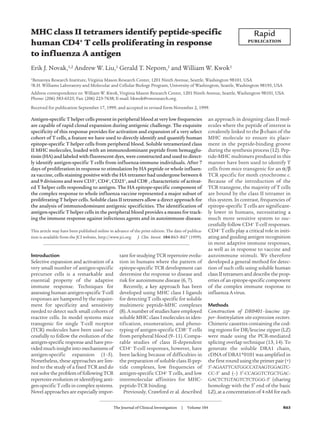
Figure 1
Specificity and HLA restriction of HA307–319 tetramer. (a) Comparison with thymidine incor-
poration at 72 hours between the T-cell clone cultured with the DRA1*0101/DRB1*0401
transfected BLS-1 pulsed with no antigen (left bar) and 10 µg/mL of HA307–319 peptide (right
bar). Error bars indicate SEM of triplicates. In b and c, clonal cells were stained with PE-
labeled tetramer for 3 hours at 37°C, washed, stained with anti-CD4, washed again, and
analyzed by flow cytometry. Staining with tetramer loaded with HA307–319 is shown in b, and
tetramer loaded with TT830-843 peptide is shown in c. In both, cells are gated on forward and
side scatter, the vertical axis shows tetramer fluorescence, and the horizontal axis shows CD4
fluorescence. Percentages shown in the margins of each panel represent the percent of total
cells present in each quadrant.
3. minute incubation, cells were washed
again and analyzed using a Becton
Dickinson FACSCalibur flow cytome-
ter (San Jose, California, USA).
Isolation, stimulation, and staining of
PBMC. PBMC from the same
DRB1*0401, DRB1*0101 individual
from which the clone was derived and a
second individual (DRB1*0401, *0401)
wereseparatedfromheparinizedvenous
blood by gradient centrifugation (Lym-
phoprep; Nycomed Pharma AS Diag-
nostics, Oslo, Norway). Cells were cul-
tured in RPMI-1640 (GIBCO BRL,
Rockville, Maryland, USA), supple-
mented with 2 mM L-glutamine, 1 mM
sodium pyruvate, 100 µg/mL peni-
cillin/streptomycin and 15% vol/vol
pooled human serum. Adherent cells
were prepared by plating out PBMC at 5
× 106 cells per well in 24-well plates for 1
hour. Nonadherent cells were removed
using a transfer pipette. Adherent cells
were incubated for 3 hours with either
10 µg/mL HA307–319 peptide, whole
influenza vaccine containing 11 µg/mL
HA (Connaught Laboratories Inc.,
Swiftwater, Pennsylvania, USA), or a
maximally stimulating dose of whole
tetanus toxoid. The nonadherent frac-
tion was passed through a nylon wool
column, washed twice with serum-free
PBS, and stained with 0.8 µM 5- (and -
6)-carboxyfluorescein diacetate succin-
imidyl ester (CFSE; Molecular Probes,
Eugene, Oregon, USA) for 10 minutes
at 37°C. Staining was stopped by
adding 100% FBS and subsequently
washing the cells twice in RPMI-1640
culture media. CFSE-stained nylon
wool–purified T cells were then added
back to the adherent cells at a density
of 2.5 × 10⁶ cells per well. Following 7
days of culture, cells were stained with
PE-labeled tetramer and combinations
of fluorochrome-labeled anti-CD3,
-CD4, -CD8, and -CD25 (PharMingen;
Becton Dickinson Immunocytometry
Systems, San Jose, California, USA) as
described above and analyzed by flow
cytometry.
Calculation of cell divisions and precur-
sor frequency. A portion of CFSE-
stained cells was stimulated with 2.5
µg/mL phytohemagglutinin (PHA)
and 10 U IL-2 and examined by FACS
on day 7. Polyclonal stimulation of T
cells with PHA and IL-2 results in cell
division with distinct CFSE fluores-
cence peaks, allowing determination
of the mean CFSE fluorescence for
each generation. These values were
used to calculate the average number
of cell divisions in cells stimulated
with antigen. Precursor frequency was
estimated by dividing the number of
tetramer-positive cells by 2x, where x is
the average number of cell divisions,
to determine the absolute number of
precursors for the tetramer-positive
cells, and then dividing this value by
the total number of cells analyzed.
The Journal of Clinical Investigation | Volume 104 R65
Figure 2
HA307–319 tetramer identification of antigen-
specific cells in relation to CFSE fluores-
cence. Nylon wool–purified T cells, labeled
with CFSE before culture with autologous
adherent cells and antigen, were stained on
day 7 with PE-labeled HA307–319 tetramer and
analyzed subsequently by flow cytometry.
Each row shows cells from a different stimu-
lating antigen for 2 different individuals: (a)
10 µg/mL HA307–319 peptide, (b) whole
influenza vaccine containing 11 µg/mL HA,
(c) whole TT at a maximally stimulating
dose. In all panels cells are gated on forward
and side scatter, the vertical axis shows PE
fluorescence of the HA307–319 tetramer, and
the horizontal axis shows CFSE fluorescence
over a 4-decade logarithmic scale. In addi-
tion, the horizontal axis shows the corre-
sponding number of cell divisions, with “P”
depicting the undivided parent population.
This scale was calculated from the distinct
CFSE fluorescence peaks produced by poly-
clonal stimulation with PHA and IL-2 as
described in Methods. Percentages shown in
the margins of each panel represent the per-
cent of total cells present in each quadrant.
The panels depict results from representative
individual experiments.
4. Results and Discussion
Earlier studies have shown that periph-
eral blood lymphocytes from individu-
als with previous exposure to influen-
za A virus generate a class II
DR-restricted T-cell proliferative
response to the HA307–319 epitope (19).
This conserved peptide can induce a
proliferative response in a number of
different DR haplotypes, including
DR1, DR4, DR5, and DR7 (20).
To detect T cells specific for the
HA307–319 peptide presented in the con-
text of DR4, we synthesized class II
DRA1*0101/DRB1*0401 tetramers
loaded with HA307–319 peptide. We test-
ed the specificity of the HA307–319
tetramer using a DRB1*0401-restricted
human T-cell clone specific for
HA307–319. As shown in Figure 1a, the
clone demonstrated antigen-specific
proliferation against HA307–319 in the
context of DRB1*0401 expressed in a
BLS-1 cell line (18). We stained
the HA307–319-specific clone using
DRB1*0401 tetramers loaded with the
HA307–319 peptide. As shown in Figure
1b, virtually all the cells stained posi-
tive for the HA307–319 tetramer and
CD4, consistent with the phenotype of
the clone. As a control we also con-
structed a DRB1*0401 tetramer
loaded with TT830-843 peptide (21). As
illustrated in Figure 1c, none of the
HA307–319 clonal cells stained positive
for the TT830-843 tetramer.
We then tested the ability of the
HA307–319 tetramer to detect antigen-
specific T cells collected from the
peripheral blood of 2 DRB1*0401
donors, including the same donor from
which the HA-specific clone was
derived. Nylon wool–purified T cells
from peripheral blood were stained
with CFSE, a fluorescent dye that stably
binds cytoskeletal actin (22). CFSE-
stained cells were cultured with autolo-
gous adherent cells pulsed with
HA307–319 peptide, whole influenza vac-
cine, or TT. After 7 days of culture,
HA307–319 tetramers were used to stain
the cells before analysis with flow
cytometry. Each time a cell divides,
CFSE is apportioned equally among
daughter cells, resulting in a halving of
CFSE fluorescence. Therefore, the
number of cell divisions can be deter-
mined by comparing the resultant
CFSE fluorescence to the original fluo-
rescence of the parent population. As
shown in Figure 2, all 3 antigens
induced cell proliferation as indicated
by populations of cells with decreased
CFSE fluorescence, shown on the hori-
zontal axis. The vertical axis shows the
fluorescence from the PE-labeled
HA307–319 tetramers, and the 2 columns
represent equivalent experiments done
on cells from the 2 donors. As shown in
the upper-left quadrants of Figure 2, a
and b, the labeled tetramer clearly iden-
tified a significant number of HA307–319-
specific cells in both the HA307–319 and
whole influenza vaccine samples in
both donors. To our knowledge, this is
the first time epitope-specific T helper
cells have been directly seen in a stimu-
lation of human lymphocytes taken
from the peripheral blood.
Examination of the cells stimulated
with HA307–319 peptide showed that in
both individuals around 90% of the
tetramer-binding cells were in the divid-
ed population, reflecting the specific
expansion of this cohort of T cells. The
observed number of cell divisions for a
given CFSE fluorescence isshownalong
the horizontal axis for all figure parts.
In donor 1, the tetramer-binding popu-
lation divided an average of 6.5 times
duringthe7-daycultureascalculatedby
CFSE fluorescence, whereas in donor
2 the tetramer-binding cells divided an
average of 9 times. This difference in the
numbers of divisions accounts for the
greater number of divided cells seen
in donor 2. The calculated precursor
frequency of DRB1*0401 tetramer-spe-
cific cells is similar for the 2 individuals,
ranging between 3 and 5 per 100,000
cells, depending on the individual
experiment.
The tetramer-binding population of
dividing cells comprised a distinct por-
tion of the total dividing cells in the
HA307–319 stimulated sample. In donor
1, some of the divided cells that are
tetramer negative likely represent T
R66 The Journal of Clinical Investigation | Volume 104
Figure 3
Phenotypic characterization of HA307–319
tetramer-specific cells. Nylon wool–purified
T cells were labeled with CFSE before culture
with autologous adherent cells and antigen.
On day 7, cells were stained with PE-labeled
tetramer and then stained with combina-
tions of fluorochrome-labeled anti-CD3,
-CD4, -CD8, and -CD25 antibodies before
flow-cytometry analysis. All panels are gated
on tetramer-positive cells. Upper panels
show CD8 versus CD4 staining in cells stim-
ulated with (a) HA307–319 peptide, and (b)
whole influenza vaccine, whereas lower pan-
els show CD25 versus CD3 staining in cells
stimulated with (c) HA307–319 peptide, and
(d) whole influenza vaccine. Boundaries for
positive and negative populations were
determined by controls. Percentages shown
in the margins of each panel represent the
percent of total cells present in each quad-
rant. The panels depict results from a repre-
sentative individual experiment.
5. cells specific for HA307–319 in the con-
text of DRB1*0101 because the donor
haplotype is DRB1*0401, DRB1*0101,
and DR1 has been described as capable
of presenting the peptide (23). In addi-
tion, background levels of prolifera-
tion of between 1% and 7% of the total
cells were seen even in control samples
where antigen was not added, depend-
ing on the individual (data not
shown). IL-2 and other cytokines lib-
erated by the antigen-specific HA307–319
cells would likely increase this back-
ground through increased bystander
activation and proliferation.
Incellsstimulatedwithwholeinfluen-
zavaccine(Figure2b),therewaslikewise
a definite population of tetramer-posi-
tive cells for both individuals, shown in
the upper-left quadrant. Therefore, even
in the context of a vigorous and com-
plex proliferative T-cell response to viral
antigens, the specific T cells correspon-
ding to an immunodominant epitope
are readily identified. There was no
tetramer labeling of cells stimulated
with TT despite vigorous proliferation
(Figure 2c). These results indicate that
the tetramer detection method is both
specific and sensitive for the population
of T cells reactive toward HA307–319.
The use of tetramer staining, togeth-
er with flow cytometry, to identify
antigen-specific cells permits simulta-
neous analysis of cells using addition-
al fluorochromes. This additional phe-
notypic analysis can provide important
information about an antigen-specific
response such as the type of T cell
involved, presence of activation or
other markers, and cytokine produc-
tion through intracellular staining. In
this study we examined surface expres-
sion levels of CD3, CD4, CD8, and
CD25 to further characterize the cells
identified by the HA307–319 tetramer.
The data in Figure 3 is gated to show
only cells identified as HA307–319-
tetramer positive. Figure 3, a and b,
shows that almost all the tetramer-
positive cells in both the HA307–319 pep-
tide–stimulated and whole influenza
vaccine–stimulated samples are CD4+
CD8–. Similarly, Figure 3, c and d,
shows that the majority of tetramer-
positive cells are CD3+ CD25+ for both
samples. We conclude that the
DRB1*0401-HA307–319 tetramer is
bound almost entirely to class
II–restricted, activated, and proliferat-
ing T cells, as expected, lending further
support to the specificity of the
tetramer detection method.
The concomitant use of peptide-
MHC class II tetramers and CFSE
staining provides a powerful tool to
assess and dissect antigen-specific T-
cell responses. Direct identification of
peptide-specific proliferation in pri-
mary cultures avoids the need for lim-
iting dilution analysis to calculate pre-
cursor frequencies and permits
simultaneous phenotypic analysis of
the antigen-specific cells using flow
cytometry. The present study demon-
strates the immunodominance of the
HA307–319 epitope in the context of the
complex response against whole
influenza vaccine and illustrates how
tetramers can be used to directly iden-
tify immunodominant antigenic speci-
ficities. The potential for loading dif-
ferent peptides into the tetramers
suggests multiple applications,
although the stability of specific pep-
tide-class II tetramers is likely to vary
among peptides. The use of class II
tetramers provides a means for under-
standing on a much more detailed level
theimmune response againstinfectious
agents and in autoimmune disease.
Acknowledgments
The authors would like to thank S.
Masewicz for expert technical assis-
tance, Å. Lernmark for constructive
comments and advice, and K. Nelson
and M. Gallagher for helpful discussion
and assistance with 4-color flow cytom-
etry analysis. This work was supported
in part by grant AI-44443 from the
National Institutes of Health. E.J.
Novak is supported by Poncin and
Acheivement Awards for College Scien-
tists predoctoral fellowships.
1. Goverman, J., et al. 1993. Transgenic mice that
express a myelin basic protein-specific T cell recep-
tor develop spontaneous autoimmunity. Cell.
72:551–560.
2. Kaye, J., et al. 1989. Selective development of CD4+
T cells in transgenic mice expressing a class II
MHC-restricted antigen receptor. Nature.
341:746–749.
3. Sagerstrom, C.G., Kerr, E.M., Allison, J.P., and
Davis, M.M. 1993. Activation and differentiation
requirements of primary T cells in vitro. Proc. Natl.
Acad. Sci. USA. 90:8987–8991.
4. Scott, B., Bluthmann, H., Teh, H.S., and von
Boehmer,H.1989.ThegenerationofmatureTcells
requiresinteractionofthealphabetaT-cellreceptor
with major histocompatibility antigens. Nature.
338:591–593.
5. Bot, A., Casares, S., Bot, S., von Boehmer, H., and
Bona, C. 1998. Cellular mechanisms involved in
protection against influenza virus infection in
transgenic mice expressing a TCR receptor specific
for class II hemagglutinin peptide in CD4+ and
CD8+ T cells. J. Immunol. 160:4500–4507.
6. Ota, K., etal. 1990. T-cell recognition of an immun-
odominant myelin basic protein epitope in multi-
ple sclerosis. Nature. 346:183–187.
7. Tuohy, V.K., Yu, M., Yin, L., Kawczak, J.A., and
Kinkel, R.P. 1999. Spontaneous regression of pri-
mary autoreactivity during chronic progression of
experimental autoimmune encephalomyelitis and
multiple sclerosis. J. Exp. Med. 189:1033–1042.
8. Altman, J.D., et al. 1996. Phenotypic analysis of
antigen-specific T lymphocytes. Science. 274:94–96.
9. Lee, P.P., et al. 1999. Characterization of circulating
T cells specific for tumor-associated antigens in
melanoma patients. Nat. Med. 5:677–685.
10. Romero, P., et al. 1998. Ex vivo staining of metasta-
tic lymph nodes by class I major histocompatibility
complextetramersrevealshighnumbersofantigen-
experiencedtumor-specificcytolyticTlymphocytes.
J.Exp.Med. 188:1641–1650.
11. Yee, C., Savage, P.A., Lee, P.P., Davis, M.M., and
Greenberg, P.D. 1999. Isolation of high avidity
melanoma-reactive CTL from heterogeneous pop-
ulations using peptide-MHC tetramers.J.Immunol.
162:2227–2234.
12. Crawford,F.,Kozono,H.,White,J.,Marrack,P.,and
Kappler, J. 1998. Detection of antigen-specific T
cells with multivalent soluble class II MHC cova-
lent peptide complexes. Immunity. 8:675–682.
13. Horton, R.M., Cai, Z.L., Ho, S.N., and Pease, L.R.
1990. Gene splicing by overlap extension: tailor-
made genes using the polymerase chain reaction.
Biotechniques. 8:528–535.
14. Chang, H.C., et al. 1994. A general method for facil-
itatingheterodimericpairingbetweentwoproteins:
application to expression of alpha and beta T-cell
receptor extracellular segments.Proc.Natl.Acad.Sci.
USA. 91:11408–11412.
15. Stern, L.J., and Wiley, D.C. 1992. The human class
II MHC protein HLA-DR1 assembles as empty
alphabetaheterodimersintheabsenceofantigenic
peptide. Cell. 68:465–477.
16. Schatz,P.J.1993.Useofpeptidelibrariestomapthe
substratespecificityofapeptide-modifyingenzyme:
a 13 residue consensus peptide specifies biotinyla-
tion in Escherichia coli. Biotechnology. 11:1138–1143.
17. Mickelson, E.M., Masewicz, S.A., Nepom, G.T.,
Martin, P.J., and Hansen, J.A. 1991. Alloreactive T-
cell clones identify multiple HLA-DQw3 variants.
Hum. Immunol. 30:32–40.
18. Kovats, S., et al. 1995. Deficient antigen-presenting
cell function in multiple genetic complementation
groupsoftypeIIbarelymphocytesyndrome. J.Clin.
Invest. 96:217–223.
19. Lamb, J.R., Eckels, D.D., Lake, P., Woody, J.N., and
Green, N. 1982. Human T-cell clones recognize
chemically synthesized peptides of influenza
haemagglutinin. Nature. 300:66–69.
20. Krieger, J.I., et al. 1991. Single amino acid changes
in DR and antigen define residues critical for pep-
tide-MHC binding and T cell recognition. J.
Immunol. 146:2331–2340.
21. Panina-Bordignon, P., et al. 1989. Universally
immunogenicTcellepitopes:promiscuousbinding
to human MHC class II and promiscuous recogni-
tion by T cells. Eur.J.Immunol. 19:2237–2242.
22. Lyons, A.B., and Parish, C.R. 1994. Determination
of lymphocyte division by flow cytometry. J.
Immunol.Methods. 171:131–137.
23. Stern, L.J., et al. 1994. Crystal structure of the
human class II MHC protein HLA-DR1 complexed
with an influenza virus peptide. Nature.
368:215–221.
The Journal of Clinical Investigation | Volume 104 R67