SlideShare a Scribd company logo
1 of 10
Download to read offline
PATHOPHYSIOLOGY AND NATURAL HISTORY
MYOCARDIAL INFARCTION
Left ventricular remodeling after myocardial
infarction: a corollary to infarct expansion
RAYMOND G. MCKAY, M.D., MARC A. PFEFFER, M.D., PH.D., RICHARD C. PASTERNAK, M.D.,
JOHN E. MARKIS, M.D., PATRICIA C. COME, M.D., SHOICHIRO NAKAO, M.D.,
JAMES D. ALDERMAN, M.D., JAMES J. FERGUSON, M.D., ROBERT D. SAFIAN, M.D.,
AND WILLIAM GROSSMAN, M.D.
ABSTRACT Dilatation of infarcted segments (infarct expansion) may occur during recovery from
myocardial infarction, but the fate of noninfarcted segments is uncertain. Accordingly, left ventricular
geometric changes were assessed by left ventricular angiography and M mode echocardiography on
admission and 2 weeks later in 30 patients with their first acute transmural myocardial infarction. All
patients demonstrated chest pain, ST segment elevation with subsequent development of Q waves (15
anterior, 15 inferior), and elevation of cardiac enzymes. Sequential left ventricular angiographic and
hemodynamic findings were available in these patients by virtue of their participation in a study of
thrombolysis in acute myocardial infarction. By that study design, all patients treated successfully with
thrombolytic therapy and demonstrating improvement of flow in an occluded coronary artery under-
went repeat cardiac catheterization. At 2 weeks there was a significant decrease in left ventricular and
pulmonary capillary wedge pressures (p < .01), whereas both left ventricular end-diastolic (LVEDV)
and end-systolic (LVESV) volume indexes increased (p < .01). The increase in LVEDV correlated
directly with the percentage of the ventriculographic silhouette that was akinetic or dyskinetic at the
initial catheterization (r = .71, p < .001). To assess regional changes in both infarcted and noninfarct-
ed segments, serial endocardial perimeter lengths of both the akinetic-dyskinetic segments (infarction
zone) and of the remainder of the cardiac silhouette (noninfarction zone) were measured in all patients
who demonstrated at least a 20% increase in their LVEDV at 2 weeks after myocardial infarction.
Notably, there was a mean increase of 13% in the endocardial perimeter length of infarcted segments
and a 19% increase in the endocardial perimeter length of noninfarcted segments. Serial M mode
echocardiographic studies showed no significant change in the wall thickness of noninfarcted myocar-
dial segments. Hemodynamic changes that occurred in this subgroup of patients included significant
decreases in left ventricular end-diastolic and pulmonary capillary wedge pressures (p < .05) and
significant increases in angiographic cardiac index (p < .01) and LVESV index (p < .01). We
conclude that in patients who manifest cardiac dilatation in the early convalescent period after myocar-
dial infarction, there is remodeling of the entire left ventricle including infarct expansion of akinetic-
dyskinetic segments and volume-overload hypertrophy of noninfarcted segments. The magnitude of
the remodeling process is directly proportional to infarct size as assessed by the extent of wall motion
abnormality present during the acute phase of infarction. Moreover, the remodeling changes that occur
are associated with hemodynamic improvement, including lower left ventricular filling pressures and
increased cardiac output, but these hemodynamic changes appear to occur at the expense of a signifi-
cant increase in left ventricular chamber volumes.
Circulation 74, No. 4, 693-702, 1986.
From the Charles A. Dana Research Institute and the Harvard-Thorn- PREVIOUS STUDIES of ventricular structure after
dike Laboratory of Beth Israel Hospital Departments of Medicine (Car- myocardial infarction have focused primarily on the
diovascular Division) and Radiology, Beth Israel Hospital and Harvard
Medical School, and from the Department of Medicine (Cardiovascular fate of infarcted segments. 8 In particular, Hutchins
Division), Brigham and Women's Hospital, Harvard Medical School, and Bulkley3 and Eaton et al.4 have described regional
Boston.
Supported in part by Research Training Grant HL07394 from the cardiac dilatation in the infarction zone and have de-
USPHS. fined the concept of "infarct expansion." According to
Address for correspondence: Raymond G. McKay, M.D., Cardio- thi decipin thi prcs per ob omo
vascular Division, Beth Israel Hospital, 330 Brookline Ave., Boston, their description, this process appears to be a form of
MA 02215. intramural myocardial rupture in which the disruption
Received April 15, 1986; revision accepted July 3, 1986. * *
m
A preliminary report was presented at the Annual Scientific Sessions t
of the American Heart Association, Miami Beach, November 1984. tion of the necrotic zone. Such changes occur almost
693
Vol. 74, No. 4, October 1986
Downloaded
from
http://ahajournals.org
by
on
March
13,
2019
McKAY et al.
exclusively in transmural infarcts and are most com-
monly seen with anterior and anteroseptal infarctions.
The process has been observed to occur clinically as
early as 3 days after infarction and may progress over
days to weeks independent of additional myocardial
necrosis or infarct extension. Most important, it is
associated with increased mortality and may be impor-
tant in the development of late aneurysm formation.
Although infarct expansion has been studied exten-
sively, only limited information is available on the fate
of noninfarcted segments during the infarction recov-
ery period.9-'3 A recent study by Weisman et al.9 re-
ported that, in the rat, there may be global remodeling
of the left ventricle immediately after infarction in-
volving structural changes in both infarcted and nonin-
farcted segments. Similarly, a study by Anversa et al.'0
found that myocardial infarction in rats results in myo-
cyte hypertrophy of remaining viable myocardium.
Studies on the fate of noninfarcted myocardium in
man, however, have been limited"; moreover, the de-
terminants and hemodynamic effects of this remodel-
ing process have not been defined. Wynne et al.12
found that regional ejection fraction in noninfarcted
segments was abnormal in 69% of patients with anteri-
or infarctions and in 30% with inferior infarctions.
Although these limited studies suggest that regional
function in noninfarcted segments may be abnormal,
the etiology of this dysfunction and the possible role
that left ventricular remodeling plays in producing
these changes remains unclear.
This study was undertaken to assess the fate of non-
infarcted myocardial segments after transmural infarc-
tion. Since patients admitted with acute transmural
infarction who elected to undergo attempted thrombol-
ysis were expected to undergo serial cardiac catheter-
izations with contrast ventriculography on admission
and, if thrombolysis was successful, 2 weeks later,
these patients were considered for enrollment and
formed the study population.
Methods
Study group. Left ventricular hemodynamic and structural
changes were assessed in a total of 30 patients. All patients
presenting with their first acute transmural myocardial infarc-
tion were considered for enrollment in this study. Entry criteria
included chest pain of at least 30 min duration, new ST segment
elevation on the electrocardiogram with the subsequent devel-
opment of Q waves in the involved leads, and elevation of
cardiac enzymes (creatine kinase MB fraction, SGOT, LDH)
during the first 24 to 72 hr of their hospitalization. Patients who
manifested persistent ischemia or recurrent infarction during
their in-hospital convalescent phase as evidenced by clinical,
electrocardiographic, or cardiac enzyme criteria were excluded
from the study. Patients with previous myocardial infarction,
694
significant valvular disease, or cardiomyopathy were not con-
sidered. Informed consent was obtained from each patient after
an appropriate explanation of risks and potential complications
of the proposed study.
Cardiac catheterization
Protocol. Cardiac catheterization was performed at the time
of admission and 2 weeks later, before discharge. For both
catheterizations, right femoral venous and right femoral arterial
sheaths were inserted after administration of local anesthesia. A
No. 7F thermodilution flow-directed catheter was passed to the
pulmonary artery-pulmonary capillary wedge position. Left
ventriculography followed by coronary angiography were per-
formed in the routine manner. Baseline hemodynamic measure-
ments included pulmonary capillary wedge and left ventricular
pressures. Recordings were inscribed with a Honeywell Elec-
tronics for Medicine (VR-16) recorder.
In each of the 30 patients, an occluded or subtotally occluded
coronary artery whose distribution corresponded to the area of
ST segment elevation by electrocardiographic criteria was
noted. Each patient was treated subsequently with either
250,000 U of intracoronary streptokinase, 1.5 million U of
intravenous streptokinase, or 80 mg of intravenous tissue-type
plasminogen activator. Only patients in whom successful
thrombolysis was achieved with dissolution of clot and im-
provement of flow in the involved coronary artery (and in whom
this improvement persisted at 2 weeks) were included, since
complete serial studies were available only in this group. After
the initial catheterization, all patients were treated with intrave-
nous heparin and intravenous lidocaine. Oral or topical nitrates,
calcium-channel blockers, and/or S-blockers were continued or
added if clinically indicated. Lidocaine was discontinued 48 to
72 hr after admission and replaced with an oral antiarrhythmic
only if significant ventricular ectopy was subsequently noted.
All other medications were continued until the time of the sec-
ond catheterization except for heparin, which was stopped 4 hr
before that catheterization.
Data analysis. Ventricular volumes were measured by the
area-length method with a regression equation developed for
single-plane angiography based on left ventricular casts for
measurement of true volume with calculation of end-diastolic
and end-systolic volumes, angiographic cardiac index, and glo-
bal ejection fraction. 14
Endocardial perimeters of akinetic-dyskinetic segments and
of the remainder of the contrast ventriculogram silhouette were
measured with a Tektronix 4956 digitizing computer. The per-
centage of the ventriculographic perimeter that was akinetic or
dyskinetic was determined on admission and at 2 weeks. In
addition, serial changes in the perimeters of akinetic-dyskinetic
segments and of the remainder of the cardiac silhouette that
occurred in the 2 week interval between the first and second
catheterization were measured with appropriate corrections for
differences in magnification.
M mode echocardiography. Echocardiography was per-
formed on admission and at 2 weeks after infarction in 19 of the
30 patients by an ATL Mark VI machine. This machine is
equipped with an M mode cursor that can be oriented perpen-
dicular to the long axis of the left ventricle for optimal M mode
recording of the septum and posterior wall. Changes in gain
were used to define the endocardial and epicardial borders. Wall
thicknesses were measured at the onset of the QRS complex on
the electrocardiogram with the leading edge-to-leading edge
technique.
Statistics. Means and standard deviations were calculated for
all variables. Paired dimensional data were analyzed with either
the paired t test or the Wilcoxon signed-rank test where appro-
priate, for parametric and nonparametric distributions. A p val-
ue of less than .05 was considered significant.
CIRCULATION
Downloaded
from
http://ahajournals.org
by
on
March
13,
2019
PATHOPHYSIOLOGY AND NATURAL HISTORY-MYOCARDIAL INFARCTION
Results
Study group. A total of 30 patients with their initial
acute myocardial infarction were included in this
study. There were 28 men and two women, with a
mean age of 55 years (range 33 to 69). Fifteen patients
presented with electrocardiographic evidence of an
anterior myocardial infarction, and each had a totally
or subtotally occluded left anterior descending coro-
nary artery. The remaining 15 patients demonstrated
electrocardiographic changes in inferior leads with to-
tal occlusion of the right coronary artery. Fifteen pa-
tients had coronary disease limited to one coronary
artery in the area of infarction; the remaining 15 had
additional stenoses, with eight patients having two-
vessel disease and seven patients having three-vessel
disease. All patients were brought to the catheteriza-
tion laboratory within 10 hr of the onset of their chest
pain. Successful thrombolysis with opening ofa totally
occluded artery or improvement in coronary flow was
achieved in all patients within 90 min with administra-
tion of either intracoronary or intravenous streptokin-
ase or intravenous tissue-type plasminogen activator.
After the initial catheterization, patients were ad-
mitted to the coronary care unit for routine monitoring.
All patients had an uncomplicated convalescent period
without evidence of recurrent infarction, development
of ventricular septal defect, or papillary muscle rup-
ture. Repeat catheterization was performed in all pa-
tients at a mean of 14. 1 days (range 9 to 18 days) after
admission. All patients manifested persistent patency
of their diseased coronary artery with a residual high-
grade stenosis.
At the time of the first catheterization, medications
included the following: nitrates, 30 patients; calcium-
channel blockers, 12 patients; ,3-blockers, six patients;
diuretics, two patients; digoxin, five patients; antiar-
rhythmics, 30 patients. At the time of the second cath-
eterization, medications included the following: ni-
trates, 30 patients; calcium-channel blockers, 15
patients; /8-blockers, six patients; diuretics, four pa-
tients; digoxin, five patients; antiarrhythmics, six pa-
tients. There were no significant differences in medi-
cations between the time of the first and second
catheterization, except for the use of antiarrhythmics
(all patients at the time of their first catheterization
were treated with intravenous lidocaine; only six pa-
tients subsequently required long-term antiarrhythmic
therapy).
Hemodynamic and ventricular volume changes. Hemo-
dynamic changes that occurred over the 2 week study
period in the 30 patients included decreases in heart
rate (80 17 to 72 + 16 beats/min; p < .03), left
Vol. 74, No. 4, October 1986
ventricular systolic pressure (122 ± 19 to 113 ± 11
mm Hg; p < .01), left ventricular end-diastolic pres-
sure (24 + 8 to 18 ± 6 mm Hg; p < .01), and
pulmonary capillary wedge pressure (18 ± 6 to 13 ±
6 mm Hg; p < .01). There was no significant change in
either angiographic cardiac index (3.3 ± 1.2 to 3.5 +
1.2 liters/min/m2; p = NS) or left ventricular ejection
fraction (55 ± 11% to 54 ± 11%; p = NS). In
addition, there were increases in left ventricular end-
diastolic volume index (76 ± 22 to 93 ± 30 m1/M2; p
< .01), end-systolic volume index (34 ± 10 to 47 ±
30 mI/M2; p < .02), and stroke volume index (42 ± 16
to 50 ± 13 ml/m2; p < .02). Table 1 summarizes
hemodynamic and angiographic data at the time of
admission and at 2 weeks for all patients.
Regional wall motion abnormalities. In 26 of the 30
patients, a discrete segment ofakinesis and/or dyskine-
sis could be identified on the contrast left ventriculo-
gram at the time of the first catheterization. This region
ofakinesis-dyskinesis was noted to persist or enlarge at
the time of the second catheterization. The percentage
of the left ventricular perimeter that was akinetic or
dyskinetic was 20 ± 14% on admission and 25 ± 14%
2 weeks later (p = NS).
Determinants of the increase in end-diastolic volume in-
dex. The increase in the end-diastolic volume index
that occurred over the 2 week period correlated signifi-
cantly with the percentage of the initial contrast ven-
triculogram that was akinetic or dyskinetic (r = .71,
p < .001) at the time ofthe first catheterization. Figure
1 summarizes the relationship between volume in-
creases and the percentage of akinesis-dyskinesis. In
addition, there was a modest inverse correlation be-
tween left ventricular ejection fraction on the first cath-
eterization and volume increases from the initial study
to the study at 2 weeks (r = -.51, p < .001).
Hemodynamic changes in patients with increased left
ventricular volumes. Since left ventricular volume
changes were related to the extent of initial infarction,
a subgroup of 17 of the 30 patients who demonstrated
at least a 20% increase in end-diastolic volume index
was identified for further evaluation. This group con-
sisted of 11 patients with anterior infarctions and six
patients with inferior infarctions. Hemodynamic and
volume changes that occurred in these patients includ-
ed decreases in left ventricular end-diastolic pressure
(23 ± 9 to 18 ± 6 mm Hg; p < .05) and pulmonary
capillary wedge pressure (17 ± 6 to 12 ± 5 mm Hg;
p < .03), with concomitant increases in angiographic
cardiac index (2.9 ± 1.0 to 3.7 ± 1.1 liters/min/m2;
p < .01), stroke volume index (36+ 13 to 49 12
ml/M2; p < .01), and end-systolic volume index (30 ±
695
Downloaded
from
http://ahajournals.org
by
on
March
13,
2019
McKAY et al.
TABLE 1
Serial hemodynamic data for patients with anterior myocardial infarction (Nos. I to 15) and inferior myocardial infarction (Nos. 16 to 30)
Patient
1 2 3 4 5 6 7 8 9 10 11 12 13 14 15
Sex M M M M M M M F M M M M m M M
Age (yr) 62 69 55 33 38 56 54 62 67 60 65 69 57 42 64
First catheterization
EDVI 82 118 70 70 79 45 48 46 64 70 72 92 140 96 87
ESVI 31 53 24 40 43 29 34 25 28 31 46 48 48 40 32
SVI 51 65 46 30 36 16 14 21 36 39 26 44 92 56 55
EF 62 55 66 43 46 36 29 46 56 56 36 48 66 58 63
HR 84 72 64 96 102 72 96 66 82 59 90 80 54 108 80
CI 4.3 4.7 2.9 2.9 3.7 1.1 1.3 1.4 3.0 2.3 2.3 3.5 5.0 6.0 4.4
LVSP 115 119 125 100 97 130 114 160 130 100 125 122 158 132 122
LVEDP 21 25 22 14 25 28 40 40 32 34 22 19 29 17 28
PCW 17 18 11 12 22 20 26 30 21 18 15 16 26 14 22
Second catheterization
EDVI 108 142 84 88 211 109 123 63 86 88 112 103 122 92 70
ESVI 38 86 30 59 183 57 83 28 43 38 55 44 49 36 32
SVI 70 56 54 29 28 52 40 35 43 50 57 59 73 56 38
EF 65 39 64 33 13 48 33 56 50 57 51 57 60 61 54
HR 66 80 68 96 102 54 84 72 66 46 60 60 96 66 72
Cl 4.6 4.5 3.7 2.8 2.9 2.8 3.4 2.5 2.8 2.3 3.4 3.5 7.0 3.7 2.7
LVSP 103 102 95 95 100 117 103 125 115 110 130 115 135 111 112
LVEDP 20 14 12 22 30 19 15 6 24 14 25 16 12 21 17
PCW 12 D 9 11 29 14 10 5 D 9 10 13 10 14 13
CI = angiographic cardiac index (I/min/m2); EDVI = left ventricular end-diastolic volume index (mi/M2); EF = left ventricular ejection fraction
(%); ESVI = left ventricular end-systolic volume index (ml/m2); HR = heart rate (bpm); LVEDP = left ventricular end-diastolic pressure (mm Hg);
LVSP = left ventricular systolic pressure (mm Hg); PCW = pulmonary capillary wedge pressure (mm Hg); SVI = left ventricular stroke volume index
(m1/m2).
Ap < .03; Bp < .02; cp < .01; D data not obtained because of technical reasons.
10 to 56 + 37 ml/mr; p < .01). There were no signifi-
cant changes in left ventricular systolic pressure (118
+ 17 to 112 + 11 mm Hg; p = NS), heart rate (83 ±
16 to 76 + 18 beats/min; p = NS), or ejection fraction
(53 ± 12% to 50 ± 14%; p = NS). Hemodynamic
and angiographic data for these patients are summa-
rized in table 2.
Serial changes in endocardial perimeters in patients with
increased volumes. To assess structural changes occur-
ring in the 17 patients who demonstrated substantial
(>-20%) increases in left ventricular end-diastolic vol-
ume, serial endocardial perimeter measurements were
made of the akinetic-dyskinetic segments (represent-
ing infarcted zones) and of the remainder ofthe cardiac
silhouette (representing noninfarcted segments) on the
left ventricular angiograms obtained on admission and
at 2 weeks. Table 3 summarizes changes in measure-
ments of endocardial perimeter length of both akinetic-
dyskinetic segments and of the remainder of the cardi-
ac silhouette. Notably, there was a mean 13 + 12%
increase in endocardial perimeter length of infarcted
segments and a 19 ± 13% increase in the endocardial
696
perimeter length of noninfarcted segments. Figure 2
shows examples of end-diastolic and end-systolic car-
diac silhouettes in four representative patients at the
time of initial presentation with acute myocardial in-
farction and at follow-up study 2 weeks later.
M mode echocardiography. Table 4 summarizes M
mode measurements of septal and posterior wall thick-
ness in 19 of 30 patients who were studied, including
12 who manifested at least a 20% increase in left ven-
tricular end-diastolic volume. Notably, patients with
anterior myocardial infarction showed no significant
change in posterior wall thickness and patients with
inferior infarction showed no change in septal wall
thickness.
Discussion
Our results demonstrate that global remodeling of
the left ventricle occurs during the early convalescent
period in certain patients with myocardial infarction.
In particular, the findings indicate that those patients
who exhibited cardiac dilatation with increased left
ventricular end-diastolic and end-systolic volumes
CIRCULATION
Downloaded
from
http://ahajournals.org
by
on
March
13,
2019
PATHOPHYSIOLOGY AND NATURAL HISTORY-MYOCARDIAL INFARCTION
TABLE 1
(Continued)
Patient
Mean ±
16 17 18 19 20 21 22 23 24 25 26 27 28 29 30 SD
M M M
40 60 51
M M M F
42 64 43 64
M M M M M M M M 28M, 2F
55 34 62 59 59 45 54 62 55 ± 10
56 60 83 67 47 55 74 75 71 103 59 90 89 101 81 76±+10
25 21 31 28 15 20 21 37 38 42 18 49 37 43 40 34+ 10
31 39 51 39 32 35 53 38 33 61 41 41 52 58 41 42+ 16
55 65 62 58 68 64 72 51 46 59 69 46 59 57 51 55 + 11
102 84 60 108 77 100 70 60 96 70 50 170 102 66 90 80± 17
3.2 3.3 3.1 4.2 2.5 3.5 3.7 2.3 3.2 4.3 2.1 2.9 5.3 3.8 3.7 3.3+1.2
137 103 103 110 130 120 120 130 105 97 120 140 130 172 92 122+ 19
13 10 20 19 16 16 26 24 20 24 15 28 21 22 22 23 ± 7
13 12 17 16 D 15 D D 14 23 5 20 21 15 14 18±5
102 103 108 94 89 72 68 83 76 111 47 105 101 90 54 97 + 30c
40 40 50 40 40 36 26 35 33 59 20 32 47 26 24 47 ±+301
62 63 58 54 49 36 42 48 43 52 27 73 54 64 30 50so13B
61 62 54 57 55 50 68 58 57 47 57 70 53 71 56 54+ 12
60 90 84 110 66 90 50 60 61 58 60 69 60 75 72 72 + 16A
3.7 5.6 4.9 5.9 3.2 3.2 2.1 2.9 2.6 3.0 1.6 5.0 3.2 4.8 2.2 3.6+ 1.2
128 117 111 125 120 112 115 130 105 105 110 121 115 130 102 114+ llc
23 20 21 18 12 11 22 20 18 21 12 22 11 12 8 17±6c
14 14 12 14 9 9 D D 13 14 6 10 10 9 6 12 +4C
during their in-hospital stay manifested changes in
both infarcted and viable segments of their ventricles.
Lengthening of endocardial perimeters of infarcted
300
250
0
> 200
0
z
0 150
z
I
100
+
+
50I
-10 0 10 20 30 40 50 60
% OF AKINESIS-DYSKINESIS
FIGURE 1. Correlation between the percentage of the left ventricular
silhouette that was akinetic-dyskinetic at the time of the first catheteriza-
tion with the subsequent increase in left ventricular end-diastolic vol-
ume index (EDVI) at the second catheterization (r = .71, p < .001).
The ordinate, "Change in EDVI," was determined by dividing the EDVI
at the 2 week catheterization by the EDVI obtained at the first catheter-
ization.
segments presumably represents infarct expansion.3
Equally important, lengthening of the endocardial pe-
rimeter of that part of the ventricle without regional
wall motion abnormalities and with no evidence of
wall thinning in these segments suggests that volume-
overload hypertrophy with a net increase in the myo-
cardial mass of these segments has occurred. These
remodeling changes occurred as hemodynamics im-
proved, including lower left ventricular filling pres-
sures and increased cardiac output, perhaps at the ex-
pense of increased chamber volumes. Moreover, the
magnitude of these remodeling changes were roughly
related to infarct size as measured by the percentage of
the ventricular silhouette exhibiting regional wall mo-
tion abnormality on admission and inversely related to
initial left ventricular pump function as measured by
ejection fraction.
Grossman and others", 16 have termed an increase in
myocardial mass without relative wall thickening as
"volume overload" hypertrophy when it occurs in the
entire left ventricle. In volume-overload hypertrophy,
as discussed below, mass increases primarily by series
addition of new sarcomeres and fiber elongation, re-
sulting in chamber enlargement. In pressure-overload
hypertrophy, in contrast, a marked increase in wall
thickness occurs as a consequence of the parallel addi-
tion of new myofibrils.16 Although usually associated
Vol. 74, No. 4, October 1986
+
+
+
697
Downloaded
from
http://ahajournals.org
by
on
March
13,
2019
McKAY et al.
TABLE 2
Serial hemodynamic and angiographic data in patients with greater than 20% increase in left ventricular end-diastolic
volume index
EDVI ESVI SVI EF HR CI LVSP LVEDP PCW
Firstcath. 67+18 31+10 36+13 53±12 83+16 2.9+1.0 118+17 23+9 18+5
Second cath. 105±33 56 37c 49± 12 50± 14 76 - 18 3.7 +1 c 112 - 11 18 6A 12 5B
Data are derived from all patients who demonstrated greater than 20% increase in end-diastolic volume index at the time of the
second catheterization, including 11 patients with anterior infarctions (Nos. 1 to 11. table 1) and six patients with inferior
infarctions (Nos. 16 to 21 table 1). Abbreviations are as in table 1.
Ap < .05; Bp < .03; cp < .01.
with some degree of wall thickening, chronic volume-
overload hypertrophy produces far less thickening than
does pressure-overload hypertrophy. Thus we believe
that the absence of thinning in the noninfarcted seg-
ments, associated with concurrent elongation, is com-
patible with volume-overload hypertrophy of these
segments.
TABLE 3
Left ventricular endocardial perimeter analysis in patients with
greater than 20% increase in left ventricular end-diastolic volume
index
Catheterization
Catheterization % Change
% Change nonin-
% Akinesis- infarcted farcted
Patient dyskinesis segment segment
Anterior
infarctions
1 24 + 6 +18
2 20 + 12 +21
3 10 + 7 +15
4 41 + 4 +33
5 46 +38 +28
6 50 + 11 +42
7 41 +20 +25
8 37 + 8 +18
9 30 +10 +10
10 30 +20 + 5
11 40 +26 + 4
Inferior
infarctions
16 32 + 8 + 16
17 18 + 9 +22
18 18 + 12 +23
19 20 - 6 + 16
20 17 +±10 +12
21 16 + 8 + 12
Mean + SD 13+12 19+ 13
Data are derived from the 1 1 patients with anterior infarctions (Nos. 1 to
1 1, table 1 ) and six patients with inferior infarctions (Nos. 16 to 21, table 1)
whose hemodynamic and angiographic findings are summarized in table 2.
698
Stimulus for volume-overload hypertrophy in the infarct-
ed ventricle. Left ventricular hypertrophy is one of the
principal adaptive mechanisms by which the heart
compensates for an increased load. The pattern of hy-
pertrophy that develops is dependent on the type of
overload and the influence of this overload on systolic
and diastolic wall stresses."5 17 According to this
hypothesis, when the primary stimulus for hyper-
trophy is pressure overload, the resultant acute in-
crease in systolic wall stress leads to parallel replica-
tion of sarcomeres, wall thickening, and concentric
hypertrophy. The wall thickening that occurs tends to
return systolic wall stress toward normal. In contrast,
when the primary stimulus to hypertrophy is a volume
overload, increased diastolic pressure causes an in-
crease in diastolic wall stress and leads to sarcomere
replication in series, fiber elogation, and chamber en-
largement. Chamber enlargement accommodates the
volume overload and returns diastolic pressure toward
normal. However, the chamber enlargement also re-
sults in an increase in systolic wall stress, which subse-
quently leads to wall thickening. The combination of
slight wall thickening and chamber enlargement return
systolic and diastolic wall stress toward normal.
Given that hypertrophy will develop in response to
increased wall stress, what evidence is there that wall
TABLE 4
Echocardiographic wall thickness measurements (mm)
Catheterization 1 Catheterization 2
Posterior Posterior
Septum wall Septum wall
Anterior
infarctions 10.8+0.8 11.8+0.9 11.5+0.9 11.4+0.9
(n = 10)
Inferior
infarctions 10.6+0.7 9.6+0.4 9.8+1.0 10.2+0.9
(n =9)
There were no significant differences between serial septal and posterior
wall thicknesses in either group.
CIRCULATION
Downloaded
from
http://ahajournals.org
by
on
March
13,
2019
PATHOPHYSIOLOGY AND NATURAL HISTORY-MYOCARDIAL INFARCTION
I'I' %
FIGURE 2. Right anterior oblique end-diastolic and end-systolic cardi-
ac silhouettes obtained in four patients on admission (left) and at 2
weeks (right). Solid lines indicate akinetic-diskinetic (infarcted) seg-
ments and stippled lines indicate noninfarcted areas. In all cases, there
has been an increase in chamber volume as well as lengthening of both
the infarcted perimeter and noninfarcted perimeter.
stresses are abnormal in patients with myocardial in-
farction during their convalescent phase? To date, this
has been a diffilcult problem to assess because of diffi-
culties in measuring wall stresses in ventricles with
regional wall abnormalities and variable wall thick-
nesses. Two recent studies, however, are notable.
Pfelfer et al.'8 examined alterations in mass, vol-
ume, and end-diastolic wall stress in rats at 2, 21, and
100 days after coronary ligation and subsequent myo-
cardial infarction and compared changes in these varia-
bles with findings in rats without coronary ligation.
Vol. 74, No. 4, October 1986
Passive pressure-volume curves were deternined and
end-diastolic stress was calculated from data on left
ventricular mass, end-diastolic pressure, and end-dia-
stolic volume. No change in mass, volume, or wall
stress was noted in any of the control rats. In rats with
myocardial infarction, however, there was a progres-
sive increase in both volume and mass, and end-dia-
stolic wall stress was elevated at all times. Of note, the
ratio of mass to volume decreased progressively, indi-
cating that left ventricular volume was increasing out
of proportion to mass and suggesting that a volume-
overload hypertrophy was occurring, perhaps in re-
sponse to the increased end-diastolic wall stress.
Pouleur et al.'9 also attempted to evaluate wall
stresses in patients with previous myocardial infarc-
tion. Using a method for calculating regional wall
stresses, 19 20 they found that both systolic and diastolic
regional wall stresses were abnormal in the infarcted
and ischemic areas and that stresses in the noninfarcted
segments were normal. Because serial studies were not
available to them in each patient, they could not com-
ment on changes in ventricular wall stress after
infarction.
Thus, although available data are limited, there is
evidence that wall stresses in healing left ventricles
may be abnormal.
A model for left ventricular remodeling after infarction.
Based on the observations that wall stresses may be
abnormal in the infarcted ventricle and the fact that
increased diastolic wall stress may initiate a volume-
overload type ofhypertrophy, a model of left ventricu-
lar remodeling after myocardial infarction may be pro-
posed. This model is summarized in figure 3. In this
model, the immediate hemodynamic consequences of
an acute myocardial infarction on ventricular function
include both systolic and diastolic dysfunction. Systol-
ic impairment secondary to loss of contractile function
of the infarcted myocardium results in a decreased
systolic ejection, increased end-systolic volume, an
increase in cardiac size, and a secondary increase in
diastolic filling pressure caused by the increase in ven-
tricular volume. Diastolic function is characterized im-
mediately by an increase in diastolic distensibility,2' 23
which minimizes the rise in filling pressure. However,
as necrotic tissue is replaced by fibrosis, a decrease in
distensibility occurs. In addition, there may be upward
shift in the diastolic pressure-volume curve secondary
to ischemia in the border zone so that filling pressure
may tend to be higher for any given volume. Thus
diastolic volume increases and diastolic pressure also
tends to increase, especially in patients with large
infarctions.
699
Downloaded
from
http://ahajournals.org
by
on
March
13,
2019
McKAY et al.
RESTORED
STROKE VOLUME
4
SEGMENTAL INFARCTION
DECREASED SYSTOLIC EJECTION
INCREASED LEFT VENTRICULAR
END-DIASTOLIC VOLUME AND PRESSURE
Frank-
INC D Starling INCREASED
INCRE ASE StrDn
SYSTOLIC
EJECTION  NON-INFARCTED SEGMENT:
REGIONAL HYPERTROPHY
DECREASED C
WALL STRESS
INFARCTED SEGMENT:
INFARCT EXPANSION
,ONTRACTILITY
LATE HEART FAILURE
FIGURE 3. Hypothesis proposed to account for the mechanisms of left ventricular remodeling.
In the presence of both systolic and diastolic dys-
function, there may be peripheral mechanisms mediat-
ed via the sympathetic nervous system and circulating
catecholamines to help maintain a normal arterial
blood pressure and cardiac output. These mechanisms
may subsequently increase ventricular preload by aug-
menting venous return and increase ventricular after-
load by causing arteriolar vasoconstriction. The result-
ing increases in ventricular radius and diastolic
pressure will, in combination, lead to an increase in
end-diastolic wall stress in all parts of the ventricle.
Simultaneous with an increase in both end-diastolic
wall stress and regional end-systolic wall stress in in-
farcted segments, there is weakening of the normal
myocardial structure needed to resist wall stress. As a
result, processes of dilatation and thinning progress in
the infarct region. At some point, because of continued
healing of the infarcted segments with increasing col-
lagenization and the production of a firm scar, the
ability of the infarction zone to resist wall stresses is
increased and infarct expansion halts. However, one
can imagine that before production of a mature scar, if
wall stresses are elevated to a sufficient degree and if
infarcted segment tensile strength has been sufficiently
reduced, myocardial rupture may occur. Finally, in
noninfarcted segments, elevation of end-diastolic wall
stress may provide the stimulus for volume-overload
hypertrophy,'5' 16 in which the combination of fiber
elongation and wall thickening result in a return of
systolic and diastolic wall stress toward normal.
Possible determinants of left ventricular remodeling.
One may predict from the preceding model that those
700
factors leading to the greatest elevation ofwall stresses
would be expected to cause the greatest amount of left
ventricular remodeling. Infarct size may be most im-
portant given that the larger infarcts would lead to
greater systolic and diastolic impairment with larger
increases in ventricular radius and filling pressure. In
this regard, our results have shown a rough correlation
between the magnitude of remodeling and the size of
the infarction (figure 1) as assessed by the percentage
of the cardiac silhouette exhibiting akinesis-dyskinesis
at initial presentation.
Infarct location is a second factor which may be
important in the production ofleft ventricular remodel-
ing after infarction. Previous studies that have evaluat-
ed infarct expansion have noted that these changes
occur predominantly in patients with anterior infarc-
tions. 1-8 In part this may be related to infarct size, since
anterior infarctions are generally associated with more
myocardial necrosis than occurs with inferior infarc-
tion. In addition, in the normal left ventricle there is a
greater degree of shortening of the anterior than of the
posterior wall, and thus similar degrees of depression
of function might be expected to result in more severe
derangements after anterior infarctions. 12 It is notable
that in this study a higher percentage of patients with
anterior than with inferior infarctions demonstrated at
least a 20% increase in end-diastolic volume index and
that the magnitude of this remodeling, as measured by
absolute changes in chamber volumes, was larger in
patients with anterior infarctions.
A third factor or set of factors that may be important
in the remodeling process after infarction are those
CIRCULATION
Downloaded
from
http://ahajournals.org
by
on
March
13,
2019
PATHOPHYSIOLOGY AND NATURAL HISTORY-MYOCARDIAL INFARCTION
variables that affect ventricular loading conditions and
thus affect ventricular wall stress. Hypertension, for
example, increases afterload and therefore systolic
wall stress and hastens the process of infarct expan-
sion. In contrast, reduction of ventricular preload and
afterload by vasodilators should lessen the tendency
for ventricular remodeling. In this regard, Pfeffer et
al.24 have shown recently that captopril may reduce the
extent of topographic changes in a rat preparation of
infarction. In Pfeffer's studies, this blunting of remod-
eling with captopril was associated with improved sur-
vival at 1 year after infarction.25
A final factor that may be important in the degree of
remodeling is the success of thrombolysis. If in fact
thrombolysis results in successful salvage of viable
myocardium, then infarct size might be reduced and
the degree of remodeling might be diminished. Since
this study considered only patients with successful
thrombolysis and since a comparable control group
was not available for concurrent analysis, we cannot
comment at present about the potential effects of
thrombolysis on the remodeling process.
Hemodynamic consequences of left ventricular remodel-
ing. The hemodynamic changes that occur in the 2
week period after myocardial infarction appear to be in
part beneficial, with improvement in cardiac output
despite lower left ventricular filling pressure. This may
explain the common clinical observation that early in
the course of an infarction, there may be mild clinical
congestive heart failure followed by spontaneous im-
provement. It is notable, however, that these hemody-
namic improvements occurred only while chamber
volumes increased significantly. Although a portion of
the increase in diastolic chamber volume may have
been related to the concomitant small decreases in
heart rate observed, similar decreases in heart rate oc-
curred both in patients with a greater than 20% in-
crease in end-diastolic volume and in patients with a
lesser increase in end-diastolic volume. Thus the de-
gree of volume change was not proportional to fall in
heart rate.
Although hemodynamic improvement may accom-
pany ventricular remodeling early after infarction, the
long-term hemodynamic consequences of remodeling
are not known. Data from at least one study have
suggested increased morbidity and mortality in postin-
farction patients with increased ventricular size.2 In
addition, a recent study from our institution has sug-
gested that ventricular dilatation after myocardial in-
farction may be progressive, continuing for months
after the original infarction.26 Perhaps a more impor-
tant clinical consequence of infarct remodeling is that
Vol. 74, No. 4, October 1986
it may lead to late decreases in left ventricular perform-
ance with depression of both global and regional con-
tractile function. This may be particularly important in
the late and often "mysterious" appearance of conges-
tive heart failure seen in patients with infarction, even
in the absence of late ischemic events.
The etiology of this late ventricular dysfunction
could be related to both infarct expansion of infarcted
segments and volume-overload hypertrophy of nonin-
farcted segments. With respect to infarcted segments,
the mechanical consequences of infarct expansion
place an unusually high burden on the residual func-
tioning myocardium.27 28 Moreover, the expanded in-
farct segment may act as a reservoir and receive blood
during systole in competition with aortic outflow, a
condition similar to mitral regurgitation. Perhaps more
important than the hemodynamic consequences of in-
farct expansion are the possible late consequences of
volume-overload hypertrophy. In most states of vol-
ume-overload hypertrophy (e.g., mitral regurgitation,
aortic insufficiency) there is an early phase of adaptive
hypertrophy in which contractile function ofthe hyper-
trophied myocardium remains normal. However, at
some point in the hypertrophy process, there follows a
transition when contractile function becomes abnor-
mal.""'7 If ventricular remodeling involves volume-
overload hypertrophy of noninfarcted segments, it is
possible that these segments may show a similar pat-
tern of initial "physiologic" hypertrophy followed by
"pathologic" hypertrophy. Since these segments must
compensate for the deleterious mechanical conse-
quences of infarct expansion, a loss of function in
these segments would be a major factor in the late
appearance of clinical congestive heart failure.
If remodeling is associated with long-term deleteri-
ous hemodynamic changes, then attempts to limit this
process should become important. The concept that
elevated wall stresses are the primary stimuli for ven-
tricular remodeling leads one to conclude that attempts
to decrease wall stress may attenuate this process, limit
progressive ventricular dilatation, and blunt any ad-
verse hemodynamic consequences that result. Of note,
some support for this notion comes from work by
Pfeffer et al. ,24 25 who have shown that administration
ofcaptopril, an angiotensin-converting enzyme inhibi-
tor that presumably reduces left ventricular systolic
wall stress, resulted in less ventricular dilatation and
prolonged survival in rats with experimentally induced
myocardial infarction in comparison with untreated
rats with infarcts of comparable size. If this concept
were applicable to humans, then maneuvers to de-
crease ventricular wall stresses by decreasing both left
701
Downloaded
from
http://ahajournals.org
by
on
March
13,
2019
McKAY et al.
ventricular preload and afterload might favorably di-
minish topographic changes after infarction and might
decrease the incidence of congestive heart failure in the
late recovery period.
Study limitations. Several important limitations of
this study should be emphasized. First, it is important
to note that hemodynamic and geometric changes that
are described in this study are in patients with acutely
reperfused infarcts. Several recent investigations have
noted that reperfusion into an infarcted region may
alter the properties of the postinfarction tissue.79 Ac-
cordingly, one needs to ask whether the remodeling
changes were caused by myocardial infarctions or by
the early reperfusion of the infarct region. However,
studies from our laboratory26 have shown that the suc-
cess or failure ofthrombolytic therapy (administered in
the same period as in the present study) has no effect
on ventricular volume changes after myocardial
infarction.
A second important criticism that might be raised is
the fact that no significant wall thinning was noted in
the area of infarction as assessed by M mode echocar-
diography. In part, this may be related to lack of ade-
quate sensitivity of the echocardiographic wall thick-
ness measurements. Alternatively, the lack of infarct
wall thinning may be related to the effect of reperfu-
sion into the infarction zone.
Finally, although we have concluded that the hemo-
dynamic changes observed in our study were conse-
quences of the remodeling process, it is possible that
other factors, such as recovery of ischemic myocardi-
um, also had an effect on hemodynamic improvement.
Only further, prospective, controlled, intervention
trials will help answer this important question
definitively.
References
1. Feild BJ, Russell RO, Dowling JT, Rackley CF: Regional left
ventricular performance in the year following myocardial infarc-
tion. Circulation 46:679, 1972
2. Kostuk WR, Kazamias TM, Gawder MP, Simon AL, Ross J: Left
ventricular size after myocardial infarction: serial changes and their
prognostic significance. Circulation 47: 1174, 1973
3 Hutchins GM, Bulkley BH: Infarct expansion versus extension:
two different complications of acute myocardial infarction. Am J
Cardiol 41: 1127, 1978
4. Eaton LW, Weiss JL, Bulkley BH, Garrison JB, Weisfeldt ML:
Regional cardiac dilatation after acute myocardial infarction. N
Engl J Med 300: 57, 1979
5. Shuster EH, Bulkley BH: Expansion of transmural myocardial
infarction: a pathophysiologic factor in cardiac rupture. Circulation
60: 1532, 1979
6. Cabin HS, Roberts WC: True left ventricular aneurysm and healed
myocardial infarction: clinical and necropsy observations including
quantification of degrees of coronary arterial narrowing. Am J
Cardiol 46: 754, 1980
7. Eaton LW, Bulkley BH: Expansion of acute myocardial infarction:
its relationship to infarct morphology in a canine model. Circ Res
49: 80, 1981
8. Weiss JL, Bulkley BH, Hutchins GM, Mason SJ: Two-dimension-
al echocardiographic recognition of myocardial injury in man:
comparison with postmortem studies. Circulation 63: 401, 1981
9. Weisman HF, Bush DE, Mannis JA, Bulkley BH: Global cardiac
remodeling after myocardial infarction: a study in the rat model. J
Am Coll Cardiol 5: 1355, 1985
10. Anversa P, Loud AV, Levicky V, Guideri G: Left ventricular
failure induced by myocardial infarction. I. Myocyte hypertrophy.
Am J Physiol 248: H876, 1985
11. Erlebacher JA, Weiss JL, Easton LW, Kallman C, Weisfeldt ML,
Buckley BH: Late effects of acute infarct dilation on heart size: a
two dimensional echocardiographic study. Am J Cardiol 49: 1120,
1982
12. Wynne J, Sayres M, Maddox DE, Idoine J, Alpert JS, Neill J,
Holman BL: Regional left ventricular function in acute myocardial
infarction: evaluation with quantitative radionuclide ventriculogra-
phy. Am J Cardiol 45: 203, 1980
13. Corday E, Kaplan L, Meerbaum S: The consequences of coronary
arterial occlusion on remote myocardium: effects of occlusion and
reperfusion. Am J Cardiol 36: 385, 1975
14. Wynne J, Green LH, Mann T, Levin D, Grossman W: Estimation
of left ventricular volumes in man from biplane cineangiograms
filmed in oblique projections. Am J Cardiol 41: 726, 1978
15. Grossman W, Jones D, McLaurin LP: Wall stress and patterns of
hypertrophy in the human left ventricle. J Clin Invest 56: 56, 1975
16. Grossman W: Cardiac hypertrophy: useful adaptation or pathologi-
cal process? Am J Med 69: 576, 1980
17. Grossman W, Carabello PA, Gunther S, Fifer MA: Ventricular
wall stress and the development of cardiac hypertrophy and failure.
In Alpert NR, editor: Myocardial hypertrophy and failure. New
York, 1983, Raven Press, pp 1-15
18. Pfeffer JM, Pfeffer MA, Mirsky I, Steinberg CR, Braunwald E:
Progressive ventricular dilatation and diastolic wall stress in rats
with myocardial infarction and failure. Circulation 66(suppl II): 11-
66, 1982 (abst)
19. PouleurH, Rousseau MF, Van Eyll C, Charlier AA: Assessment of
regional left ventricular relaxation in patients with coronary artery
disease: importance of geometric factors and changes in wall thick-
ness. Circulation 69: 696, 1984
20. Janz RF: Estimation of local myocardial stress. Am J Physiol 242:
H875, 1982
21. Tyberg JV, Forrester JS, Wyatt HL, Goldner SJ, Parmley WW,
Swan HJC: An analysis of segmental ischemic dysfunction utiliz-
ing the pressure-length loop. Circulation 49: 748, 1974
22. Serizawa T, Vogel WM, Apstein CS, Grossman W: Comparison of
acute alterations in left ventricular relaxation and diastolic chamber
stiffness induced by hypoxia and ischemia. J Clin Invest 68: 91,
1981
23. ForresterJS, Diamond G, Parmley WW, Swan HJC: Early increase
in left ventricular compliance after myocardial infarction. J Clin
Invest 51: 598, 1972
24. Pfeffer JM, Pfeffer MA, Braunwald E: Influence of chronic capto-
pril therapy on the infarcted left ventricle of the rat. Circ Res 57:
84, 1985
25. Pfeffer MA, Pfeffer JM, Steinbert C, Finn P: Survival after an
experimental infarction: beneficial effects of long-term therapy
with captopril. Circulation 72: 406, 1985
26. Warren SE, McKay RG, Royal HD, Parker JA, Ransil BJ, Markis
JE, Grossman W: Time course of left ventricular dilatation follow-
ing acute myocardial infarction. Circulation 72(suppl III): 111-439,
1985 (abst)
27. Klein MD, Herman MV, Gorlin R: A hemodynamic study of left
ventricular aneurysm. Circulation 35: 614, 1967
28. Herman MV, Heinle RA, Klein MD, Gorlin R: Localized disorders
in myocardial contraction: asynergy and its role in congestive heart
failure. N Engl J Med 277: 222, 1967
29. Connelly CM, Vogel VM, Wiegner AW, Osmers EL, Bing OH,
Kloner RA, Dunn DM, Franzblau C, Apstein C: Effects ofreperfu-
sion after coronary artery occlusion on post-infarction scar tissue.
Circ Res 57: 562, 1985
CIRCULATION
702
Downloaded
from
http://ahajournals.org
by
on
March
13,
2019

More Related Content

What's hot

Revascularization in heart faliure seminar
Revascularization in heart faliure seminarRevascularization in heart faliure seminar
Revascularization in heart faliure seminarAnkit Jain
 
STICH (Surgical Treatment for Ischemic Heart Failure)
STICH (Surgical Treatment for Ischemic Heart Failure)STICH (Surgical Treatment for Ischemic Heart Failure)
STICH (Surgical Treatment for Ischemic Heart Failure)theheart.org
 
Cardiocase
CardiocaseCardiocase
Cardiocaserjt0013
 
Lyons SCCM poster Feb 07
Lyons SCCM poster Feb 07Lyons SCCM poster Feb 07
Lyons SCCM poster Feb 07Melanie Lyons
 
Primary Percutaneus coronary intervention
Primary Percutaneus coronary interventionPrimary Percutaneus coronary intervention
Primary Percutaneus coronary interventionRamachandra Barik
 
Cabg is superior to pci in heart failure patients with multivessel disease co...
Cabg is superior to pci in heart failure patients with multivessel disease co...Cabg is superior to pci in heart failure patients with multivessel disease co...
Cabg is superior to pci in heart failure patients with multivessel disease co...drucsamal
 
Effect of Cardioplegic Infusion of Antegrade Aortic Root and Bypass Graft Com...
Effect of Cardioplegic Infusion of Antegrade Aortic Root and Bypass Graft Com...Effect of Cardioplegic Infusion of Antegrade Aortic Root and Bypass Graft Com...
Effect of Cardioplegic Infusion of Antegrade Aortic Root and Bypass Graft Com...M. Tuğrul Göncü
 
12.3.61 colonic ischemia in evar &amp; open repair aaa
12.3.61 colonic ischemia in evar &amp; open repair aaa12.3.61 colonic ischemia in evar &amp; open repair aaa
12.3.61 colonic ischemia in evar &amp; open repair aaaMai Parachy
 
Cardiogenic shock following acute MI
Cardiogenic shock following acute MICardiogenic shock following acute MI
Cardiogenic shock following acute MIDr. Lokesh Khandelwal
 
Anticoagulación, actualización chest 2012 resumen
Anticoagulación, actualización chest 2012 resumenAnticoagulación, actualización chest 2012 resumen
Anticoagulación, actualización chest 2012 resumenAnderson David
 
Coronary Artery Bypass Graft (CABG) - Desun Hospital Health Insight
Coronary Artery Bypass Graft (CABG) - Desun Hospital Health InsightCoronary Artery Bypass Graft (CABG) - Desun Hospital Health Insight
Coronary Artery Bypass Graft (CABG) - Desun Hospital Health InsightDESUN Hospital
 
Balloon sizing for percutaneus balloon mitral valvotomy
Balloon sizing for percutaneus balloon mitral valvotomy Balloon sizing for percutaneus balloon mitral valvotomy
Balloon sizing for percutaneus balloon mitral valvotomy Ramachandra Barik
 
Ischaemic cardiomyopathy revascularisation how when and why
Ischaemic cardiomyopathy  revascularisation how when and whyIschaemic cardiomyopathy  revascularisation how when and why
Ischaemic cardiomyopathy revascularisation how when and whycardiositeindia
 
Management of HOCM
Management of HOCMManagement of HOCM
Management of HOCMRohitWalse2
 
Right Ventricular Infarction
Right Ventricular InfarctionRight Ventricular Infarction
Right Ventricular InfarctionVinod Namana
 

What's hot (18)

Revascularization in heart faliure seminar
Revascularization in heart faliure seminarRevascularization in heart faliure seminar
Revascularization in heart faliure seminar
 
STICH (Surgical Treatment for Ischemic Heart Failure)
STICH (Surgical Treatment for Ischemic Heart Failure)STICH (Surgical Treatment for Ischemic Heart Failure)
STICH (Surgical Treatment for Ischemic Heart Failure)
 
Cardiocase
CardiocaseCardiocase
Cardiocase
 
Bs va
Bs vaBs va
Bs va
 
Post mi vsd
Post mi vsdPost mi vsd
Post mi vsd
 
Lyons SCCM poster Feb 07
Lyons SCCM poster Feb 07Lyons SCCM poster Feb 07
Lyons SCCM poster Feb 07
 
Primary Percutaneus coronary intervention
Primary Percutaneus coronary interventionPrimary Percutaneus coronary intervention
Primary Percutaneus coronary intervention
 
Cabg is superior to pci in heart failure patients with multivessel disease co...
Cabg is superior to pci in heart failure patients with multivessel disease co...Cabg is superior to pci in heart failure patients with multivessel disease co...
Cabg is superior to pci in heart failure patients with multivessel disease co...
 
Myocardial viability
Myocardial viabilityMyocardial viability
Myocardial viability
 
Effect of Cardioplegic Infusion of Antegrade Aortic Root and Bypass Graft Com...
Effect of Cardioplegic Infusion of Antegrade Aortic Root and Bypass Graft Com...Effect of Cardioplegic Infusion of Antegrade Aortic Root and Bypass Graft Com...
Effect of Cardioplegic Infusion of Antegrade Aortic Root and Bypass Graft Com...
 
12.3.61 colonic ischemia in evar &amp; open repair aaa
12.3.61 colonic ischemia in evar &amp; open repair aaa12.3.61 colonic ischemia in evar &amp; open repair aaa
12.3.61 colonic ischemia in evar &amp; open repair aaa
 
Cardiogenic shock following acute MI
Cardiogenic shock following acute MICardiogenic shock following acute MI
Cardiogenic shock following acute MI
 
Anticoagulación, actualización chest 2012 resumen
Anticoagulación, actualización chest 2012 resumenAnticoagulación, actualización chest 2012 resumen
Anticoagulación, actualización chest 2012 resumen
 
Coronary Artery Bypass Graft (CABG) - Desun Hospital Health Insight
Coronary Artery Bypass Graft (CABG) - Desun Hospital Health InsightCoronary Artery Bypass Graft (CABG) - Desun Hospital Health Insight
Coronary Artery Bypass Graft (CABG) - Desun Hospital Health Insight
 
Balloon sizing for percutaneus balloon mitral valvotomy
Balloon sizing for percutaneus balloon mitral valvotomy Balloon sizing for percutaneus balloon mitral valvotomy
Balloon sizing for percutaneus balloon mitral valvotomy
 
Ischaemic cardiomyopathy revascularisation how when and why
Ischaemic cardiomyopathy  revascularisation how when and whyIschaemic cardiomyopathy  revascularisation how when and why
Ischaemic cardiomyopathy revascularisation how when and why
 
Management of HOCM
Management of HOCMManagement of HOCM
Management of HOCM
 
Right Ventricular Infarction
Right Ventricular InfarctionRight Ventricular Infarction
Right Ventricular Infarction
 

Similar to Lv remodelling and ami

Fragmented QRS Complex is associated with the Left Ventricular Remodeling in ...
Fragmented QRS Complex is associated with the Left Ventricular Remodeling in ...Fragmented QRS Complex is associated with the Left Ventricular Remodeling in ...
Fragmented QRS Complex is associated with the Left Ventricular Remodeling in ...submissionclinmedima
 
FragmentedQRSComplexisassociatedwiththeLeftVentricular Remodeling in Patients...
FragmentedQRSComplexisassociatedwiththeLeftVentricular Remodeling in Patients...FragmentedQRSComplexisassociatedwiththeLeftVentricular Remodeling in Patients...
FragmentedQRSComplexisassociatedwiththeLeftVentricular Remodeling in Patients...semualkaira
 
A Complex Case Of Polianeurysmatic Disease
A Complex Case Of Polianeurysmatic DiseaseA Complex Case Of Polianeurysmatic Disease
A Complex Case Of Polianeurysmatic DiseaseSalvatore Ronsivalle
 
First in man endovascular treatementaodinchildrenbec-2017 [autosaved]
First in man endovascular treatementaodinchildrenbec-2017 [autosaved]First in man endovascular treatementaodinchildrenbec-2017 [autosaved]
First in man endovascular treatementaodinchildrenbec-2017 [autosaved]Ivo Petrov
 
Tratamiento de Estenosis de Válvula Mitral
Tratamiento de Estenosis de Válvula MitralTratamiento de Estenosis de Válvula Mitral
Tratamiento de Estenosis de Válvula MitralChristian Jiménez
 
Cpc.0921presentation
Cpc.0921presentationCpc.0921presentation
Cpc.0921presentationcalaf0618
 
Abstract world congress
Abstract world congressAbstract world congress
Abstract world congressSergio Pinski
 
Efek varicocelectomy tentang aliran arteri antara laaproskopik dengan mikro
Efek varicocelectomy tentang aliran arteri antara laaproskopik dengan mikroEfek varicocelectomy tentang aliran arteri antara laaproskopik dengan mikro
Efek varicocelectomy tentang aliran arteri antara laaproskopik dengan mikroandilania
 
Harb griffin2017 article-mitral_valvediseasea_comprehensi
Harb griffin2017 article-mitral_valvediseasea_comprehensiHarb griffin2017 article-mitral_valvediseasea_comprehensi
Harb griffin2017 article-mitral_valvediseasea_comprehensigisa_legal
 
Konno rastan procedure combined with manougian root enlargement for small aor...
Konno rastan procedure combined with manougian root enlargement for small aor...Konno rastan procedure combined with manougian root enlargement for small aor...
Konno rastan procedure combined with manougian root enlargement for small aor...Clinical Surgery Research Communications
 
Management of left ventricular aneurysm
Management of left ventricular aneurysmManagement of left ventricular aneurysm
Management of left ventricular aneurysmAbdulsalam Taha
 
Faisal al atawi study2
Faisal al atawi study2Faisal al atawi study2
Faisal al atawi study2alatawi2
 
RỐI LOẠN NHỊP THẤT TRONG NHỒI MÁU CƠ TIM
RỐI LOẠN NHỊP THẤT TRONG NHỒI MÁU CƠ TIMRỐI LOẠN NHỊP THẤT TRONG NHỒI MÁU CƠ TIM
RỐI LOẠN NHỊP THẤT TRONG NHỒI MÁU CƠ TIMSoM
 

Similar to Lv remodelling and ami (20)

Fragmented QRS Complex is associated with the Left Ventricular Remodeling in ...
Fragmented QRS Complex is associated with the Left Ventricular Remodeling in ...Fragmented QRS Complex is associated with the Left Ventricular Remodeling in ...
Fragmented QRS Complex is associated with the Left Ventricular Remodeling in ...
 
FragmentedQRSComplexisassociatedwiththeLeftVentricular Remodeling in Patients...
FragmentedQRSComplexisassociatedwiththeLeftVentricular Remodeling in Patients...FragmentedQRSComplexisassociatedwiththeLeftVentricular Remodeling in Patients...
FragmentedQRSComplexisassociatedwiththeLeftVentricular Remodeling in Patients...
 
A Complex Case Of Polianeurysmatic Disease
A Complex Case Of Polianeurysmatic DiseaseA Complex Case Of Polianeurysmatic Disease
A Complex Case Of Polianeurysmatic Disease
 
Shock Trial
Shock TrialShock Trial
Shock Trial
 
First in man endovascular treatementaodinchildrenbec-2017 [autosaved]
First in man endovascular treatementaodinchildrenbec-2017 [autosaved]First in man endovascular treatementaodinchildrenbec-2017 [autosaved]
First in man endovascular treatementaodinchildrenbec-2017 [autosaved]
 
Tratamiento de Estenosis de Válvula Mitral
Tratamiento de Estenosis de Válvula MitralTratamiento de Estenosis de Válvula Mitral
Tratamiento de Estenosis de Válvula Mitral
 
Cpc.0921presentation
Cpc.0921presentationCpc.0921presentation
Cpc.0921presentation
 
Ojchd.000532
Ojchd.000532Ojchd.000532
Ojchd.000532
 
JET surgical substrates
JET surgical substratesJET surgical substrates
JET surgical substrates
 
CCTGA dobutamine
CCTGA dobutamineCCTGA dobutamine
CCTGA dobutamine
 
Abstract world congress
Abstract world congressAbstract world congress
Abstract world congress
 
Efek varicocelectomy tentang aliran arteri antara laaproskopik dengan mikro
Efek varicocelectomy tentang aliran arteri antara laaproskopik dengan mikroEfek varicocelectomy tentang aliran arteri antara laaproskopik dengan mikro
Efek varicocelectomy tentang aliran arteri antara laaproskopik dengan mikro
 
Diary of Practical Training
Diary of Practical Training Diary of Practical Training
Diary of Practical Training
 
Harb griffin2017 article-mitral_valvediseasea_comprehensi
Harb griffin2017 article-mitral_valvediseasea_comprehensiHarb griffin2017 article-mitral_valvediseasea_comprehensi
Harb griffin2017 article-mitral_valvediseasea_comprehensi
 
Rhabdomyoma in neonate WJPCHS
Rhabdomyoma in neonate WJPCHSRhabdomyoma in neonate WJPCHS
Rhabdomyoma in neonate WJPCHS
 
Konno rastan procedure combined with manougian root enlargement for small aor...
Konno rastan procedure combined with manougian root enlargement for small aor...Konno rastan procedure combined with manougian root enlargement for small aor...
Konno rastan procedure combined with manougian root enlargement for small aor...
 
Management of left ventricular aneurysm
Management of left ventricular aneurysmManagement of left ventricular aneurysm
Management of left ventricular aneurysm
 
Sliide repair IAA WJPCHS
Sliide repair IAA WJPCHSSliide repair IAA WJPCHS
Sliide repair IAA WJPCHS
 
Faisal al atawi study2
Faisal al atawi study2Faisal al atawi study2
Faisal al atawi study2
 
RỐI LOẠN NHỊP THẤT TRONG NHỒI MÁU CƠ TIM
RỐI LOẠN NHỊP THẤT TRONG NHỒI MÁU CƠ TIMRỐI LOẠN NHỊP THẤT TRONG NHỒI MÁU CƠ TIM
RỐI LOẠN NHỊP THẤT TRONG NHỒI MÁU CƠ TIM
 

Recently uploaded

Artifacts in Nuclear Medicine with Identifying and resolving artifacts.
Artifacts in Nuclear Medicine with Identifying and resolving artifacts.Artifacts in Nuclear Medicine with Identifying and resolving artifacts.
Artifacts in Nuclear Medicine with Identifying and resolving artifacts.MiadAlsulami
 
Call Girls Service in Bommanahalli - 7001305949 with real photos and phone nu...
Call Girls Service in Bommanahalli - 7001305949 with real photos and phone nu...Call Girls Service in Bommanahalli - 7001305949 with real photos and phone nu...
Call Girls Service in Bommanahalli - 7001305949 with real photos and phone nu...narwatsonia7
 
Sonagachi Call Girls Services 9907093804 @24x7 High Class Babes Here Call Now
Sonagachi Call Girls Services 9907093804 @24x7 High Class Babes Here Call NowSonagachi Call Girls Services 9907093804 @24x7 High Class Babes Here Call Now
Sonagachi Call Girls Services 9907093804 @24x7 High Class Babes Here Call NowRiya Pathan
 
VIP Call Girls Pune Vrinda 9907093804 Short 1500 Night 6000 Best call girls S...
VIP Call Girls Pune Vrinda 9907093804 Short 1500 Night 6000 Best call girls S...VIP Call Girls Pune Vrinda 9907093804 Short 1500 Night 6000 Best call girls S...
VIP Call Girls Pune Vrinda 9907093804 Short 1500 Night 6000 Best call girls S...Miss joya
 
Hi,Fi Call Girl In Mysore Road - 7001305949 | 24x7 Service Available Near Me
Hi,Fi Call Girl In Mysore Road - 7001305949 | 24x7 Service Available Near MeHi,Fi Call Girl In Mysore Road - 7001305949 | 24x7 Service Available Near Me
Hi,Fi Call Girl In Mysore Road - 7001305949 | 24x7 Service Available Near Menarwatsonia7
 
Bangalore Call Girls Majestic 📞 9907093804 High Profile Service 100% Safe
Bangalore Call Girls Majestic 📞 9907093804 High Profile Service 100% SafeBangalore Call Girls Majestic 📞 9907093804 High Profile Service 100% Safe
Bangalore Call Girls Majestic 📞 9907093804 High Profile Service 100% Safenarwatsonia7
 
Russian Call Girls in Bangalore Manisha 7001305949 Independent Escort Service...
Russian Call Girls in Bangalore Manisha 7001305949 Independent Escort Service...Russian Call Girls in Bangalore Manisha 7001305949 Independent Escort Service...
Russian Call Girls in Bangalore Manisha 7001305949 Independent Escort Service...narwatsonia7
 
Call Girl Bangalore Nandini 7001305949 Independent Escort Service Bangalore
Call Girl Bangalore Nandini 7001305949 Independent Escort Service BangaloreCall Girl Bangalore Nandini 7001305949 Independent Escort Service Bangalore
Call Girl Bangalore Nandini 7001305949 Independent Escort Service Bangalorenarwatsonia7
 
High Profile Call Girls Jaipur Vani 8445551418 Independent Escort Service Jaipur
High Profile Call Girls Jaipur Vani 8445551418 Independent Escort Service JaipurHigh Profile Call Girls Jaipur Vani 8445551418 Independent Escort Service Jaipur
High Profile Call Girls Jaipur Vani 8445551418 Independent Escort Service Jaipurparulsinha
 
Aspirin presentation slides by Dr. Rewas Ali
Aspirin presentation slides by Dr. Rewas AliAspirin presentation slides by Dr. Rewas Ali
Aspirin presentation slides by Dr. Rewas AliRewAs ALI
 
CALL ON ➥9907093804 🔝 Call Girls Hadapsar ( Pune) Girls Service
CALL ON ➥9907093804 🔝 Call Girls Hadapsar ( Pune)  Girls ServiceCALL ON ➥9907093804 🔝 Call Girls Hadapsar ( Pune)  Girls Service
CALL ON ➥9907093804 🔝 Call Girls Hadapsar ( Pune) Girls ServiceMiss joya
 
Call Girls Doddaballapur Road Just Call 7001305949 Top Class Call Girl Servic...
Call Girls Doddaballapur Road Just Call 7001305949 Top Class Call Girl Servic...Call Girls Doddaballapur Road Just Call 7001305949 Top Class Call Girl Servic...
Call Girls Doddaballapur Road Just Call 7001305949 Top Class Call Girl Servic...narwatsonia7
 
Call Girls Service Pune Vaishnavi 9907093804 Short 1500 Night 6000 Best call ...
Call Girls Service Pune Vaishnavi 9907093804 Short 1500 Night 6000 Best call ...Call Girls Service Pune Vaishnavi 9907093804 Short 1500 Night 6000 Best call ...
Call Girls Service Pune Vaishnavi 9907093804 Short 1500 Night 6000 Best call ...Miss joya
 
Russian Call Girls in Pune Tanvi 9907093804 Short 1500 Night 6000 Best call g...
Russian Call Girls in Pune Tanvi 9907093804 Short 1500 Night 6000 Best call g...Russian Call Girls in Pune Tanvi 9907093804 Short 1500 Night 6000 Best call g...
Russian Call Girls in Pune Tanvi 9907093804 Short 1500 Night 6000 Best call g...Miss joya
 
CALL ON ➥9907093804 🔝 Call Girls Baramati ( Pune) Girls Service
CALL ON ➥9907093804 🔝 Call Girls Baramati ( Pune)  Girls ServiceCALL ON ➥9907093804 🔝 Call Girls Baramati ( Pune)  Girls Service
CALL ON ➥9907093804 🔝 Call Girls Baramati ( Pune) Girls ServiceMiss joya
 
Low Rate Call Girls Ambattur Anika 8250192130 Independent Escort Service Amba...
Low Rate Call Girls Ambattur Anika 8250192130 Independent Escort Service Amba...Low Rate Call Girls Ambattur Anika 8250192130 Independent Escort Service Amba...
Low Rate Call Girls Ambattur Anika 8250192130 Independent Escort Service Amba...narwatsonia7
 
Call Girls Yelahanka Just Call 7001305949 Top Class Call Girl Service Available
Call Girls Yelahanka Just Call 7001305949 Top Class Call Girl Service AvailableCall Girls Yelahanka Just Call 7001305949 Top Class Call Girl Service Available
Call Girls Yelahanka Just Call 7001305949 Top Class Call Girl Service Availablenarwatsonia7
 
VIP Mumbai Call Girls Hiranandani Gardens Just Call 9920874524 with A/C Room ...
VIP Mumbai Call Girls Hiranandani Gardens Just Call 9920874524 with A/C Room ...VIP Mumbai Call Girls Hiranandani Gardens Just Call 9920874524 with A/C Room ...
VIP Mumbai Call Girls Hiranandani Gardens Just Call 9920874524 with A/C Room ...Garima Khatri
 
Low Rate Call Girls Pune Esha 9907093804 Short 1500 Night 6000 Best call girl...
Low Rate Call Girls Pune Esha 9907093804 Short 1500 Night 6000 Best call girl...Low Rate Call Girls Pune Esha 9907093804 Short 1500 Night 6000 Best call girl...
Low Rate Call Girls Pune Esha 9907093804 Short 1500 Night 6000 Best call girl...Miss joya
 

Recently uploaded (20)

Artifacts in Nuclear Medicine with Identifying and resolving artifacts.
Artifacts in Nuclear Medicine with Identifying and resolving artifacts.Artifacts in Nuclear Medicine with Identifying and resolving artifacts.
Artifacts in Nuclear Medicine with Identifying and resolving artifacts.
 
Call Girls Service in Bommanahalli - 7001305949 with real photos and phone nu...
Call Girls Service in Bommanahalli - 7001305949 with real photos and phone nu...Call Girls Service in Bommanahalli - 7001305949 with real photos and phone nu...
Call Girls Service in Bommanahalli - 7001305949 with real photos and phone nu...
 
Sonagachi Call Girls Services 9907093804 @24x7 High Class Babes Here Call Now
Sonagachi Call Girls Services 9907093804 @24x7 High Class Babes Here Call NowSonagachi Call Girls Services 9907093804 @24x7 High Class Babes Here Call Now
Sonagachi Call Girls Services 9907093804 @24x7 High Class Babes Here Call Now
 
VIP Call Girls Pune Vrinda 9907093804 Short 1500 Night 6000 Best call girls S...
VIP Call Girls Pune Vrinda 9907093804 Short 1500 Night 6000 Best call girls S...VIP Call Girls Pune Vrinda 9907093804 Short 1500 Night 6000 Best call girls S...
VIP Call Girls Pune Vrinda 9907093804 Short 1500 Night 6000 Best call girls S...
 
Hi,Fi Call Girl In Mysore Road - 7001305949 | 24x7 Service Available Near Me
Hi,Fi Call Girl In Mysore Road - 7001305949 | 24x7 Service Available Near MeHi,Fi Call Girl In Mysore Road - 7001305949 | 24x7 Service Available Near Me
Hi,Fi Call Girl In Mysore Road - 7001305949 | 24x7 Service Available Near Me
 
Bangalore Call Girls Majestic 📞 9907093804 High Profile Service 100% Safe
Bangalore Call Girls Majestic 📞 9907093804 High Profile Service 100% SafeBangalore Call Girls Majestic 📞 9907093804 High Profile Service 100% Safe
Bangalore Call Girls Majestic 📞 9907093804 High Profile Service 100% Safe
 
Russian Call Girls in Bangalore Manisha 7001305949 Independent Escort Service...
Russian Call Girls in Bangalore Manisha 7001305949 Independent Escort Service...Russian Call Girls in Bangalore Manisha 7001305949 Independent Escort Service...
Russian Call Girls in Bangalore Manisha 7001305949 Independent Escort Service...
 
Call Girl Bangalore Nandini 7001305949 Independent Escort Service Bangalore
Call Girl Bangalore Nandini 7001305949 Independent Escort Service BangaloreCall Girl Bangalore Nandini 7001305949 Independent Escort Service Bangalore
Call Girl Bangalore Nandini 7001305949 Independent Escort Service Bangalore
 
High Profile Call Girls Jaipur Vani 8445551418 Independent Escort Service Jaipur
High Profile Call Girls Jaipur Vani 8445551418 Independent Escort Service JaipurHigh Profile Call Girls Jaipur Vani 8445551418 Independent Escort Service Jaipur
High Profile Call Girls Jaipur Vani 8445551418 Independent Escort Service Jaipur
 
Aspirin presentation slides by Dr. Rewas Ali
Aspirin presentation slides by Dr. Rewas AliAspirin presentation slides by Dr. Rewas Ali
Aspirin presentation slides by Dr. Rewas Ali
 
CALL ON ➥9907093804 🔝 Call Girls Hadapsar ( Pune) Girls Service
CALL ON ➥9907093804 🔝 Call Girls Hadapsar ( Pune)  Girls ServiceCALL ON ➥9907093804 🔝 Call Girls Hadapsar ( Pune)  Girls Service
CALL ON ➥9907093804 🔝 Call Girls Hadapsar ( Pune) Girls Service
 
Call Girls Doddaballapur Road Just Call 7001305949 Top Class Call Girl Servic...
Call Girls Doddaballapur Road Just Call 7001305949 Top Class Call Girl Servic...Call Girls Doddaballapur Road Just Call 7001305949 Top Class Call Girl Servic...
Call Girls Doddaballapur Road Just Call 7001305949 Top Class Call Girl Servic...
 
Call Girls Service Pune Vaishnavi 9907093804 Short 1500 Night 6000 Best call ...
Call Girls Service Pune Vaishnavi 9907093804 Short 1500 Night 6000 Best call ...Call Girls Service Pune Vaishnavi 9907093804 Short 1500 Night 6000 Best call ...
Call Girls Service Pune Vaishnavi 9907093804 Short 1500 Night 6000 Best call ...
 
Russian Call Girls in Pune Tanvi 9907093804 Short 1500 Night 6000 Best call g...
Russian Call Girls in Pune Tanvi 9907093804 Short 1500 Night 6000 Best call g...Russian Call Girls in Pune Tanvi 9907093804 Short 1500 Night 6000 Best call g...
Russian Call Girls in Pune Tanvi 9907093804 Short 1500 Night 6000 Best call g...
 
CALL ON ➥9907093804 🔝 Call Girls Baramati ( Pune) Girls Service
CALL ON ➥9907093804 🔝 Call Girls Baramati ( Pune)  Girls ServiceCALL ON ➥9907093804 🔝 Call Girls Baramati ( Pune)  Girls Service
CALL ON ➥9907093804 🔝 Call Girls Baramati ( Pune) Girls Service
 
Low Rate Call Girls Ambattur Anika 8250192130 Independent Escort Service Amba...
Low Rate Call Girls Ambattur Anika 8250192130 Independent Escort Service Amba...Low Rate Call Girls Ambattur Anika 8250192130 Independent Escort Service Amba...
Low Rate Call Girls Ambattur Anika 8250192130 Independent Escort Service Amba...
 
Call Girls Yelahanka Just Call 7001305949 Top Class Call Girl Service Available
Call Girls Yelahanka Just Call 7001305949 Top Class Call Girl Service AvailableCall Girls Yelahanka Just Call 7001305949 Top Class Call Girl Service Available
Call Girls Yelahanka Just Call 7001305949 Top Class Call Girl Service Available
 
VIP Mumbai Call Girls Hiranandani Gardens Just Call 9920874524 with A/C Room ...
VIP Mumbai Call Girls Hiranandani Gardens Just Call 9920874524 with A/C Room ...VIP Mumbai Call Girls Hiranandani Gardens Just Call 9920874524 with A/C Room ...
VIP Mumbai Call Girls Hiranandani Gardens Just Call 9920874524 with A/C Room ...
 
Low Rate Call Girls Pune Esha 9907093804 Short 1500 Night 6000 Best call girl...
Low Rate Call Girls Pune Esha 9907093804 Short 1500 Night 6000 Best call girl...Low Rate Call Girls Pune Esha 9907093804 Short 1500 Night 6000 Best call girl...
Low Rate Call Girls Pune Esha 9907093804 Short 1500 Night 6000 Best call girl...
 
Escort Service Call Girls In Sarita Vihar,, 99530°56974 Delhi NCR
Escort Service Call Girls In Sarita Vihar,, 99530°56974 Delhi NCREscort Service Call Girls In Sarita Vihar,, 99530°56974 Delhi NCR
Escort Service Call Girls In Sarita Vihar,, 99530°56974 Delhi NCR
 

Lv remodelling and ami

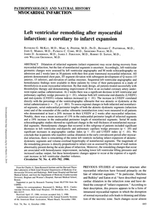
  • 1. PATHOPHYSIOLOGY AND NATURAL HISTORY MYOCARDIAL INFARCTION Left ventricular remodeling after myocardial infarction: a corollary to infarct expansion RAYMOND G. MCKAY, M.D., MARC A. PFEFFER, M.D., PH.D., RICHARD C. PASTERNAK, M.D., JOHN E. MARKIS, M.D., PATRICIA C. COME, M.D., SHOICHIRO NAKAO, M.D., JAMES D. ALDERMAN, M.D., JAMES J. FERGUSON, M.D., ROBERT D. SAFIAN, M.D., AND WILLIAM GROSSMAN, M.D. ABSTRACT Dilatation of infarcted segments (infarct expansion) may occur during recovery from myocardial infarction, but the fate of noninfarcted segments is uncertain. Accordingly, left ventricular geometric changes were assessed by left ventricular angiography and M mode echocardiography on admission and 2 weeks later in 30 patients with their first acute transmural myocardial infarction. All patients demonstrated chest pain, ST segment elevation with subsequent development of Q waves (15 anterior, 15 inferior), and elevation of cardiac enzymes. Sequential left ventricular angiographic and hemodynamic findings were available in these patients by virtue of their participation in a study of thrombolysis in acute myocardial infarction. By that study design, all patients treated successfully with thrombolytic therapy and demonstrating improvement of flow in an occluded coronary artery under- went repeat cardiac catheterization. At 2 weeks there was a significant decrease in left ventricular and pulmonary capillary wedge pressures (p < .01), whereas both left ventricular end-diastolic (LVEDV) and end-systolic (LVESV) volume indexes increased (p < .01). The increase in LVEDV correlated directly with the percentage of the ventriculographic silhouette that was akinetic or dyskinetic at the initial catheterization (r = .71, p < .001). To assess regional changes in both infarcted and noninfarct- ed segments, serial endocardial perimeter lengths of both the akinetic-dyskinetic segments (infarction zone) and of the remainder of the cardiac silhouette (noninfarction zone) were measured in all patients who demonstrated at least a 20% increase in their LVEDV at 2 weeks after myocardial infarction. Notably, there was a mean increase of 13% in the endocardial perimeter length of infarcted segments and a 19% increase in the endocardial perimeter length of noninfarcted segments. Serial M mode echocardiographic studies showed no significant change in the wall thickness of noninfarcted myocar- dial segments. Hemodynamic changes that occurred in this subgroup of patients included significant decreases in left ventricular end-diastolic and pulmonary capillary wedge pressures (p < .05) and significant increases in angiographic cardiac index (p < .01) and LVESV index (p < .01). We conclude that in patients who manifest cardiac dilatation in the early convalescent period after myocar- dial infarction, there is remodeling of the entire left ventricle including infarct expansion of akinetic- dyskinetic segments and volume-overload hypertrophy of noninfarcted segments. The magnitude of the remodeling process is directly proportional to infarct size as assessed by the extent of wall motion abnormality present during the acute phase of infarction. Moreover, the remodeling changes that occur are associated with hemodynamic improvement, including lower left ventricular filling pressures and increased cardiac output, but these hemodynamic changes appear to occur at the expense of a signifi- cant increase in left ventricular chamber volumes. Circulation 74, No. 4, 693-702, 1986. From the Charles A. Dana Research Institute and the Harvard-Thorn- PREVIOUS STUDIES of ventricular structure after dike Laboratory of Beth Israel Hospital Departments of Medicine (Car- myocardial infarction have focused primarily on the diovascular Division) and Radiology, Beth Israel Hospital and Harvard Medical School, and from the Department of Medicine (Cardiovascular fate of infarcted segments. 8 In particular, Hutchins Division), Brigham and Women's Hospital, Harvard Medical School, and Bulkley3 and Eaton et al.4 have described regional Boston. Supported in part by Research Training Grant HL07394 from the cardiac dilatation in the infarction zone and have de- USPHS. fined the concept of "infarct expansion." According to Address for correspondence: Raymond G. McKay, M.D., Cardio- thi decipin thi prcs per ob omo vascular Division, Beth Israel Hospital, 330 Brookline Ave., Boston, their description, this process appears to be a form of MA 02215. intramural myocardial rupture in which the disruption Received April 15, 1986; revision accepted July 3, 1986. * * m A preliminary report was presented at the Annual Scientific Sessions t of the American Heart Association, Miami Beach, November 1984. tion of the necrotic zone. Such changes occur almost 693 Vol. 74, No. 4, October 1986 Downloaded from http://ahajournals.org by on March 13, 2019
  • 2. McKAY et al. exclusively in transmural infarcts and are most com- monly seen with anterior and anteroseptal infarctions. The process has been observed to occur clinically as early as 3 days after infarction and may progress over days to weeks independent of additional myocardial necrosis or infarct extension. Most important, it is associated with increased mortality and may be impor- tant in the development of late aneurysm formation. Although infarct expansion has been studied exten- sively, only limited information is available on the fate of noninfarcted segments during the infarction recov- ery period.9-'3 A recent study by Weisman et al.9 re- ported that, in the rat, there may be global remodeling of the left ventricle immediately after infarction in- volving structural changes in both infarcted and nonin- farcted segments. Similarly, a study by Anversa et al.'0 found that myocardial infarction in rats results in myo- cyte hypertrophy of remaining viable myocardium. Studies on the fate of noninfarcted myocardium in man, however, have been limited"; moreover, the de- terminants and hemodynamic effects of this remodel- ing process have not been defined. Wynne et al.12 found that regional ejection fraction in noninfarcted segments was abnormal in 69% of patients with anteri- or infarctions and in 30% with inferior infarctions. Although these limited studies suggest that regional function in noninfarcted segments may be abnormal, the etiology of this dysfunction and the possible role that left ventricular remodeling plays in producing these changes remains unclear. This study was undertaken to assess the fate of non- infarcted myocardial segments after transmural infarc- tion. Since patients admitted with acute transmural infarction who elected to undergo attempted thrombol- ysis were expected to undergo serial cardiac catheter- izations with contrast ventriculography on admission and, if thrombolysis was successful, 2 weeks later, these patients were considered for enrollment and formed the study population. Methods Study group. Left ventricular hemodynamic and structural changes were assessed in a total of 30 patients. All patients presenting with their first acute transmural myocardial infarc- tion were considered for enrollment in this study. Entry criteria included chest pain of at least 30 min duration, new ST segment elevation on the electrocardiogram with the subsequent devel- opment of Q waves in the involved leads, and elevation of cardiac enzymes (creatine kinase MB fraction, SGOT, LDH) during the first 24 to 72 hr of their hospitalization. Patients who manifested persistent ischemia or recurrent infarction during their in-hospital convalescent phase as evidenced by clinical, electrocardiographic, or cardiac enzyme criteria were excluded from the study. Patients with previous myocardial infarction, 694 significant valvular disease, or cardiomyopathy were not con- sidered. Informed consent was obtained from each patient after an appropriate explanation of risks and potential complications of the proposed study. Cardiac catheterization Protocol. Cardiac catheterization was performed at the time of admission and 2 weeks later, before discharge. For both catheterizations, right femoral venous and right femoral arterial sheaths were inserted after administration of local anesthesia. A No. 7F thermodilution flow-directed catheter was passed to the pulmonary artery-pulmonary capillary wedge position. Left ventriculography followed by coronary angiography were per- formed in the routine manner. Baseline hemodynamic measure- ments included pulmonary capillary wedge and left ventricular pressures. Recordings were inscribed with a Honeywell Elec- tronics for Medicine (VR-16) recorder. In each of the 30 patients, an occluded or subtotally occluded coronary artery whose distribution corresponded to the area of ST segment elevation by electrocardiographic criteria was noted. Each patient was treated subsequently with either 250,000 U of intracoronary streptokinase, 1.5 million U of intravenous streptokinase, or 80 mg of intravenous tissue-type plasminogen activator. Only patients in whom successful thrombolysis was achieved with dissolution of clot and im- provement of flow in the involved coronary artery (and in whom this improvement persisted at 2 weeks) were included, since complete serial studies were available only in this group. After the initial catheterization, all patients were treated with intrave- nous heparin and intravenous lidocaine. Oral or topical nitrates, calcium-channel blockers, and/or S-blockers were continued or added if clinically indicated. Lidocaine was discontinued 48 to 72 hr after admission and replaced with an oral antiarrhythmic only if significant ventricular ectopy was subsequently noted. All other medications were continued until the time of the sec- ond catheterization except for heparin, which was stopped 4 hr before that catheterization. Data analysis. Ventricular volumes were measured by the area-length method with a regression equation developed for single-plane angiography based on left ventricular casts for measurement of true volume with calculation of end-diastolic and end-systolic volumes, angiographic cardiac index, and glo- bal ejection fraction. 14 Endocardial perimeters of akinetic-dyskinetic segments and of the remainder of the contrast ventriculogram silhouette were measured with a Tektronix 4956 digitizing computer. The per- centage of the ventriculographic perimeter that was akinetic or dyskinetic was determined on admission and at 2 weeks. In addition, serial changes in the perimeters of akinetic-dyskinetic segments and of the remainder of the cardiac silhouette that occurred in the 2 week interval between the first and second catheterization were measured with appropriate corrections for differences in magnification. M mode echocardiography. Echocardiography was per- formed on admission and at 2 weeks after infarction in 19 of the 30 patients by an ATL Mark VI machine. This machine is equipped with an M mode cursor that can be oriented perpen- dicular to the long axis of the left ventricle for optimal M mode recording of the septum and posterior wall. Changes in gain were used to define the endocardial and epicardial borders. Wall thicknesses were measured at the onset of the QRS complex on the electrocardiogram with the leading edge-to-leading edge technique. Statistics. Means and standard deviations were calculated for all variables. Paired dimensional data were analyzed with either the paired t test or the Wilcoxon signed-rank test where appro- priate, for parametric and nonparametric distributions. A p val- ue of less than .05 was considered significant. CIRCULATION Downloaded from http://ahajournals.org by on March 13, 2019
  • 3. PATHOPHYSIOLOGY AND NATURAL HISTORY-MYOCARDIAL INFARCTION Results Study group. A total of 30 patients with their initial acute myocardial infarction were included in this study. There were 28 men and two women, with a mean age of 55 years (range 33 to 69). Fifteen patients presented with electrocardiographic evidence of an anterior myocardial infarction, and each had a totally or subtotally occluded left anterior descending coro- nary artery. The remaining 15 patients demonstrated electrocardiographic changes in inferior leads with to- tal occlusion of the right coronary artery. Fifteen pa- tients had coronary disease limited to one coronary artery in the area of infarction; the remaining 15 had additional stenoses, with eight patients having two- vessel disease and seven patients having three-vessel disease. All patients were brought to the catheteriza- tion laboratory within 10 hr of the onset of their chest pain. Successful thrombolysis with opening ofa totally occluded artery or improvement in coronary flow was achieved in all patients within 90 min with administra- tion of either intracoronary or intravenous streptokin- ase or intravenous tissue-type plasminogen activator. After the initial catheterization, patients were ad- mitted to the coronary care unit for routine monitoring. All patients had an uncomplicated convalescent period without evidence of recurrent infarction, development of ventricular septal defect, or papillary muscle rup- ture. Repeat catheterization was performed in all pa- tients at a mean of 14. 1 days (range 9 to 18 days) after admission. All patients manifested persistent patency of their diseased coronary artery with a residual high- grade stenosis. At the time of the first catheterization, medications included the following: nitrates, 30 patients; calcium- channel blockers, 12 patients; ,3-blockers, six patients; diuretics, two patients; digoxin, five patients; antiar- rhythmics, 30 patients. At the time of the second cath- eterization, medications included the following: ni- trates, 30 patients; calcium-channel blockers, 15 patients; /8-blockers, six patients; diuretics, four pa- tients; digoxin, five patients; antiarrhythmics, six pa- tients. There were no significant differences in medi- cations between the time of the first and second catheterization, except for the use of antiarrhythmics (all patients at the time of their first catheterization were treated with intravenous lidocaine; only six pa- tients subsequently required long-term antiarrhythmic therapy). Hemodynamic and ventricular volume changes. Hemo- dynamic changes that occurred over the 2 week study period in the 30 patients included decreases in heart rate (80 17 to 72 + 16 beats/min; p < .03), left Vol. 74, No. 4, October 1986 ventricular systolic pressure (122 ± 19 to 113 ± 11 mm Hg; p < .01), left ventricular end-diastolic pres- sure (24 + 8 to 18 ± 6 mm Hg; p < .01), and pulmonary capillary wedge pressure (18 ± 6 to 13 ± 6 mm Hg; p < .01). There was no significant change in either angiographic cardiac index (3.3 ± 1.2 to 3.5 + 1.2 liters/min/m2; p = NS) or left ventricular ejection fraction (55 ± 11% to 54 ± 11%; p = NS). In addition, there were increases in left ventricular end- diastolic volume index (76 ± 22 to 93 ± 30 m1/M2; p < .01), end-systolic volume index (34 ± 10 to 47 ± 30 mI/M2; p < .02), and stroke volume index (42 ± 16 to 50 ± 13 ml/m2; p < .02). Table 1 summarizes hemodynamic and angiographic data at the time of admission and at 2 weeks for all patients. Regional wall motion abnormalities. In 26 of the 30 patients, a discrete segment ofakinesis and/or dyskine- sis could be identified on the contrast left ventriculo- gram at the time of the first catheterization. This region ofakinesis-dyskinesis was noted to persist or enlarge at the time of the second catheterization. The percentage of the left ventricular perimeter that was akinetic or dyskinetic was 20 ± 14% on admission and 25 ± 14% 2 weeks later (p = NS). Determinants of the increase in end-diastolic volume in- dex. The increase in the end-diastolic volume index that occurred over the 2 week period correlated signifi- cantly with the percentage of the initial contrast ven- triculogram that was akinetic or dyskinetic (r = .71, p < .001) at the time ofthe first catheterization. Figure 1 summarizes the relationship between volume in- creases and the percentage of akinesis-dyskinesis. In addition, there was a modest inverse correlation be- tween left ventricular ejection fraction on the first cath- eterization and volume increases from the initial study to the study at 2 weeks (r = -.51, p < .001). Hemodynamic changes in patients with increased left ventricular volumes. Since left ventricular volume changes were related to the extent of initial infarction, a subgroup of 17 of the 30 patients who demonstrated at least a 20% increase in end-diastolic volume index was identified for further evaluation. This group con- sisted of 11 patients with anterior infarctions and six patients with inferior infarctions. Hemodynamic and volume changes that occurred in these patients includ- ed decreases in left ventricular end-diastolic pressure (23 ± 9 to 18 ± 6 mm Hg; p < .05) and pulmonary capillary wedge pressure (17 ± 6 to 12 ± 5 mm Hg; p < .03), with concomitant increases in angiographic cardiac index (2.9 ± 1.0 to 3.7 ± 1.1 liters/min/m2; p < .01), stroke volume index (36+ 13 to 49 12 ml/M2; p < .01), and end-systolic volume index (30 ± 695 Downloaded from http://ahajournals.org by on March 13, 2019
  • 4. McKAY et al. TABLE 1 Serial hemodynamic data for patients with anterior myocardial infarction (Nos. I to 15) and inferior myocardial infarction (Nos. 16 to 30) Patient 1 2 3 4 5 6 7 8 9 10 11 12 13 14 15 Sex M M M M M M M F M M M M m M M Age (yr) 62 69 55 33 38 56 54 62 67 60 65 69 57 42 64 First catheterization EDVI 82 118 70 70 79 45 48 46 64 70 72 92 140 96 87 ESVI 31 53 24 40 43 29 34 25 28 31 46 48 48 40 32 SVI 51 65 46 30 36 16 14 21 36 39 26 44 92 56 55 EF 62 55 66 43 46 36 29 46 56 56 36 48 66 58 63 HR 84 72 64 96 102 72 96 66 82 59 90 80 54 108 80 CI 4.3 4.7 2.9 2.9 3.7 1.1 1.3 1.4 3.0 2.3 2.3 3.5 5.0 6.0 4.4 LVSP 115 119 125 100 97 130 114 160 130 100 125 122 158 132 122 LVEDP 21 25 22 14 25 28 40 40 32 34 22 19 29 17 28 PCW 17 18 11 12 22 20 26 30 21 18 15 16 26 14 22 Second catheterization EDVI 108 142 84 88 211 109 123 63 86 88 112 103 122 92 70 ESVI 38 86 30 59 183 57 83 28 43 38 55 44 49 36 32 SVI 70 56 54 29 28 52 40 35 43 50 57 59 73 56 38 EF 65 39 64 33 13 48 33 56 50 57 51 57 60 61 54 HR 66 80 68 96 102 54 84 72 66 46 60 60 96 66 72 Cl 4.6 4.5 3.7 2.8 2.9 2.8 3.4 2.5 2.8 2.3 3.4 3.5 7.0 3.7 2.7 LVSP 103 102 95 95 100 117 103 125 115 110 130 115 135 111 112 LVEDP 20 14 12 22 30 19 15 6 24 14 25 16 12 21 17 PCW 12 D 9 11 29 14 10 5 D 9 10 13 10 14 13 CI = angiographic cardiac index (I/min/m2); EDVI = left ventricular end-diastolic volume index (mi/M2); EF = left ventricular ejection fraction (%); ESVI = left ventricular end-systolic volume index (ml/m2); HR = heart rate (bpm); LVEDP = left ventricular end-diastolic pressure (mm Hg); LVSP = left ventricular systolic pressure (mm Hg); PCW = pulmonary capillary wedge pressure (mm Hg); SVI = left ventricular stroke volume index (m1/m2). Ap < .03; Bp < .02; cp < .01; D data not obtained because of technical reasons. 10 to 56 + 37 ml/mr; p < .01). There were no signifi- cant changes in left ventricular systolic pressure (118 + 17 to 112 + 11 mm Hg; p = NS), heart rate (83 ± 16 to 76 + 18 beats/min; p = NS), or ejection fraction (53 ± 12% to 50 ± 14%; p = NS). Hemodynamic and angiographic data for these patients are summa- rized in table 2. Serial changes in endocardial perimeters in patients with increased volumes. To assess structural changes occur- ring in the 17 patients who demonstrated substantial (>-20%) increases in left ventricular end-diastolic vol- ume, serial endocardial perimeter measurements were made of the akinetic-dyskinetic segments (represent- ing infarcted zones) and of the remainder ofthe cardiac silhouette (representing noninfarcted segments) on the left ventricular angiograms obtained on admission and at 2 weeks. Table 3 summarizes changes in measure- ments of endocardial perimeter length of both akinetic- dyskinetic segments and of the remainder of the cardi- ac silhouette. Notably, there was a mean 13 + 12% increase in endocardial perimeter length of infarcted segments and a 19 ± 13% increase in the endocardial 696 perimeter length of noninfarcted segments. Figure 2 shows examples of end-diastolic and end-systolic car- diac silhouettes in four representative patients at the time of initial presentation with acute myocardial in- farction and at follow-up study 2 weeks later. M mode echocardiography. Table 4 summarizes M mode measurements of septal and posterior wall thick- ness in 19 of 30 patients who were studied, including 12 who manifested at least a 20% increase in left ven- tricular end-diastolic volume. Notably, patients with anterior myocardial infarction showed no significant change in posterior wall thickness and patients with inferior infarction showed no change in septal wall thickness. Discussion Our results demonstrate that global remodeling of the left ventricle occurs during the early convalescent period in certain patients with myocardial infarction. In particular, the findings indicate that those patients who exhibited cardiac dilatation with increased left ventricular end-diastolic and end-systolic volumes CIRCULATION Downloaded from http://ahajournals.org by on March 13, 2019
  • 5. PATHOPHYSIOLOGY AND NATURAL HISTORY-MYOCARDIAL INFARCTION TABLE 1 (Continued) Patient Mean ± 16 17 18 19 20 21 22 23 24 25 26 27 28 29 30 SD M M M 40 60 51 M M M F 42 64 43 64 M M M M M M M M 28M, 2F 55 34 62 59 59 45 54 62 55 ± 10 56 60 83 67 47 55 74 75 71 103 59 90 89 101 81 76±+10 25 21 31 28 15 20 21 37 38 42 18 49 37 43 40 34+ 10 31 39 51 39 32 35 53 38 33 61 41 41 52 58 41 42+ 16 55 65 62 58 68 64 72 51 46 59 69 46 59 57 51 55 + 11 102 84 60 108 77 100 70 60 96 70 50 170 102 66 90 80± 17 3.2 3.3 3.1 4.2 2.5 3.5 3.7 2.3 3.2 4.3 2.1 2.9 5.3 3.8 3.7 3.3+1.2 137 103 103 110 130 120 120 130 105 97 120 140 130 172 92 122+ 19 13 10 20 19 16 16 26 24 20 24 15 28 21 22 22 23 ± 7 13 12 17 16 D 15 D D 14 23 5 20 21 15 14 18±5 102 103 108 94 89 72 68 83 76 111 47 105 101 90 54 97 + 30c 40 40 50 40 40 36 26 35 33 59 20 32 47 26 24 47 ±+301 62 63 58 54 49 36 42 48 43 52 27 73 54 64 30 50so13B 61 62 54 57 55 50 68 58 57 47 57 70 53 71 56 54+ 12 60 90 84 110 66 90 50 60 61 58 60 69 60 75 72 72 + 16A 3.7 5.6 4.9 5.9 3.2 3.2 2.1 2.9 2.6 3.0 1.6 5.0 3.2 4.8 2.2 3.6+ 1.2 128 117 111 125 120 112 115 130 105 105 110 121 115 130 102 114+ llc 23 20 21 18 12 11 22 20 18 21 12 22 11 12 8 17±6c 14 14 12 14 9 9 D D 13 14 6 10 10 9 6 12 +4C during their in-hospital stay manifested changes in both infarcted and viable segments of their ventricles. Lengthening of endocardial perimeters of infarcted 300 250 0 > 200 0 z 0 150 z I 100 + + 50I -10 0 10 20 30 40 50 60 % OF AKINESIS-DYSKINESIS FIGURE 1. Correlation between the percentage of the left ventricular silhouette that was akinetic-dyskinetic at the time of the first catheteriza- tion with the subsequent increase in left ventricular end-diastolic vol- ume index (EDVI) at the second catheterization (r = .71, p < .001). The ordinate, "Change in EDVI," was determined by dividing the EDVI at the 2 week catheterization by the EDVI obtained at the first catheter- ization. segments presumably represents infarct expansion.3 Equally important, lengthening of the endocardial pe- rimeter of that part of the ventricle without regional wall motion abnormalities and with no evidence of wall thinning in these segments suggests that volume- overload hypertrophy with a net increase in the myo- cardial mass of these segments has occurred. These remodeling changes occurred as hemodynamics im- proved, including lower left ventricular filling pres- sures and increased cardiac output, perhaps at the ex- pense of increased chamber volumes. Moreover, the magnitude of these remodeling changes were roughly related to infarct size as measured by the percentage of the ventricular silhouette exhibiting regional wall mo- tion abnormality on admission and inversely related to initial left ventricular pump function as measured by ejection fraction. Grossman and others", 16 have termed an increase in myocardial mass without relative wall thickening as "volume overload" hypertrophy when it occurs in the entire left ventricle. In volume-overload hypertrophy, as discussed below, mass increases primarily by series addition of new sarcomeres and fiber elongation, re- sulting in chamber enlargement. In pressure-overload hypertrophy, in contrast, a marked increase in wall thickness occurs as a consequence of the parallel addi- tion of new myofibrils.16 Although usually associated Vol. 74, No. 4, October 1986 + + + 697 Downloaded from http://ahajournals.org by on March 13, 2019
  • 6. McKAY et al. TABLE 2 Serial hemodynamic and angiographic data in patients with greater than 20% increase in left ventricular end-diastolic volume index EDVI ESVI SVI EF HR CI LVSP LVEDP PCW Firstcath. 67+18 31+10 36+13 53±12 83+16 2.9+1.0 118+17 23+9 18+5 Second cath. 105±33 56 37c 49± 12 50± 14 76 - 18 3.7 +1 c 112 - 11 18 6A 12 5B Data are derived from all patients who demonstrated greater than 20% increase in end-diastolic volume index at the time of the second catheterization, including 11 patients with anterior infarctions (Nos. 1 to 11. table 1) and six patients with inferior infarctions (Nos. 16 to 21 table 1). Abbreviations are as in table 1. Ap < .05; Bp < .03; cp < .01. with some degree of wall thickening, chronic volume- overload hypertrophy produces far less thickening than does pressure-overload hypertrophy. Thus we believe that the absence of thinning in the noninfarcted seg- ments, associated with concurrent elongation, is com- patible with volume-overload hypertrophy of these segments. TABLE 3 Left ventricular endocardial perimeter analysis in patients with greater than 20% increase in left ventricular end-diastolic volume index Catheterization Catheterization % Change % Change nonin- % Akinesis- infarcted farcted Patient dyskinesis segment segment Anterior infarctions 1 24 + 6 +18 2 20 + 12 +21 3 10 + 7 +15 4 41 + 4 +33 5 46 +38 +28 6 50 + 11 +42 7 41 +20 +25 8 37 + 8 +18 9 30 +10 +10 10 30 +20 + 5 11 40 +26 + 4 Inferior infarctions 16 32 + 8 + 16 17 18 + 9 +22 18 18 + 12 +23 19 20 - 6 + 16 20 17 +±10 +12 21 16 + 8 + 12 Mean + SD 13+12 19+ 13 Data are derived from the 1 1 patients with anterior infarctions (Nos. 1 to 1 1, table 1 ) and six patients with inferior infarctions (Nos. 16 to 21, table 1) whose hemodynamic and angiographic findings are summarized in table 2. 698 Stimulus for volume-overload hypertrophy in the infarct- ed ventricle. Left ventricular hypertrophy is one of the principal adaptive mechanisms by which the heart compensates for an increased load. The pattern of hy- pertrophy that develops is dependent on the type of overload and the influence of this overload on systolic and diastolic wall stresses."5 17 According to this hypothesis, when the primary stimulus for hyper- trophy is pressure overload, the resultant acute in- crease in systolic wall stress leads to parallel replica- tion of sarcomeres, wall thickening, and concentric hypertrophy. The wall thickening that occurs tends to return systolic wall stress toward normal. In contrast, when the primary stimulus to hypertrophy is a volume overload, increased diastolic pressure causes an in- crease in diastolic wall stress and leads to sarcomere replication in series, fiber elogation, and chamber en- largement. Chamber enlargement accommodates the volume overload and returns diastolic pressure toward normal. However, the chamber enlargement also re- sults in an increase in systolic wall stress, which subse- quently leads to wall thickening. The combination of slight wall thickening and chamber enlargement return systolic and diastolic wall stress toward normal. Given that hypertrophy will develop in response to increased wall stress, what evidence is there that wall TABLE 4 Echocardiographic wall thickness measurements (mm) Catheterization 1 Catheterization 2 Posterior Posterior Septum wall Septum wall Anterior infarctions 10.8+0.8 11.8+0.9 11.5+0.9 11.4+0.9 (n = 10) Inferior infarctions 10.6+0.7 9.6+0.4 9.8+1.0 10.2+0.9 (n =9) There were no significant differences between serial septal and posterior wall thicknesses in either group. CIRCULATION Downloaded from http://ahajournals.org by on March 13, 2019
  • 7. PATHOPHYSIOLOGY AND NATURAL HISTORY-MYOCARDIAL INFARCTION I'I' % FIGURE 2. Right anterior oblique end-diastolic and end-systolic cardi- ac silhouettes obtained in four patients on admission (left) and at 2 weeks (right). Solid lines indicate akinetic-diskinetic (infarcted) seg- ments and stippled lines indicate noninfarcted areas. In all cases, there has been an increase in chamber volume as well as lengthening of both the infarcted perimeter and noninfarcted perimeter. stresses are abnormal in patients with myocardial in- farction during their convalescent phase? To date, this has been a diffilcult problem to assess because of diffi- culties in measuring wall stresses in ventricles with regional wall abnormalities and variable wall thick- nesses. Two recent studies, however, are notable. Pfelfer et al.'8 examined alterations in mass, vol- ume, and end-diastolic wall stress in rats at 2, 21, and 100 days after coronary ligation and subsequent myo- cardial infarction and compared changes in these varia- bles with findings in rats without coronary ligation. Vol. 74, No. 4, October 1986 Passive pressure-volume curves were deternined and end-diastolic stress was calculated from data on left ventricular mass, end-diastolic pressure, and end-dia- stolic volume. No change in mass, volume, or wall stress was noted in any of the control rats. In rats with myocardial infarction, however, there was a progres- sive increase in both volume and mass, and end-dia- stolic wall stress was elevated at all times. Of note, the ratio of mass to volume decreased progressively, indi- cating that left ventricular volume was increasing out of proportion to mass and suggesting that a volume- overload hypertrophy was occurring, perhaps in re- sponse to the increased end-diastolic wall stress. Pouleur et al.'9 also attempted to evaluate wall stresses in patients with previous myocardial infarc- tion. Using a method for calculating regional wall stresses, 19 20 they found that both systolic and diastolic regional wall stresses were abnormal in the infarcted and ischemic areas and that stresses in the noninfarcted segments were normal. Because serial studies were not available to them in each patient, they could not com- ment on changes in ventricular wall stress after infarction. Thus, although available data are limited, there is evidence that wall stresses in healing left ventricles may be abnormal. A model for left ventricular remodeling after infarction. Based on the observations that wall stresses may be abnormal in the infarcted ventricle and the fact that increased diastolic wall stress may initiate a volume- overload type ofhypertrophy, a model of left ventricu- lar remodeling after myocardial infarction may be pro- posed. This model is summarized in figure 3. In this model, the immediate hemodynamic consequences of an acute myocardial infarction on ventricular function include both systolic and diastolic dysfunction. Systol- ic impairment secondary to loss of contractile function of the infarcted myocardium results in a decreased systolic ejection, increased end-systolic volume, an increase in cardiac size, and a secondary increase in diastolic filling pressure caused by the increase in ven- tricular volume. Diastolic function is characterized im- mediately by an increase in diastolic distensibility,2' 23 which minimizes the rise in filling pressure. However, as necrotic tissue is replaced by fibrosis, a decrease in distensibility occurs. In addition, there may be upward shift in the diastolic pressure-volume curve secondary to ischemia in the border zone so that filling pressure may tend to be higher for any given volume. Thus diastolic volume increases and diastolic pressure also tends to increase, especially in patients with large infarctions. 699 Downloaded from http://ahajournals.org by on March 13, 2019
  • 8. McKAY et al. RESTORED STROKE VOLUME 4 SEGMENTAL INFARCTION DECREASED SYSTOLIC EJECTION INCREASED LEFT VENTRICULAR END-DIASTOLIC VOLUME AND PRESSURE Frank- INC D Starling INCREASED INCRE ASE StrDn SYSTOLIC EJECTION NON-INFARCTED SEGMENT: REGIONAL HYPERTROPHY DECREASED C WALL STRESS INFARCTED SEGMENT: INFARCT EXPANSION ,ONTRACTILITY LATE HEART FAILURE FIGURE 3. Hypothesis proposed to account for the mechanisms of left ventricular remodeling. In the presence of both systolic and diastolic dys- function, there may be peripheral mechanisms mediat- ed via the sympathetic nervous system and circulating catecholamines to help maintain a normal arterial blood pressure and cardiac output. These mechanisms may subsequently increase ventricular preload by aug- menting venous return and increase ventricular after- load by causing arteriolar vasoconstriction. The result- ing increases in ventricular radius and diastolic pressure will, in combination, lead to an increase in end-diastolic wall stress in all parts of the ventricle. Simultaneous with an increase in both end-diastolic wall stress and regional end-systolic wall stress in in- farcted segments, there is weakening of the normal myocardial structure needed to resist wall stress. As a result, processes of dilatation and thinning progress in the infarct region. At some point, because of continued healing of the infarcted segments with increasing col- lagenization and the production of a firm scar, the ability of the infarction zone to resist wall stresses is increased and infarct expansion halts. However, one can imagine that before production of a mature scar, if wall stresses are elevated to a sufficient degree and if infarcted segment tensile strength has been sufficiently reduced, myocardial rupture may occur. Finally, in noninfarcted segments, elevation of end-diastolic wall stress may provide the stimulus for volume-overload hypertrophy,'5' 16 in which the combination of fiber elongation and wall thickening result in a return of systolic and diastolic wall stress toward normal. Possible determinants of left ventricular remodeling. One may predict from the preceding model that those 700 factors leading to the greatest elevation ofwall stresses would be expected to cause the greatest amount of left ventricular remodeling. Infarct size may be most im- portant given that the larger infarcts would lead to greater systolic and diastolic impairment with larger increases in ventricular radius and filling pressure. In this regard, our results have shown a rough correlation between the magnitude of remodeling and the size of the infarction (figure 1) as assessed by the percentage of the cardiac silhouette exhibiting akinesis-dyskinesis at initial presentation. Infarct location is a second factor which may be important in the production ofleft ventricular remodel- ing after infarction. Previous studies that have evaluat- ed infarct expansion have noted that these changes occur predominantly in patients with anterior infarc- tions. 1-8 In part this may be related to infarct size, since anterior infarctions are generally associated with more myocardial necrosis than occurs with inferior infarc- tion. In addition, in the normal left ventricle there is a greater degree of shortening of the anterior than of the posterior wall, and thus similar degrees of depression of function might be expected to result in more severe derangements after anterior infarctions. 12 It is notable that in this study a higher percentage of patients with anterior than with inferior infarctions demonstrated at least a 20% increase in end-diastolic volume index and that the magnitude of this remodeling, as measured by absolute changes in chamber volumes, was larger in patients with anterior infarctions. A third factor or set of factors that may be important in the remodeling process after infarction are those CIRCULATION Downloaded from http://ahajournals.org by on March 13, 2019
  • 9. PATHOPHYSIOLOGY AND NATURAL HISTORY-MYOCARDIAL INFARCTION variables that affect ventricular loading conditions and thus affect ventricular wall stress. Hypertension, for example, increases afterload and therefore systolic wall stress and hastens the process of infarct expan- sion. In contrast, reduction of ventricular preload and afterload by vasodilators should lessen the tendency for ventricular remodeling. In this regard, Pfeffer et al.24 have shown recently that captopril may reduce the extent of topographic changes in a rat preparation of infarction. In Pfeffer's studies, this blunting of remod- eling with captopril was associated with improved sur- vival at 1 year after infarction.25 A final factor that may be important in the degree of remodeling is the success of thrombolysis. If in fact thrombolysis results in successful salvage of viable myocardium, then infarct size might be reduced and the degree of remodeling might be diminished. Since this study considered only patients with successful thrombolysis and since a comparable control group was not available for concurrent analysis, we cannot comment at present about the potential effects of thrombolysis on the remodeling process. Hemodynamic consequences of left ventricular remodel- ing. The hemodynamic changes that occur in the 2 week period after myocardial infarction appear to be in part beneficial, with improvement in cardiac output despite lower left ventricular filling pressure. This may explain the common clinical observation that early in the course of an infarction, there may be mild clinical congestive heart failure followed by spontaneous im- provement. It is notable, however, that these hemody- namic improvements occurred only while chamber volumes increased significantly. Although a portion of the increase in diastolic chamber volume may have been related to the concomitant small decreases in heart rate observed, similar decreases in heart rate oc- curred both in patients with a greater than 20% in- crease in end-diastolic volume and in patients with a lesser increase in end-diastolic volume. Thus the de- gree of volume change was not proportional to fall in heart rate. Although hemodynamic improvement may accom- pany ventricular remodeling early after infarction, the long-term hemodynamic consequences of remodeling are not known. Data from at least one study have suggested increased morbidity and mortality in postin- farction patients with increased ventricular size.2 In addition, a recent study from our institution has sug- gested that ventricular dilatation after myocardial in- farction may be progressive, continuing for months after the original infarction.26 Perhaps a more impor- tant clinical consequence of infarct remodeling is that Vol. 74, No. 4, October 1986 it may lead to late decreases in left ventricular perform- ance with depression of both global and regional con- tractile function. This may be particularly important in the late and often "mysterious" appearance of conges- tive heart failure seen in patients with infarction, even in the absence of late ischemic events. The etiology of this late ventricular dysfunction could be related to both infarct expansion of infarcted segments and volume-overload hypertrophy of nonin- farcted segments. With respect to infarcted segments, the mechanical consequences of infarct expansion place an unusually high burden on the residual func- tioning myocardium.27 28 Moreover, the expanded in- farct segment may act as a reservoir and receive blood during systole in competition with aortic outflow, a condition similar to mitral regurgitation. Perhaps more important than the hemodynamic consequences of in- farct expansion are the possible late consequences of volume-overload hypertrophy. In most states of vol- ume-overload hypertrophy (e.g., mitral regurgitation, aortic insufficiency) there is an early phase of adaptive hypertrophy in which contractile function ofthe hyper- trophied myocardium remains normal. However, at some point in the hypertrophy process, there follows a transition when contractile function becomes abnor- mal.""'7 If ventricular remodeling involves volume- overload hypertrophy of noninfarcted segments, it is possible that these segments may show a similar pat- tern of initial "physiologic" hypertrophy followed by "pathologic" hypertrophy. Since these segments must compensate for the deleterious mechanical conse- quences of infarct expansion, a loss of function in these segments would be a major factor in the late appearance of clinical congestive heart failure. If remodeling is associated with long-term deleteri- ous hemodynamic changes, then attempts to limit this process should become important. The concept that elevated wall stresses are the primary stimuli for ven- tricular remodeling leads one to conclude that attempts to decrease wall stress may attenuate this process, limit progressive ventricular dilatation, and blunt any ad- verse hemodynamic consequences that result. Of note, some support for this notion comes from work by Pfeffer et al. ,24 25 who have shown that administration ofcaptopril, an angiotensin-converting enzyme inhibi- tor that presumably reduces left ventricular systolic wall stress, resulted in less ventricular dilatation and prolonged survival in rats with experimentally induced myocardial infarction in comparison with untreated rats with infarcts of comparable size. If this concept were applicable to humans, then maneuvers to de- crease ventricular wall stresses by decreasing both left 701 Downloaded from http://ahajournals.org by on March 13, 2019
  • 10. McKAY et al. ventricular preload and afterload might favorably di- minish topographic changes after infarction and might decrease the incidence of congestive heart failure in the late recovery period. Study limitations. Several important limitations of this study should be emphasized. First, it is important to note that hemodynamic and geometric changes that are described in this study are in patients with acutely reperfused infarcts. Several recent investigations have noted that reperfusion into an infarcted region may alter the properties of the postinfarction tissue.79 Ac- cordingly, one needs to ask whether the remodeling changes were caused by myocardial infarctions or by the early reperfusion of the infarct region. However, studies from our laboratory26 have shown that the suc- cess or failure ofthrombolytic therapy (administered in the same period as in the present study) has no effect on ventricular volume changes after myocardial infarction. A second important criticism that might be raised is the fact that no significant wall thinning was noted in the area of infarction as assessed by M mode echocar- diography. In part, this may be related to lack of ade- quate sensitivity of the echocardiographic wall thick- ness measurements. Alternatively, the lack of infarct wall thinning may be related to the effect of reperfu- sion into the infarction zone. Finally, although we have concluded that the hemo- dynamic changes observed in our study were conse- quences of the remodeling process, it is possible that other factors, such as recovery of ischemic myocardi- um, also had an effect on hemodynamic improvement. Only further, prospective, controlled, intervention trials will help answer this important question definitively. References 1. Feild BJ, Russell RO, Dowling JT, Rackley CF: Regional left ventricular performance in the year following myocardial infarc- tion. Circulation 46:679, 1972 2. Kostuk WR, Kazamias TM, Gawder MP, Simon AL, Ross J: Left ventricular size after myocardial infarction: serial changes and their prognostic significance. Circulation 47: 1174, 1973 3 Hutchins GM, Bulkley BH: Infarct expansion versus extension: two different complications of acute myocardial infarction. Am J Cardiol 41: 1127, 1978 4. Eaton LW, Weiss JL, Bulkley BH, Garrison JB, Weisfeldt ML: Regional cardiac dilatation after acute myocardial infarction. N Engl J Med 300: 57, 1979 5. Shuster EH, Bulkley BH: Expansion of transmural myocardial infarction: a pathophysiologic factor in cardiac rupture. Circulation 60: 1532, 1979 6. Cabin HS, Roberts WC: True left ventricular aneurysm and healed myocardial infarction: clinical and necropsy observations including quantification of degrees of coronary arterial narrowing. Am J Cardiol 46: 754, 1980 7. Eaton LW, Bulkley BH: Expansion of acute myocardial infarction: its relationship to infarct morphology in a canine model. Circ Res 49: 80, 1981 8. Weiss JL, Bulkley BH, Hutchins GM, Mason SJ: Two-dimension- al echocardiographic recognition of myocardial injury in man: comparison with postmortem studies. Circulation 63: 401, 1981 9. Weisman HF, Bush DE, Mannis JA, Bulkley BH: Global cardiac remodeling after myocardial infarction: a study in the rat model. J Am Coll Cardiol 5: 1355, 1985 10. Anversa P, Loud AV, Levicky V, Guideri G: Left ventricular failure induced by myocardial infarction. I. Myocyte hypertrophy. Am J Physiol 248: H876, 1985 11. Erlebacher JA, Weiss JL, Easton LW, Kallman C, Weisfeldt ML, Buckley BH: Late effects of acute infarct dilation on heart size: a two dimensional echocardiographic study. Am J Cardiol 49: 1120, 1982 12. Wynne J, Sayres M, Maddox DE, Idoine J, Alpert JS, Neill J, Holman BL: Regional left ventricular function in acute myocardial infarction: evaluation with quantitative radionuclide ventriculogra- phy. Am J Cardiol 45: 203, 1980 13. Corday E, Kaplan L, Meerbaum S: The consequences of coronary arterial occlusion on remote myocardium: effects of occlusion and reperfusion. Am J Cardiol 36: 385, 1975 14. Wynne J, Green LH, Mann T, Levin D, Grossman W: Estimation of left ventricular volumes in man from biplane cineangiograms filmed in oblique projections. Am J Cardiol 41: 726, 1978 15. Grossman W, Jones D, McLaurin LP: Wall stress and patterns of hypertrophy in the human left ventricle. J Clin Invest 56: 56, 1975 16. Grossman W: Cardiac hypertrophy: useful adaptation or pathologi- cal process? Am J Med 69: 576, 1980 17. Grossman W, Carabello PA, Gunther S, Fifer MA: Ventricular wall stress and the development of cardiac hypertrophy and failure. In Alpert NR, editor: Myocardial hypertrophy and failure. New York, 1983, Raven Press, pp 1-15 18. Pfeffer JM, Pfeffer MA, Mirsky I, Steinberg CR, Braunwald E: Progressive ventricular dilatation and diastolic wall stress in rats with myocardial infarction and failure. Circulation 66(suppl II): 11- 66, 1982 (abst) 19. PouleurH, Rousseau MF, Van Eyll C, Charlier AA: Assessment of regional left ventricular relaxation in patients with coronary artery disease: importance of geometric factors and changes in wall thick- ness. Circulation 69: 696, 1984 20. Janz RF: Estimation of local myocardial stress. Am J Physiol 242: H875, 1982 21. Tyberg JV, Forrester JS, Wyatt HL, Goldner SJ, Parmley WW, Swan HJC: An analysis of segmental ischemic dysfunction utiliz- ing the pressure-length loop. Circulation 49: 748, 1974 22. Serizawa T, Vogel WM, Apstein CS, Grossman W: Comparison of acute alterations in left ventricular relaxation and diastolic chamber stiffness induced by hypoxia and ischemia. J Clin Invest 68: 91, 1981 23. ForresterJS, Diamond G, Parmley WW, Swan HJC: Early increase in left ventricular compliance after myocardial infarction. J Clin Invest 51: 598, 1972 24. Pfeffer JM, Pfeffer MA, Braunwald E: Influence of chronic capto- pril therapy on the infarcted left ventricle of the rat. Circ Res 57: 84, 1985 25. Pfeffer MA, Pfeffer JM, Steinbert C, Finn P: Survival after an experimental infarction: beneficial effects of long-term therapy with captopril. Circulation 72: 406, 1985 26. Warren SE, McKay RG, Royal HD, Parker JA, Ransil BJ, Markis JE, Grossman W: Time course of left ventricular dilatation follow- ing acute myocardial infarction. Circulation 72(suppl III): 111-439, 1985 (abst) 27. Klein MD, Herman MV, Gorlin R: A hemodynamic study of left ventricular aneurysm. Circulation 35: 614, 1967 28. Herman MV, Heinle RA, Klein MD, Gorlin R: Localized disorders in myocardial contraction: asynergy and its role in congestive heart failure. N Engl J Med 277: 222, 1967 29. Connelly CM, Vogel VM, Wiegner AW, Osmers EL, Bing OH, Kloner RA, Dunn DM, Franzblau C, Apstein C: Effects ofreperfu- sion after coronary artery occlusion on post-infarction scar tissue. Circ Res 57: 562, 1985 CIRCULATION 702 Downloaded from http://ahajournals.org by on March 13, 2019