1. The Modulation of Provisional Matrix in Nitrogen Mustard Skin Wound by Connexin 43 Antisense Therapy
Enoch Yue, Hui-Ying Chang, Peihong, Zhou, Rita Hahn, Marion K Gordon, Donald R. Gerecke, and Yoke-Chen Chang
Department of Pharm & Tox, Ernest Mario School of Pharmacy, Rutgers University, Piscataway, NJ
Abstract
Sulfur mustard(SM, Bis(2-chloroethyl) sulfide) and its analog Nitrogen Mustard (NM, Bis(2-chloroethyl)
methylamine) are cytotoxic alkylating agents that cause adverse skin reactions including edema and fluid
filled blisters. It can cause necrosis and apoptosis of cells, induce strong inflammatory-stress responses,
delayed wound healing, and possibly result in extensive scarring. Studies showed that Cx43 antisense
oligodeoxynucleotide (asODN) topical treatment accelerated wound closure and re-epithialization in
diabetic skin wounds. Mice deficient in Cx43 improved wound repair and modulated the extracellular
matrix. If the downregulation of Cx43 accelerated re-epithialization, it may modulate the provisional matrix
to promote wound repair. A time course study (up to 10 days) of NM exposed hairless mouse (SKH-1) skin
with treatment of Cx43-asODN was performed. Provisional matrix proteins including, Tenascin-C (TNC, an
anti-adhesive glycoprotein), and fibronectin (FN, a cell adhesive glycoprotein), were examined. The RNA of
murine back skin was extracted and was converted into cDNA by reverse transcription. PCR was performed
to insure cDNA quality. Gene expression assays with TaqMan probes targeting TNC and FN were performed
by Real-Time PCR. H&E histology showed characteristic time dependent NM induced skin wounds. After the
treatment with Cx43-asODN, apparent acceleration of re-epithelialization and wound healing progression
was observed. The mRNA gene expression analyses indicated elevated expression of TNC and reduced
expression of FN in NM injured skin samples, compared to the unexposed control samples. After the
treatment with CX43-asODN, we saw a down-regulation in TNC expression and an upregulation in FN
expression in the NM plus Cx43-asODN treated group, compared to the NM exposed alone group . This
suggests that the treatment of Cx43-asODN may play a role in modulating the provisional matrix in NM
induced skin wound repair.
Grant funding supports by ES005022, EY09056, T32ES007148, and NIAMS U54AR055073
Introduction
Sulfur mustard was used in the Iraq-Iran War in 1988 as a chemical warfare agent. Mustard agents,
such as sulfur mustard, and its analog nitrogen mustard, can target the basement membrane zone which
induce a) separation of epidermis and the dermis below, b) prolonged inflammatory response, and c)
delayed wound repair process. Gap junction communication is tightly regulated in skin wound healing.
Studies showed connexin 43 antisense oligonucleotide (Cx43-asODN) topical treatment improves wound
closure and re-epithialization in diabetic and incisional cutaneous wounds (1). Connexin 43 deficiency
accelerates skin wound healing and modulates extracellular matrix remodeling in mice (2). In our
laboratory, we treated Cx43 antisense oligodeoxynucleotides (Cx43-asODN) on NM exposed SKH-1 mouse
dorsal skin to evaluate the wound healing response. Mice treated with Cx43-asODN after NM exposure
had an accelerated wound rate and improved re-epithelialization (3). Two matricellular proteins, including
Tenascin C (TNC) and Fibronectin(FN), play important role in provisional matrix during wound repair are
examined in this study. TNC, an anti-adhesive glycoprotein in the matrix, is associated with cell migration
and can recruit of inflammatory cells and fibroblasts to the provisional matrix upon injury. Prolonged
deposition of TNC in the provisional matrix may elongate wound repair process. Fibronectin is another
glycoprotein in the matrix that mediates cell adhesion and modulates the spreading of fibroblasts that
interact with cellular receptors. In this experiment, we look at how nitrogen mustard exposure, along with
anti-sense therapy, affects the mRNA expression of these matricellular proteins via TaqMan Cx43 gene
expression assays.
References:
1. Mori et al. (2006). J Cell Sci. 119, 5193-5203.
2. Cogliati et al (2015) J Dermatol Sci. 79(1):50-6.
3. Chang et al. (2014) FASEB J 28 (1) S.216.6.
Experimental Design
Mouse Back Skin Extracted RNA cDNA
Check quality of cDNA
using 18S primer mix.Check TNC and FN mRNA gene expression
Transcription
Traditional PCR
Gene Expression Assay: targeted TNC and FN using TaqMan Probe in Real Time PCR
Traditional PCR: Amplification of gene to check the quality of cDNA.
PCR (using 18S primer mix) reaction program as follows.
1) Denaturation: 94 – 98 º C – breaking double stranded DNA to single stranded.
2) Annealing: 50-65º C – allows hybridization of primers and binding to
the single-stranded DNA template.
3) Elongation: 72º C – cDNA replicated and synthesized.
4) Final PCR product run on agarose gel for gel electrophoresis.
Real- Time PCR: Different from traditional PCR in that it can monitor PCR reaction while its
replicating and not only when the reaction is finished. Quantitative PCR analyses were performed by
normalized the target gene (TNC,FN) with house keeping gene (GAPDH). Fold changes of
unexposed and untreated control were considered as 1; data were presented as fold changes ± SD.
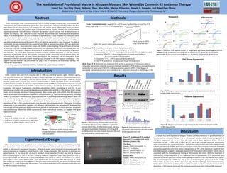
Methods
Figure 1. The phases of skin wound repair
include hemostasis, inflammation, proliferation,
and maturation.
Figure 2. H&E histology showed NM induced skin
wound including separation of dermal-epidermal
junction (DEJ), influx of inflammatory cells, necrosis,
and severe hyperplasia. Treatment of Cx43-asODN
on NM exposed mice skin showed improved DEJ and
less infiltration of inflammatory cells.
Figure 3. Western blots showed treatment
of Cx43-asODN reduced Cx43 protein
expression in NM exposed mice skin.
SKH-1 female hairless mice aged 6-10 weeks purchased from Charles River Laboratories (Wilmington, MA)
were used as an in vivo animal model to evaluate the effectiveness of the potential countermeasure Cx43-
asODN to NM induced skin wound formation. All animal studies were conducted in accordance with the
protocol approved by Laboratory Animal Services (LAS), Rutgers University. NM (5μmoles in acetone) was
applied topically to the dorsal skin of mice. A standard circular template (15 mm) was used to ensure a
uniform exposure area of NM on all mice. Following exposure Elizabethan collars were placed on mice to
prevent grooming of the wound site. A single topical application (150 µl of 1 µM in 30% Pluronic F-127 Gel) of
Cx43-asODN or sense control ODN (scODN) were applied to the wound 2 hours after NM exposure. (1 µM of
Cx43-asODN was found to be most effective.) The mice were euthanized by CO2 gas and punch biopsies were
collected at 1, 3, 7 and 10 days after NM exposure. The samples were collected for histological evaluation and
for RNA and protein extractions.
Results
Figure 5. Traditional PCR method
detected PCR product with serial
dilution (primers:18s).
Top Row:
Sample: NM+Cx43-asODN 7d
Well # 2: No dilution;
Well #3 :Dilution factor1:2.5
Well #4: Dilution factor 1:5
Bottom Row:
Sample: NM+Cx43asODN 10d
Well #4: No dilution;
Well #5: Dilution Factor 1:2.5
Well #6: Dilution Factor 1:5
Cx43
GapDH
50 kDa
37 kDa
Discussion
Animals that were exposed to nitrogen mustard showed alterations to gene expression of
the matricellular proteins (TNC and FN). In NM exposed skin, our study showed upregulation of
TNC gene expression (+2 to +3.5 folds at 3-10d) and a down-regulation of the FN gene
expression (-9 fold , -3 fold, and -1.5 fold at 1, 3, and 10 day post NM exposure, respectively)
when compared to the unexposed control. Animals that were treated with Cx43-asODN showed
a down-regulation of the TNC gene and up-regulation of the FN gene when compared to the NM
exposed group at the same time points. Upregulation of TNC gene expression (up to 10 days
post NM exposure) suggests a prolonged deposition of provisional matrix protein, which may be
associated with the delayed wound healing in NM induced skin injury. With the treatment of
Cx43-asODN, a down-regulation in TNC expression and an upregulation in FN expression were
observed in the NM plus Cx43-asODN treated group, compared to the NM exposed alone group.
The treatment of Cx43-asODN may play a role in modulating the provisional matrix in NM
induced skin wound repair. Protein expression (eg., Western blottings, immunohistochemistry,
ELISA) of the matricellular proteins will be studied to further clarify the role of provisional matrix
deposition in NM induced injury and the improved wound repair with Cx43-asODN treatment.
Tenascin C Fibronectin
A
B
D
C
E
F
G H
I J
naïve 1d 3d 7d 10d
nm 1.00 0.11 0.323110489 0.930950903 0.653030808
nm+as 0.23225429 0.339551315 1.1143882 0.715104856
0.00
0.20
0.40
0.60
0.80
1.00
1.20
1.40
FN Gene Expression
nm nm+as
naïve 1d 3d 7d 10d
NM 1 0.421017512 2.028593962 3.463884737 1.696208862
NM+AS 0.264146015 1.630962246 2.833238058 1.160472615
0
0.5
1
1.5
2
2.5
3
3.5
4
TNC Gene Expression
NM NM+AS
Figure 6. Real-time PCR reaction curves of target gene and house keeping gene: GAPDH.
Tenascin C: A) Unexposed Control; B) NM 1d; C) NM+As 1d; D) NM 3d ;E) NM+As 3d
Fibronectin: F) Unexposed Control; G) NM 1d ; H) NM+As 1d ;I) NM 3d; J) NM+As 3d
Figure 7. TNC gene expression down-regulated with the treatment of Cx43-
asODN in NM exposed skin.
Figure 8. FN gene expression up-regulated with the treatment of Cx43-asODN
in NM exposed skin.Figure 4. Gene expression of Cx43 was
decreased with treatment of Cx43-asODN
in NM exposed mice skin.