
Recommended
More Related Content
What's hot
What's hot (19)
Similar to Hypothermia ecg 2019
Similar to Hypothermia ecg 2019 (20)
More from EmogeneAldridge
More from EmogeneAldridge (20)
Recently uploaded
Recently uploaded (20)
Hypothermia ecg 2019
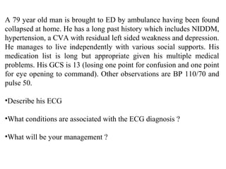
- 1. A 79 year old man is brought to ED by ambulance having been found collapsed at home. He has a long past history which includes NIDDM, hypertension, a CVA with residual left sided weakness and depression. He manages to live independently with various social supports. His medication list is long but appropriate given his multiple medical problems. His GCS is 13 (losing one point for confusion and one point for eye opening to command). Other observations are BP 110/70 and pulse 50. •Describe his ECG •What conditions are associated with the ECG diagnosis ? •What will be your management ?
- 3. The ECG rhythm is slow AF. Although it is tempting to call the presence of P waves in V1 these aren’t completely regular and vary in morphology. The other marked ECG feature is an extra positive deflection immediately after the main QRS seen most obviously in leads V3-V6 and leads II & III. These are J or Osborne waves. Put together with slow AF the ECG pattern is one of moderate to severe hypothermia. This patient’s temperature measured at 29.50 C True “environmental hypothermia” (eg a fisherman lost off a boat)is rare. Most cases we see have an underlying cause which leaves the patient in a situation where they can’t rewarm themselves such as: •Trauma – hip fracture, wrist fracture, head injury •Neurological – stroke, post-ictal •Drugs – alcohol, sedative overdose •Endocrine – especially hypoglycaemia •Systemic illness – sepsis eg pneumonia, urinary tract infection As well as treating the underlying illness the focus is rewarming. Use a forced air rewarming blanket on the highest setting. Give glucose as this is required as the energy substrate to generate heat (by mouth eg sweet drinks if tolerated or IV as 5% or 10% dextrose if not)