A seminar presentation on gross anatomy of the large intestine
•Download as PPTX, PDF•
5 likes•348 views

A seminar presentation on gross anatomy of the large intestine

Recommended
More Related Content
What's hot
What's hot (20)
Similar to A seminar presentation on gross anatomy of the large intestine
Similar to A seminar presentation on gross anatomy of the large intestine (20)
Recently uploaded
Recently uploaded (20)
A seminar presentation on gross anatomy of the large intestine
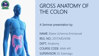
- 1. NAME: Ebere Uchenna Emmanuel REG. NO.: 2017/MD/6106 DEPT.: Anatomy COURSE CODE: ANA 441 SUPERVISOR: Dr. Ezemagu GROSS ANATOMY OF THE COLON A Seminar presentation by:
- 2. OUTLINE 01 02 03 04 05 OVERVIEW OF THE LARGE INTESTINE DIVISIONS OF THE LARGE INTESTINE GENERAL FEATURES OF THE COLON THE CAECUM THE VERMIFORM APPENDIX 06 THE ASCENDING COLON 07 08 09 10 11 TRANSVERSE COLON DESCENDING COLON SIGMOID COLON VASCULAR SUPPLY NERVE AND LYMPHATICS 12 CLINICAL ANATOMY
- 3. − The large intestine measures 1.5m in the adult. − Extends from the ileoceacal junction with the appendix vermiformis (in the right iliac fossa) to the anus. Functions − Absorbs fluid from the indigestible chyme − Converts the chyme to semi-solid stool − Temporarily store and allow to accumulate until defecation occurs. 01| OVERVIEW OF THE LARGE INTESTINE
- 4. 02| DIVISIONS OF THE LARGE INTESTINE • Caecum (6cm) • Appendix vermiformis (9cm) • Ascending colon (15cm) • Transverse colon (50cm) • Descending colon (25cm) • Sigmoid colon (50cm) © 2015-2017 TeachMeAnatomy.com [CC-BY-NC-ND 4.0]
- 5. 1. Appendices epiploicae: Fat-filled peritoneal processes present over the surface of caecum, AC, TC, DC, and the SC. 2. Taenia coli: These are three distinct longitudinal bands: mesocolic omental tenia, and libera taenia. 3. Haustra: Sacculations of the wall of the colon between the taenia. 03| GENERAL FEATURES OF THE COLON © 2015-2017 TeachMeAnatomy.com [CC-BY-NC-ND 4.0]
- 6. 04| THE CAECUM The caecum, is the first part of the large intestine and it is a blind pouch. − Lies in the right iliac fossa, − Between caecum and ileum is the ileocecal valve - prevents reflux during peristalsis. − May be palpable through the ant. lat. abd. wall if distended with feces. − Continuous with the AC. − The vermiform appendix is attached. © 2015-2017 TeachMeAnatomy.com [CC-BY-NC-ND 4.0]
- 7. 05| THE VERMIFORM APPENDIX The appendix vermiformis, (L. wormlike) is a blind intestinal diverticulum attached at the posteromedial aspect of the caecum. − Contains large amount of lymphoid tissue - has no vital function. − Variable position © 2015-2017 TeachMeAnatomy.com [CC-BY-NC-ND 4.0]
- 8. 06| THE ASCENDING COLON The ascending colon, is the second part of large intestine. − Passes superiorly on the right side of the abdominal cavity from the caecum to the right lobe of the liver − It forms the right colic flexure at the right lobe of the liver. − It is retroperitoneal. ANATOMICAL RELATIONS: − ANTERIOLY: − Small intestine, − Greater omentum, − Ant. Abd. Wall − POSTERIORLY: − Iliacus, − Right kidney, − Quadratus lumborum − ANTERO-MEDIALLY: − Fundus of gallbladder
- 9. 07| THE TRANSVERSE COLON The transverse colon, is the third part of large intestine. − Extends from the right colic flexure to the spleen. − From the spleen, it turns 90o degrees to point inferiorly forming the left colic flexure. − The TC is the least fixed and has a variable position (can dip into the pelvis in tall, thin individuals) − The TC is the only part which is intraperitoneal. ANATOMICAL RELATIONS: − ANTERIOLY: − Greater omentum, − Ant. Abd. Wall − POSTERIORLY: − Duodenum, − Head of pancreas − Spleen
- 10. 08| THE DESCENDING COLON The descending colon, is the fourth part of large intestine. − After the left colic flexure, the colon moves down towards the pelvis – forms the DC. − It is retroperitoneal. ANATOMICAL RELATIONS: − ANTERIOLY: − Small intestine, − Greater omentum, − Ant. Abd. Wall − POSTERIORLY: − Iliacus, − Quadratus lumborum, − Left kidney
- 11. 09| THE SIGMOID COLON The sigmoid colon, is the fifth part of large intestine. − When the DC turns medially, it becomes the SC. − Located in the left lower quadrant. − Extends from left iliac fossa to S3. ANATOMICAL RELATIONS: − SUPERIORLY: − Bladder, − Uterus − INFERIORLY: − Anus
- 13. 11| NERVE AND LYMPHATICS INNERVATION − Sympathetic and parasympathetic nerves: From superior mesenteric plexus. LYMHATICS − Ascending and transverse colon: Drains into the superior mesenteric nodes. − Descending and sigmoid colon: Drains into the inferior mesenteric nodes.
- 14. 12| CLINICAL ANATOMY • Colitis: Inflammation of the colon. • Caecal volvulus: Twisting of the caecum – represents 10% of intestinal volvuluses. • Colon cancer: Cancer of colon affects more than 100,000 people each year, though it is usually preventable through regular screening that is colon biopsies. • Appendicitis: Inflammation of the appendix. • Appendectomy: Surgical removal of the inflamed appendix. • Marginal Artery of Drummond: The marginal artery of Drummond is a clinically important vessel that provides collateral supply to the colon – thereby maintaining arterial supply in the case of occlusion or stenosis of one of the major vessels. © 2014 WebMD, LLC. All rights reserved
- 15. 13| REFERNCES • Susan Standring (2015). Gray's Anatomy - The Anatomical Basis of Clinical Practic e 41st Edition. MSCambo Elsevier • Singh I. Blood Vessels of Stomach, Intestines, Liver, Pancreas and Spleen. In: Textb ook of Human Anatomy (Textbook of Anatomy), 5th ed. Jaypee Brothers Medical Publishers(P)Ltd. New Delhi; 2015.p. 583-585. • BD Chaurasia’s Human Anatomy, Volume 2 – Lower Limb, Abdomen and Pelvis, 6th Edition. P .278-295 • Moore K.L., Arthur F.D., Anne M.R.A. Bones of Upper limb. Clinically Oriented An atomy, 8th ed. Lippincott Williams and Wilkins Publishers.Philadelphia;2018. p.153- 154. • Netter F., Machado C., Hassen J. Atlas of Human Anatomy,7th ed. Elsevier: Saund ers.Philadelphia;2014. p.416. • https://teachmeanatomy.info/abdomen/gi-tract/cecum • https://teachmeanatomy.info/abdomen/gi-tract/appendix • https://en.m.wikipedia.org/wiki/colon
Editor's Notes
- Functions Absorbs fluid from the indigestible chyme Converts the chyme to semi-solid stool Temporarily store and allow to accumulate until defecation occurs
- Appendices epiploicae: The caecum, AC, TC, DC, and the SC all have fat-filled peritoneal processes present over the surface. Taenia coli: (MOL):These are three distinct longitudinal bands: mesocolic taenia (TMC and SMC attach here), omental tenia (appendices epiploicae attach here), and libera taenia (free; nothing attaches here). Haustra: Sacculations of the wall of the colon between the taenia. Diameter: A much greater diameter.
- Lies in the right iliac fossa, inferior to the ileocecal junction. The cecum served as a site for cellulose digestion in our ancestors, but now the cecum simply acts as a reservoir for chyme which it receives from the ileum.
- The position of the free-end of the appendix vermiformis is highly variable. Of clinical relevance - The sympathetic afferent fibres of the appendix arise from T10 of the spinal cord – thus explaining why the visceral pain of early appendicitis is felt centrally within the abdomen. Post-ileal: Posterior to the terminal ileum. Pre-ileal: Anterior to the terminal ileum. Sub-ileal: Parallel with the terminal ileum. Pelvic: Descending over the pelvic brim. Paracecal: Alongside the lateral border of the cecum. Retrocecal: Behind the cecum.
- At the left colic flexure, the colon is attached to the diaphragm by the phrenicocolic ligament.
- The neurovascular supply of the colon is linked to the embryological origin: ARTERIAL SUPPLY Superior mesenteric artery: The caecum, appendix, AC and proximal 2/3rd of the TC (midgut derivatives) are supplied from ileocolic, right colic and middle colic branches of the SMA. Inferior mesenteric artery: The distal 1/3rd of the TC, DC, SC, rectum and upper anal canal (hindgut derivatives) are supplied IMA via the left colic, sigmoid and superior rectal arteries. VEINOUS DRAINAGE The venous drainage is via the accompanying veins. INNERVATION Sympathetic and parasympathetic nerves: From superior mesenteric plexus.
- - The superior mesenteric plexus is a continuation of the lower part of the celiac plexus (arise from preganglionic splanchnic nerves). Receiving a branch from the junction of the right vagus nerve with the plexus. - Most of the lymph from the superior mesenteric and inferior mesenteric nodes passes into the intestinal lymph trunk, and on to the cisternae chyli – where it ultimately empties into the thoracic duct.
- Appendicitis: Is caused by blockage of the appendix by poop, a foreign body, trauma to the abdomen, stool, parasites, etc.