Posterior Vitreous Detachment Treatment.pptx

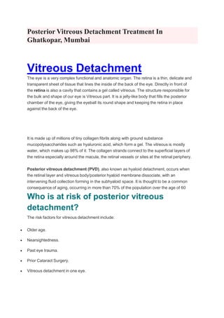
Vitreous Detachment The eye is a very complex functional and anatomic organ. The retina is a thin, delicate and transparent sheet of tissue that lines the inside of the back of the eye. Directly in front of the retina is also a cavity that contains a gel called vitreous. The structure responsible for the bulk and shape of our eye is Vitreous part. It is a jelly-like body that fills the posterior chamber of the eye, giving the eyeball its round shape and keeping the retina in place against the back of the eye. It is made up of millions of tiny collagen fibrils along with ground substance mucopolysaccharides such as hyaluronic acid, which form a gel. The vitreous is mostly water, which makes up 98% of it. The collagen strands connect to the superficial layers of the retina especially around the macula, the retinal vessels or sites at the retinal periphery. Posterior vitreous detachment (PVD), also known as hyaloid detachment, occurs when the retinal layer and vitreous body/posterior hyaloid membrane dissociate, with an intervening fluid collection forming in the subhyaloid space. It is thought to be a common consequence of aging, occurring in more than 70% of the population over the age of 60 Who is at risk of posterior vitreous detachment? The risk factors for vitreous detachment include: Older age. Nearsightedness. Past eye trauma. Prior Cataract Surgery. Vitreous detachment in one eye.

Recommended
More Related Content
Similar to Posterior Vitreous Detachment Treatment.pptx
Similar to Posterior Vitreous Detachment Treatment.pptx (20)
Recently uploaded
Recently uploaded (20)
Posterior Vitreous Detachment Treatment.pptx
- 1. Posterior Vitreous Detachment Treatment In Ghatkopar, Mumbai Vitreous Detachment The eye is a very complex functional and anatomic organ. The retina is a thin, delicate and transparent sheet of tissue that lines the inside of the back of the eye. Directly in front of the retina is also a cavity that contains a gel called vitreous. The structure responsible for the bulk and shape of our eye is Vitreous part. It is a jelly-like body that fills the posterior chamber of the eye, giving the eyeball its round shape and keeping the retina in place against the back of the eye. It is made up of millions of tiny collagen fibrils along with ground substance mucopolysaccharides such as hyaluronic acid, which form a gel. The vitreous is mostly water, which makes up 98% of it. The collagen strands connect to the superficial layers of the retina especially around the macula, the retinal vessels or sites at the retinal periphery. Posterior vitreous detachment (PVD), also known as hyaloid detachment, occurs when the retinal layer and vitreous body/posterior hyaloid membrane dissociate, with an intervening fluid collection forming in the subhyaloid space. It is thought to be a common consequence of aging, occurring in more than 70% of the population over the age of 60 Who is at risk of posterior vitreous detachment? The risk factors for vitreous detachment include: Older age. Nearsightedness. Past eye trauma. Prior Cataract Surgery. Vitreous detachment in one eye.
- 2. What are symptoms of Vitreous Detachment? Posterior vitreous detachment (PVD) doesn’t cause pain or permanent vision loss, but you might experience other symptoms. They include: Flashes. These small flashes of light are comparable to “seeing stars” after hitting your head. They can last a few seconds or minutes and tend to stop, or occur less often, once detachment is complete. Floaters. These floating spots in your field of vision can resemble tiny specks, dust, dots, or cobweb-like shadows. They typically occur in the first few weeks of Posterior vitreous detachment (PVD) and are most noticeable when looking at a light surface, such as a white wall or the sky. Cobweb effect. You may begin to see the outer edge of the vitreous as it separates from the retina. It can feel like you’re looking through a cobweb. This is temporary and goes away once detachment is complete. How Vitreous Detachment Develops? In normal eyes, the vitreous is attached to the surface of the retina through millions of tiny, intertwined fibers. Your vitreous gel is mostly made of water. As we age, the vitreous slowly shrinks, and these fibers pull on the retina's surface. If the fibers break, the vitreous can shrink further and separate from the retina, causing a vitreous detachment. Most people get Posterior vitreous detachment (PVD) at age 60 or older and it's very common after 80. It happens to men and women equally. What other problems can vitreous detachment cause? Vitreous detachment can sometimes lead to more serious eye conditions: Retinal tear. Sometimes, the vitreous fibers tear a hole in the retina when they pull away. If you don’t get treatment quickly, this can lead to retinal detachment. Retinal detachment. Sometimes vitreous detachment pulls the entire retina away from its normal position at the back of the eye. This can be a medical emergency. Learn more about retinal detachment. Macular hole. Sometimes vitreous detachment tears a hole in the macula (the part of the retina that controls your central vision). This can happen before or after the vitreous detaches enough to cause floaters or flashes of light. Learn more about macular hole. Macular pucker. Sometimes vitreous detachment causes a thin layer of scar tissue to grow over the macula. This usually happens slowly in the months or years after vitreous detachment. Learn more about macular pucker.
- 3. These conditions can cause vision loss but treatment may help preserve your vision. Tell your eye doctor right away if you notice symptoms of vitreous detachment so they can check for these more serious problems. Diagnosis A Routine eye examination can help your doctor to spot problems like Posterior vitreous detachment (PVD) early and that can help protect your vision. Your doctor may use special eye drops to make your pupils (the holes in the center of your eyes) bigger and use a slit-lamp test to look for signs of Posterior vitreous detachment (PVD). This is done with a microscope that looks through the front of your eye. It is helpful in detecting, if Posterior vitreous detachment (PVD) has caused bleeding, a torn retina, or something else that could harm your eyesight. Your doctor also may use other tests to make sure the gel hasn't pulled away from your retina. These include: Optical coherence tomography (OCT)- a 3-D scan of the inside of your eye Ocular ultrasound - a test that uses sound waves to show the inside of your eye Treatment Posterior Vitreous Detachment usually doesn’t require treatment. Complete detachment typically takes no longer than three months. If you continue to see floaters after detachment is complete, discuss treatment options with your doctor. When complications occur and your ophthalmologist recommends treatment, there are a number of options available, including: Laser or cryosurgery: This can be done in the office with no anesthesia. Your doctor repairs the holes or tears in your retina, which prevents further progression of the condition. Scleral buckle: This involves placing a band around the outside of the eye to counterbalance the force of the vitreous pulling on the retina. Fluid is then drained from behind your retina, allowing it to return to its proper position. This is usually done as outpatient surgery. Pneumatic retinopexy: This surgery is sometimes done in the office. Your doctor injects a gas bubble into the vitreous behind your eye. The bubble pushes the tear against the back wall of the eye and closes it. Then your doctor uses laser or cryosurgery to secure the retina where it belongs. After surgery, you may need to keep your head in a certain position for a few days. The gas bubble dissipates over time. Follow up of Vitreous Detachment
- 4. Once it has been confirmed that the vitreous detachment is isolated, follow-up examinations are recommended at regular intervals thereafter. The period between examinations depends, of course, on the presence of blood in the vitreous or other signs which could increase the likelihood of retinal detachment. Thus the first re-visit may be after a week or a month, according to the nature of the detachment. Important Reminder: This information is only intended to provide guidance, not a definitive medical advice. Please consult eye doctor about your specific condition. Only a trained, experienced board certified eye doctor can determine an accurate diagnosis and proper treatment. To schedule an appointment with our experts for Posterior vitreous detachment (PVD) Treatment In Ghatkopar, please call us at +91 8451045935, +91-8451045934 or visit our clinic at Address. Tag : best eye specialist in ghatkopar, eye clinic in ghatkopar east, Retina Detachment Treatment In Ghatkopar For more information : https://www.mumbaieyecare.com/