Hematology analyzer channels-modes- handling errors
•
2 likes•229 views

Hematology analyzer channels-modes- handling errors For Lab.students

Recommended
More Related Content
What's hot
What's hot (20)
Similar to Hematology analyzer channels-modes- handling errors
Similar to Hematology analyzer channels-modes- handling errors (20)
More from DrAbdulrazzaqAlagbar
More from DrAbdulrazzaqAlagbar (17)
Recently uploaded
Recently uploaded (20)
Hematology analyzer channels-modes- handling errors
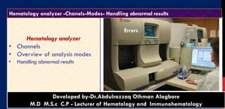
- 1. Hematology analyzer • Channels • Overview of analysis modes • Handling abnormal results 1 Diagnostic Hematology - Dr.Alagbare BACK to contentDeveloped by-Dr.Abdulrazzaq Othman Alagbare M.D M.S.c C.P - Lecturer of Hematology and Immunohematology Hematology analyzer -Chanels-Modes- Handling abnormal results Errors
- 2. 2 Hematology analyzer Channels Channel for Red blood cells and platelets analyzing , RBCs and platelets using the same dilution Channel for WBC and hemoglobin measurement: Lytic agents lyse red cells first before analysis. Channel for WBC differential count. Channel for Reticulocyte count. Diagnostic Hematology - Dr.Alagbare
- 3. 3 Other channels: A. nucleated red blood cell (NRBC) channel, B. separate hemoglobin (Hb) channel, C. WBC/basophil counting channel, D. and immature granulocyte counting channel. Diagnostic Hematology - Dr.Alagbare
- 4. Small volume of EDTA blood sample is aspirated and diluted in an electrically conductive diluent. 1. RBC/PLT chamber 2. WBC/Hgb chamber • RBCs lysed, Hgb directly measured by spectrophotometry and WBCs counted 1. Mixing chamber – differential 2. Reticulocyte dilution chamber 4 Diagnostic Hematology - Dr.Alagbare
- 5. 5 In the channel of RBC/PLT counting • Particles between 2 and 20 fL are counted as platelets, • and particles larger than 35 fL are counted as RBCs. Diagnostic Hematology - Dr.Alagbare They fall in the overlap area between platelets and RBCs, generating a warning flag. Why
- 6. Errors in platelet counts 6 Falsely high platelet counts Microcytic red cells Red cell fragments White cell fragments of leukaemic blast cells Contaminated blood sample (Bacteria Malaria, fungus)
- 7. 7 Microcytic red cells that notched in IDA, if the RBC have very low volume fall in the platelets counting zone and they counted as platelets, for that we have slight increase of platelets count in IDA
- 8. Errors in platelet counts Falsely low platelet counts 8 Diagnostic Hematology - Dr.Alagbare giant plateletsclumped platelets
- 9. 9 Diagnostic Hematology - Dr.Alagbare
- 10. Needs 30-40 µl This is the mode of analyzing collected blood sample in the whole blood status 10 USES For Paediatric specimens. This mode is used in analyzing a minute amount of child’s blood, for example, collected from the earlobe or fingertip. Diagnostic Hematology - Dr.Alagbare Overview of analysis modes 1-Whole blood mode 2-Pre-diluted mode HOW? In this mode, blood sample diluted into 1:26 before analysis is used. The sample aspiration procedure is the same as in the whole blood mode.
- 11. 1-Plts < 40,000 How solved? 1. Check the integrity of the specimen (look for clots, short draw, etc.) 2. Confirm count with smear review for clumps, RBC fragments, giant platelets, very small RBCs HANDLING ABNORMAL RESULTS 11 Diagnostic Hematology - Dr.Alagbare
- 12. 2-Plt ++++ How solved? HANDLING ABNORMAL RESULTS 12 Diagnostic Hematology - Dr.Alagbare If present, o perform plt. estimate. o If they do not agree, perform manual plt count. If not present, o dilute specimen 1:2 with Isoton or further until count is within linearity, multiply diluted result by dilution factor. Do PBS Check smear for RBC fragments or microcytes.
- 13. Diagnostic Hematology - Dr.Alagbare 13 CASE A 44-year-old woman comes in for a complete blood count (CBC) as part of a routine physical exam. The results from the hematology analyzer,, are: PLEASE answer the following 1. What is abnormal about her CBC? 2. Which parts can be reported? 3. What procedures can be done regarding the abnormal result? 4-Which cells included in MID and Gran?
- 14. 14 Questions 1. What is abnormal about the blood count? 2. Which parts of the CBC can be reported? 3. What would you do to investigate the abnormal result? 4-write the situation which cause low false PLT count?
- 15. Diagnostic Hematology - Dr.Alagbare 15 1 2 3 Answer the following What called each image? Describe and write a small paragraph for each image how developed, and what effect on the platelets count? How solved each problem of them if present?
- 16. 3-WBC ++++ How solved? Dilute 1:2 with Isoton or further until count is within linearity (for final result, multiply diluted result by dilution factor. Plt counts are not affected by high WBC. Add comment, “Unable to report Hgb, MCH, MCHC due to high WBC.” HANDLING ABNORMAL RESULTS 16 Diagnostic Hematology - Dr.Alagbare
- 17. 17 In some printouts use the following symbols UWBC represents uncorrected WBC count WBC represents corrected WBC count In some printouts use the following symbols “&”WBC represents corrected WBC count Plt & represents corrected PLT count Diagnostic Hematology - Dr.Alagbare
- 18. 18 Diagnostic Hematology - Dr.Alagbare
- 19. HANDLING ABNORMAL RESULTS 4-RBC > 7.0 How solved? 1. Dilute 1:2 with Isoton or further until count is within linearity, multiply dilution result by dilution factor; 2. perform spun Hct, 3. review Hgb, 4. recalculate MCH, MCHC 19 Diagnostic Hematology - Dr.Alagbare
- 20. 20 Normal White blood cells volume Cells with volume Designated as 35-90 fl Lymphocytes, 90-160 fl Monocyte, Eos, BAS 160-450 Neutrophils, Diagnostic Hematology - Dr.Alagbare
- 21. 21 هلل الحمد رب العالمين Diagnostic Hematology - Dr.Alagbare العمل بهذا ابتغاء ارجو هللا مرضات