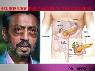
Neuroendocrine tumors of the pancreas
•Download as PPTX, PDF•
3 likes•136 views

By Dr Dipesh K.K General Surgeon

Recommended
Recommended
More Related Content
What's hot
What's hot (20)
Similar to Neuroendocrine tumors of the pancreas
Similar to Neuroendocrine tumors of the pancreas (20)
More from Dr Dipesh K.K
More from Dr Dipesh K.K (11)
Recently uploaded
Recently uploaded (20)
Neuroendocrine tumors of the pancreas
- 2. Rudolf Heidenhain discovered neuroendocrine cells in 1870 The first report of a PNET was done by Albert Nicholls in 1902
- 3. Pancreatic endocrine tumors (PETs) are rare neoplasms that comprise 2% to 4% of all clinically detected pancreatic tumors.
- 4. The origin of these tumors is not completely understood. Based on the most recent evidence, it has been suggested that these tumors arise from an endocrine cell–derived gastrointestinal epithelium.
- 5. 1 case per 100,000 individuals per year. Represent 2 – 4% of pancreatic tumors. Forth to sixth decade of life. Most pancreatic NETs are sporadic, but they can be associated with hereditary endocrinopathies. MEN1. 80 – 100% Von Hipple Lindau (VHL). 20% Neurofibromatosis 1 (NF-1). 10% Tuberous sclerosis complex (TSC). 1%
- 6. Well-Differentiated Endocrine Tumor Type 1: Benign Behavior Confined to the pancreas <2 cm in diameter <2 mitoses per high-powered field <2% Ki-67–positive cells No vascular or perineural invasion Well-Differentiated Endocrine Carcinoma Low-grade malignant Gross local invasion Metastases Type 2: Uncertain Behavior Confined to the pancreas and one of the following: >2 cm in diameter >2 mitoses per high-powered field >2% Ki-67–positive cells Vascular or perineural invasion Poorly Differentiated Carcinoma High-grade malignant >10 mitoses per high-powered field
- 7. T: Primary Tumor T0: No evidence of cancer Tis: Carcinoma in situ T2: Tumor limited to the pancreas, size >2 cm T3: Tumor extends beyond the pancreas but does not involve the celiac axis or superior mesenteric artery T4: Tumor involves celiac axis or superiormesenteric artery (unresectable primary tumor) N: Regional Lymph Nodes N0: No regional lymph nodes involved N1: Regional lymph nodes involved M: Distant Metastases Stages 0: Tis N0 M0 IA: T1 N0 M0 IB: T2 N0 M0 IIA: T3 N0 M0 IIB: T1 N1 M0, T2 N1 M0, T3 N1 M0 III: T4, any N, M0 IV: Any T, any N, M1
- 8. Based on functionality – i.e. syndrome produced – 1.Functional (Detected early due to symptoms produced due to hormone excess) • Insulinoma • Gastrinoma • VIPoma • Glucagonoma • Somatostatinoma
- 9. Cell Type Hormone Produced Endocrine Tumor/Syndrome Alpha (A) Glucagon Glucagonoma Beta (B) Insulin Insulinoma Delta (D) Somatostatin Somatostatinoma D2 VIP VIPoma G Gastrin Gastrinoma
- 10. Most common type of functioning pancreatic endocrine tumor. Women (2:1) 50 - 60 years old. Benign. Solitary and small. Can occur sporadically or in association with the hereditary MEN-1.
- 11. WIPPLE TRIAD Whipple described a triad of signs and symptoms associated with insulinomas Symptoms of hypoglycemia. Blood glucose < 45 mg/dL. Relief of symptoms with Glucose
- 12. SYMPTOMS OF HYPOGLYCEMIA: Altered mental state Weakness. Diplopia. Seizures. Coma. Sweating. Palpitations. Anxiety.
- 13. Monitored fast for <48 hours with documented blood glucose <50 mg/dL with hypoglycemic symptoms Relief of symptoms after oral glucose load. Elevated insulin > 5 – 10 μU/mL. Increase proinsulin level > 22 pmol. Absence of sulfonylureas in plasma or urine. Elevated C peptide levels.
- 14. CT has been shown to be more sensitive for detecting small insulinomas. Most insulinomas are vascular and can be visualized on arterial phase imaging. One series comparing the use of CT, EUS, and the two in combination found the sensitivity of CT with EUS was superior to either modality done separately MRI is considered a second-line modality in the evaluation of insulinomas because of its greater expense and more limited availability.
- 16. Fig. 1. Contrast-enhanced CT of the abdomen demonstrates a well-circumscribed, round insulinoma (arrow) in the body of the pancreas with homogeneous enhancement
- 17. Fig:-Homogenous hypoechoic mass lesion in the head of the pancreas adjacent to the common bile duct (CBD, above) and portal vein (PV, below) without invasion of these structures
- 18. Pathological specimen demonstrates characteristic pale well-defined mass consistent with an
- 19. The definitive treatment for patients with insulinomas is resection of the tumor, Presurgical therapy to alleviate the symptoms and neurologic affects of hypoglycemia should be instituted. A number of insulin antisecretagogues can be used, such as - Diazoxide -Verapamil -Octreotide -Dilantin
- 20. Stabilize the glucose level with diet and/or diazoxide. If the tumour is exophytic or pheripheral(head,distal) by imaging Tumour enucleation,consider laparoscopic resection If deeper and invasive tumour and those in proximity to MPD Head-pancreaticoduodenectomy Distal-distal pancreatectomy, consider laparoscopic resection Metastatic disease: -If resectable then resection with octreotide and chemotherapy -If unresectable then palliative treatment
- 21. 2nd most common functioning islet cell tumor of the pancreas. 0.5 to 3 per million population per year Peak age of onset is 50 years20-30% associated with MEN1 Zollinger Ellison syndrome – ectopic gastrin secretion - excessive gastric acid secretion - PUD, GERD and diarrhea.
- 22. Peptic ulcer disease with diarrhea. Ulcers in unusual locations. Refractory to medication ulcers. Young age ulcer with complications. Abdominal pain. Chronic diarrhea. Heartburn. Nausea,Vomiting. Bleeding. Esophageal strictures. Pyloric or duodenal scarring. Prominent gastric folds
- 23. Differential Diagnosis of Hypergastrinemia High Acid Output Zollinger-Ellison syndrome G-cell hyperplasia Retained gastric antrum Gastric outlet obstruction Normal Acid Production Atrophic gastritis Proton pump inhibitors Postvagotomy syndrome Renal failure
- 24. Diagnostic Criteria for Gastrinoma Fasting gastrin level: > 100 pg/mL or >10 times higher than upper limit. Secretin stimulation test: Gastrin > 200 pg/mL. Calcium infusion provocative testing: Gastrin > 395 pg/dL. Basic acid output level > 15 mEq/L
- 25. Once the biochemical diagnosis of ZES has been established,the next step is to localize the primary lesions and determine the presence or extent of tumor spread by -CT scan -Endoscopic ultrasound. -Somatostatin Receptor Scintigraphy. -Intraoperative palpation and Ultrasound.
- 27. Gastrinoma Triangle: More than 80% of gastrinomas are located within this triangle
- 28. The primary goal of treatment is To control acid production, Remove the primary tumor, and Prevent malignant progression.
- 29. TREATMENT 1) PPI- to control acid production 2) Surgical HEAD:- If exophytic or pheripheral tumour by imaging Enucleation of tumour +periduodenal node dessection If deeper and invasive tumour and those in proximity to MPD pancreaticoduodenectomy DISTAL:- Distal pancreatectomy +/- splenectomy
- 30. DUODENAL TUMOUR:- Duodenotomy and intraoperative ultrasonogram;local resection/enucleation of tumour + periduodenal node dissection. Metastatic disease: -If resectable then resection with octreotide and chemotherapy -If unresectable then palliative treatment
- 31. MEN-1 patients with ZES:- The operative role and appropriate procedure in MEN-1 patients with ZES is controversial. (because more than 50% of these patients are initially seen with evidence of metastases; Thus MEN-1 patients are rarely cured by surgery. The goal of surgery in MEN-1 patients is not cure but prevention of metastatic disease
- 32. The first patient with a glucagonoma was described in 1942 by Becker and colleagues. Male = Female. Age 50 – 60. Malignant 60 – 70 % Association with MEN-1 is rare.
- 33. Clinical presentation: Diabetes. Necrolytic migratory erythema. Deep vein thrombosis. Depression. Weight loss. Stomatitis. Fig.Necrolytic migratory erythema of glucagonoma
- 34. DIAGNOSIS: Fasting serum glucagon > 1000 pg/dL. Increase glucose. Decrease plasma amino acids Normochromic normocytic anemia,
- 35. CT scan is sufficient for localization and has been reported to detect 86% of tumors. EUS is usually unnecessary for localization, but it can be useful for US-guided needle biopsy. SRS has been used more for long-term follow-up of these patients and can demonstrate metastatic disease.
- 38. Correction of metabolic deficits. Somatostatin analogues should be considered to diminish circulating levels of glucagon The majority of tumors are located in the body or tail Distal pancreatectomy with peripancreatic lymphnode dissection with spleenectomy Metastatic disease: -If resectable then resection with octreotide and chemotherapy -If unresectable then palliative treatment
- 39. Male > Female. Mean age 48. Benign 50% Rare with incidence- 1 per 10 million Over 70% patients have metastatic disease at the time of presentation Solitary, large and are usually diagnosed at >3 cm in size 10% associated with MEN1
- 41. Diagnosis Fasting serum VIP level should be greater than 200pg/mL; average levels are close to 1000 pg/mL in patients with VIP tumors. Location: The majority of pancreatic VIP tumors are located within the tail of the pancreas (72%) CT scan of theabdomen and pelvis. for localizing these lesions approaches 100%.(Because of their relatively large size)
- 42. Treatment: Correction of electrolyte imbalance. Somatostatin Stabilize glucose level The majority of tumors are located in tail Distal pancreatectomy with peripancreatic lymphnode dissection with spleenectomy
- 43. If tumor located in head:- pancreatoduodenectomy with peripancreatic lymphnode dissection Metastatic disease: -If resectable then resection with octreotide and chemotherapy -If unresectable then palliative treatment
- 44. Rare, only 1% of the Neuroendocrine tumors. Mean age: 50 years. Men = Women. lesions are solitary and generally average between 5 and 6 cm. Malignant: 60 – 70 %
- 45. Clinical presentation: Hyperglycemia. Cholelitiasis. Steatorrhea. Diarrhea. Gastric hypochloridia. Jaundice. Diagnosis: Fasting somatostatin > 30 pg/mL
- 46. Most tumors located in the head of the pancreas are large. Localization of pancreatic somatostatinomas often can be accomplished by CT or US, whereas EUS, MRI, and SRS play a role in localization of smaller or metastatic tumors.
- 48. Surgical resection is the curative treatment, Debulking can provide symptomatic relief cholecystectomy should be performed at the time of operation because of the high incidence of cholelithiasis. In unresectable disease, octreotide and interferon-alfa may improve symptoms
- 49. In patients without metastatic disease, the mean 5-year survival is 100%. Those patients with metastatic disease who undergo radical resection or debulking procedures have a mean 5-year survival of 60%
- 50. NON FUNCTIONAL NEUROENDOCRINE TUMOR - They do not present clinically with a hormonal syndrome as compared with their functional counterparts - They often present later in the course of the disease with symptoms of local compression or metastatic disease
- 51. They represent more than 75% of all pancreatic endocrine tumors. lack a clinical syndrome of hormone overproduction Women (2:1) , Mean age: 45yr 60 – 80% are metastatic at diagnosis. 60% are malignant
- 52. These lesions are typically discovered on routine radiographic imaging done for nonspecific abdominal complaints. As these tumors grow larger and begin to compress surrounding structures, patients may develop symptoms of pain or obstruction.
- 53. Blood testing for tumor markers include pancreatic polypeptide, neurotensin, protein S, neuron-specific enolase,and chromogranin A. Measurement of these levels prior to resection may help establish a baseline for tumor burden and provide a possible marker for follow-up for tumor
- 54. Localization of the primary tumor and establishment of the extent of metastatic disease is best achieved with a triplephase— arterial, portal, and venous phase—CT scan. Because these tumors are typically large, EUS is only necessary for biopsy or to identify subcentimeter disease.
- 60. Tricia A, Moo-Young and Richard A. Endocrine tumors of the pancreas: clinical picture, diagnosis, and therapy. In: Blumgart's Surgery of the Liver, Biliary Tract, and Pancreas. 5th edition. Chapter61. Pp 934 – 944. Ladner D and Norton J. Neuroendocrine Tumors of the Pancreas. In: Shackelford’s Surgery of the Alimentary Tract. 7th edition. Pp 1206 – 1216. http://www.uptodate.com/contents/classification-epidemiology-clinical- presentationlocalization-and-staging-of-pancreatic-neuroendocrine- tumors-islet-cell-tumors http://www.uptodate.com/contents/insulinoma?source=see_link http://www.uptodate.com/contents/zollinger-ellison-syndrome- gastrinoma-clinicalmanifestations-and-diagnosis?source=see_link