
Recommended
More Related Content
Similar to Thyroid Anatomy.ppt
Similar to Thyroid Anatomy.ppt (20)
Recently uploaded
Recently uploaded (20)
Thyroid Anatomy.ppt
- 2. Background What: brownish-red, highly vascular gland Location: ant neck at C5-T1, overlays 2nd – 4th tracheal rings Avg width: 12-15 mm (each lobe) Avg height: 50-60 mm long Avg weight: 25-30 g in adults (slightly more in women) **enlarges during menstruation and pregnancy**
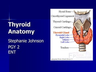
- 3. Background Pyramidal lobe: often ascends from the isthmus or the adjacent part of either lobe (usu L) up to the hyoid bone may be attached by a fibrous/fibromuscular band “levator” of the thyroid gland
- 4. Transverse view: relationship to other NB structures in neck
- 5. Structure Under middle layer of deep cervical fascia (pretracheal) thyroid inner true capsule thin and closely adherent to the gland capsule extensions within the gland form septae, dividing it into lobes and lobules lobules are composed of follicles = structural units of the gland layer epithelium enclosing a colloid-filled cavity colloid (pink on H&E stain) contains an iodinated glycoprotein, iodothyroglobulin (precursor of thyroid hormones).
- 6. Structure Follicles = variable size surrounded by dense plexuses of fenestrated capillaries, lymphatic vessels, and sympathetic nerves.
- 7. Structure Epithelial cells = 2 types: principal (ie: follicular) – formation of colloid (iodothyroglobulin) parafollicular (ie: C cells -clear, light), lie adjacent to follicles w/in basal lamina produce calcitonin
- 8. Relation w/ Strap muscles Lateral - sternothyroid Anterior - omohyoid muscle - sternohyoid Inferior - SCM (lower portion) ** careful - motor nerve supply from the ansa cervicalis enters these muscles inferiorly.
- 9. Recurrent laryngeal nerve Recall: innervates all larynx except cricothyroid Closely assoc with ITA (see next slides for details) NB: ‘non recurrent LN’ ~5/1000 pt’s on R side – When retroesophageal R SCA from dorsal aortic arch – NRLN - branches fr X at ~ cricoid cartilage – directly enters the larynx without looping around SC – L sided - only when R aortic arch and ligamentum arteriosum concurrent w/ L retroesophageal subclavian artery.
- 10. Vascular Anatomy ARTERIAL: superior and inferior thyroid arteries (occ thyroidea ima) ++ collateral anastomoses (ipsi and contralaterally) thyroid ima (when pres) originates from aortic arch or innominate artery, enters the thyroid at inferior border of isthmus.
- 11. Vascular Anatomy
- 12. Vascular Anatomy SUPERIOR THYROID ARTERY first anterior branch ECA descends laterally to the larynx under the omohyoid and sternohyoid muscles runs superficially on the anterior border of the lateral lobe, sending a branch deep into the gland before curving toward the isthmus where it anastomoses with the contralateral artery
- 13. Vascular anatomy SUPERIOR THYROID ARTERY: Relationship with SLN: Cephalad to the superior pole, ext branch of SLN runs w/ STA before turning medially supply cricothyroid muscle **careful when ligating artery**
- 14. Vascular anatomy INFERIOR THYROID ARTERY SCA thyrocervical trunk ITA ITA ascends vertically and then curves medially to enter the tracheoesophageal groove (posterior to carotid sheath) Branches penetrate the posterior aspect of the lateral lobe
- 15. Vascular anatomy Relationship with RLN: RLN ascends in the TE groove and enters the larynx b/w the inferior cornu of the thyroid cartilage and the arch of the cricoid RLN can be found after it emerges from the superior thoracic outlet: – Sup: thyroid lobe – Lat: common carotid artery – Medial: trachea
- 16. Vascular anatomy **Careful - relationship between RLN and ITA highly variable (Redd, 1943 – described 28 variations) Examples: Deep to ITA (40%) superficial (20%) b/w branches of the artery (35%) **also – only 17% of the time is the nerve/artery relationship the same on both sides **at level ITA – extralaryngeal branches RLN present 5% of the time
- 17. Vascular anatomy VENOUS: 3 pairs of veins: 1) STV – asc along STA and becomes a tributary of the IJV 2) MTV – directly lateral IJV 3) ITV (variable): – R – passes ant to innominate a R BCV or ant trachea L BCV – L – drainage L BCV **occ – both inf veins form a common trunk “thyroid ima vein” empties into L BCV
- 18. Lymphatics Extensive, multidirectional flow periglandular prelaryngeal (Delphian) pretracheal paratracheal (along RLN) brachiocephalic (sup mediastinum) deep cervical thoracic duct NB: regional mets of thyroid carcinoma are superior and lateral, along IJV ie: invasion of the pretracheal and paratracheal LNs and obstruction of normal lymph flow.
- 19. Lymphatics
- 20. Innervation Principally from ANS Parasympathetic fibers – from vagus Sympathetic fibers – from superior, middle, and inferior ganglia of the sympathetic trunk Enter the gland along with the blood vessels.
- 21. References: 1) Schwartz 2) www.emedicine.com 3) www.utdol.com 4) Gray’s anatomy 5) http://www.ncbi.nlm.nih.gov/books/b v.fcgi?rid=endocrin.box.330 6) Netter’s anatomy