
Recommended
More Related Content
Similar to 002 TRIGEMINAL NERVE.pptx
Similar to 002 TRIGEMINAL NERVE.pptx (20)
More from DeniseMathre1
More from DeniseMathre1 (20)
Recently uploaded
Recently uploaded (20)
002 TRIGEMINAL NERVE.pptx
- 2. NERVE: PHYSIOLOGY COMPONENTS OF NERVE PHYSIOLOGY OF NERVE CONDUCTION CRANIAL NERVES TRIGEMINAL NERVE: INTRODUCTION OPTHALMIC NERVE MAXILLARY NERVE MANDIBULAR NERVE TRIGEMINAL NEURALGIA APPLIED ASPECT 2
- 3. NERVE: NEURON – NERVE CELL 3
- 4. COMPONENTS OF THE NERVE 4
- 5. PHYSIOLOGY OF NERVE CONDUCTION: . 5
- 6. NERVOUS SYSTEM GUYTON AND HALL, TEXTBOOK OF MEDICAL PHYSIOLOGY, 11TH EDITION 6
- 8. CRIBRIFORM PLATE OLFACTORY NERVE(I) OPTIC FORAMEN OPTIC NERVE (II) SUPERIOR ORBITAL FISSURE OCCULOMOTOR (III) TROCHLEAR (V) ABDUCENS (VI) OPTHALMIC DIV OF TRIGEMINAL (V1) FORAMEN ROTANDUM MAXILLARY DIV OF TRIGEMINAL (V2) FORAMEN OVALE MANDIBULAR DIV OF TRIGEMINAL (V3) INTERNAL AUDITORYCANAL FACIAL (VII) VESTIBULOCOCHLEAR (VIII) JUGULAR FORAMEN GLOSSOPHARYNGEAL (IX) VAGUS (X) ACCESSORY (XI) HYPOGLOSSAL CANAL HYPOGLOSSAL (XII) EXIT OF THE CRANIAL NERVES: 8 SNELL’S ANATOMY, 7TH EDITION, RICHARD S. SNELL.
- 9. 9 TRIGEMINAL NERVE – WHY? SNELL’S ANATOMY, 7TH EDITION, RICHARD S. SNELL.
- 10. INTRODUCTION largest cranial nerve Fifth cranial nerve. Mixed nerve ( sensory and motor ) Sensory to – Skin of face Motor to – Mucosa of cranial viscera Except base of tongue and pharynx Muscles of Mastication Tensor ville palatini,Tensor tympany Anterior belly of digastric Mylohyoid SNELL’S ANATOMY, 7TH EDITION, RICHARD S. SNELL.
- 11. SENSORY ROOT Face, Scalp, Teeth, Gingiva, Oral, Nasal, Cavities, Para nasal sinus, Conjunctiva and Cornea Pain, temp, light touch touch, pressure proprioception Trigeminal gang. Bypasses trigem gang. sensory root. Spinal Principal sensory Mesencephalic CNS SNELL’S ANATOMY, 7TH EDITION, RICHARD S. SNELL.
- 12. Tensor tympani Tensor palatini Muscles of mastication Masseter Lateral & Medial Pterygoids Temporalis CNS MOTOR NUCLEUS MOTOR ROOT MANDIBULAR NERVE MOTOR ROOT SNELL’S ANATOMY, 7TH EDITION, RICHARD S. SNELL.
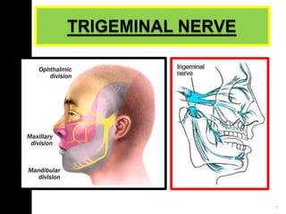
- 13. DIVISIONS OF TRIGEMINAL NERVE 1. Ophthalmic nerve 2. Maxillary nerve 3. Mandibular nerve SNELL’S ANATOMY, 7TH EDITION, RICHARD S. SNELL.
- 14. OPTHALMIC NERVE SNELL’S ANATOMY, 7TH EDITION, RICHARD S. SNELL. 14
- 15. OPTHALMIC NERVE Superior division of the V nerve Entirely Sensory Has 3 branches. All 3 of them pass through the sup. orbital fissure into the orbit. They are; 1.Lacrimal nerve 2.Frontal nerve 3.Nasocilliary nerve SNELL’S ANATOMY, 7TH EDITION, SNELL’S ANATOMY, 7TH EDITION,RICHARR I DC H A SR .D SS N. S E N L E L L L . . 15
- 16. 16 COURSE Emerges from trigeminal ganglion Lateral wall cavernous sinus 3 branches in ant part of cavernous sinus lacrimal, nasocilliary, frontal Superior orbital fissure Orbit SNELL’S ANATOMY, 7TH EDITION, RICH S A N E R L D L ’ S S A N . A S T O N M E Y L ,7 L T . H EDITION, RICHARD S. SNELL.
- 17. BRANCHES: 1. Lacrimal nerve: Smallest Sensory Supplies : lacrimal gland & the conjuntiva , skin of the upper eyelid. 17 SNELL’S ANATOMY, 7TH EDITION, RICHARD S. SNELL.
- 18. 2) Frontal nerve: largest branch It enters the orbit through the superior orbital fissure divides into 2 branches. i. The supra orbital branch: It is larger ,arises from the orbit through the supraorbital foramen. Supplies : skin of the forehead & scalp mucous membrane of the frontal sinus ii. The supra trochlear branch: It is smaller & from the orbit above the trochlea. Supplies : skin of the upper eyelid lower part of the forehead. SNELL’S ANATOMY, 7TH EDITION, RICHARD S. SNELL. 18
- 19. 3) Nasocilliary nerve: It enters the orbit through middle of superior orbital fissure and ends in anterior ethmoidal foramen Branches: i. Communicating branch to cilliary ganglion: sensory . ii. Long ciliary nerve: Iris & Cornea. SNELL’S ANATOMY, 7TH EDITION, RICHARD S. SNELL. 19
- 20. iii. Posterior ethmoidal nerve: mucous membrane lining of the Post. Etmoidal & Sphenoidal paranasal sinus. iv. Anterior ethmoidal nerve: Ant.ethmoidal & frontal paranasal air cells. In the upper part of the nasal cavity, further divides : 1) Internal nasal branches: medial and lateral mucosa of nose. 2) External nasal branches: lower border of the nasal bone. v.Infra trochlear nerve: medial end of eyelid , conjunctiva, lacrimal sacs and upper half of nose. SNELL’S ANATOMY, 7TH EDITION, RICHARD S. SNELL. 20
- 21. SNELL’S ANATOMY, 7TH EDITION, RICHARD S. SNELL. 21 APPLIED ASPECT: Loss of Corneal Reflex. HERPES ZOSTER OPHTHALMICUS 1. Recurrent neuro-cutaneous inf. 2. In ophthalmic Division of trigeminal dermatome, most freq. affecting nasociliary branch. 3. HHV3 / vericella zoster
- 22. MAXILLARY NERVE SNELL’S ANATOMY, 7TH EDITION, RICHARD S. SNELL. 21
- 23. MAXILLARY NERVE Second division of trigeminal nerve Pure sensory Supplies: derivatives of maxillary process and frontonasal process SNELL’S ANATOMY, 7TH EDITION, RICHARD S. SNELL. 22
- 24. COURSE: SNELL’S ANATOMY, 7TH EDITION, RICHARD S. SNELL. 24
- 25. BRANCHES: 1) In the cranium: 2) In the pterygopalatine fossa: 3) In the infra orbital canal: 4) On the face: Meningeal Ganglionic, Zygomatic, Post.superior alveolar Middle sup. alveolar, Anterior superior/ Greater alveolar Palpebral, nasal, superior labial SNELL’S ANATOMY, 7TH EDITION, RICHARD S. SNELL. 25
- 26. I. In the cranium: SNELL’S ANATOMY, 7TH EDITION, RICHARD S. SNELL. 26 Meningeal branch: It is given off near the foramen rotundum. Supplies: duramater of the anterior & middle cranial fossae. II. In the pterygopalatine fossa. 1.The ganglionic branches: Connect the maxillary nerve to the pterygopalatine ganglion. Contain secretomotor fibres to the lacrimal gland. Provide sensory fibres to the orbital periosteum & mucous membrane of the nose, palate & pharynx.
- 27. 2.The zygomatic nerve: In the Inferior orbital fissure it divides into 2 branches. The Zygomaticofacial nerve : the skin over the zygomatic bone. The Zygomaticotemporal nerve : the skin over the anterior temporal fossa region. SNELL’S ANATOMY, 7TH EDITION, RICHARD S. SNELL. 27
- 28. 3. Posterior superior alveolar nerve: 3 branches , emerge through the pterygomaxillary fissure. 2 branches enter the posterior wall of the maxilla above the tuberosity Supply the molar teeth(except the mesiobuccal root of first molar). THE THIRD BRANCH pierces the buccinator Supplies : adjoining part of the gingiva & cheek along the buccal side of the upper molar teeth. SNELL’S ANATOMY, 7TH EDITION, RICHARD S. SNELL. 28
- 29. III. SNELL’S ANATOMY, 7TH EDITION, RICHARD S. SNELL. 29 Branches in the Infraorbital canals 1.Middle superior alveolar nerve: runs downwards & forwards along the infraorbital groove along the lateral wall of the maxillary sinus. Supply the maxillary premolars & mesiobuccal root of the first molar teeth.
- 30. 2.Anterior superior alveolar nerve: It runs in the anterior wall of the maxillary antrum. It runs inferiorly & divides into the branches, which supply the canine & incisors. A nasal branch from this nerve, given off from the superior dental plexus supplies the mucous membrane of the anterior part of the lateral wall & floor of the nasal cavity. It ends in the nasal septum. SNELL’S ANATOMY, 7TH EDITION, RICHARD S. SNELL. 30
- 31. IV. Branches given on the face: 1. The palpebral branches: They arise deep to the orbicularis oculi & pierce the muscle, supplying the skin over the lower eyelid& lateral angle of the eye along with the Zygomaticofacial & Facial nerves. SNELL’S ANATOMY, 7TH EDITION, RICHARD S. SNELL. 31
- 32. 2.The nasal branches: They supply the skin of the nose & tip of the nasal septum & join the External nasal branch of the anterior ethmoidal nerve. SNELL’S ANATOMY, 7TH EDITION, RICHARD S. SNELL. 32
- 33. 3.The superior labial branches: These are large & numerous. They supply the skin over the anterior part of the cheek & upper lip including the mucous membrane & labial glands. They are joined by the facial nerve & form the infraorbital plexus. SNELL’S ANATOMY, 7TH EDITION, RICHARD S. SNELL. 33
- 34. APPLIED ASPECT: Maxillary Nerve Blocks: SNELL’S ANATOMY, 7TH EDITION, RICHARD S. SNELL. 34
- 35. INFRAORBITAL NERVE BLOCK SNELL’S ANATOMY, 7TH EDITION, RICHARD S. SNELL. 35
- 36. SNELL’S ANATOMY, 7TH EDITION, RICHARD S. SNELL. 36
- 37. LOSS OF SENSATION OF THE ENTIRE FACE SNELL’S ANATOMY, 7TH EDITION, RICHARD S. SNELL. 37
- 38. MANDIBULAR NERVE SNELL’S ANATOMY, 7TH EDITION, RICHARD S. SNELL. 38
- 39. MANDIBULAR NERVE Third & largest division large sensory root - foramen ovale small motor root which passes deep to the ganglion, & unites with the sensory root in the infratemporal fossa .. SNELL’S ANATOMY, 7TH EDITION, RICHARD S. SNELL. 39
- 40. BRANCHES: ANTERIOR DIVISION undivided nerve 1.Meningeal branch / Nervous spinosus Nerve to the medial pterygoid divided nerve 1.Buccal nerve Masseteric nerve Deep temporal nerve Nerve to the lateral pterygoid SNELL’S ANATOMY, 7TH EDITION, RICHARD S. SNELL. 40
- 41. POSTERIOR DIVISION Auriculotemporal nerve Lingual nerve Inferior alveolar nerve SNELL’S ANATOMY, 7TH EDITION, RICHARD S. SNELL. 41
- 42. BRANCHES FROM THE UNDIVIDED BRANCH: 1.Nervous spinosus or Meningeal branch of Mandibular nerve: Enters cranial cavity through foramen spinosus along with middle meningial artery Supply : Dura matter of middle cranial fossa SNELL’S ANATOMY, 7TH EDITION, RICHARD S. SNELL. 42
- 43. 2.Nerve to medial Pterygoid Supplies medial pterygoid Through Otic ganglion to supply the medial pterygoid muscle. SNELL’S ANATOMY, 7TH EDITION, RICHARD S. SNELL. 43
- 44. BRANCHES OF THE DIVIDED NERVE I. Anterior division 1.The buccal nerve: It passes between the 2 heads of the lateral pterygoid & it supplies the buccinator muscle and it is a sensory branch SNELL’S ANATOMY, 7TH EDITION, RICHARD S. SNELL. 44
- 45. 2.The massetric nerve: ~arises from the upper border lateral pterygoid muscle ~It suplies the masseter muscle. ~It also gives a branch to the TMJ. SNELL’S ANATOMY, 7TH EDITION, RICHARD S. SNELL. 45
- 46. 3.The deep temporal nerves: ~2 in number. ~supply the temporalis muscle. 4.The nerve to the lateral pterygoid. ~2 in number; one supplying each muscle head. SNELL’S ANATOMY, 7TH EDITION, RICHARD S. SNELL. 46
- 47. Branches Of Posterior Division SNELL’S ANATOMY, 7TH EDITION, RICHARD S. SNELL. 47
- 48. 1. Auriculotemporal nerve- Arises from 2 roots which run backwards and encircle the middle meningeal artery and form single trunk The trunk passes backward to lateral pterygoid between neck of mandible and sphenomandibular ligament. Lies behind the TMJ close to the parotid gland 2 branches-auricular and temporal branches SNELL’S ANATOMY, 7TH EDITION, RICHARD S. SNELL. 47
- 49. Branches Of Auriculotemporal Nerve 1. Auricular branches- supply tragus ,tympanic membrane,pinna 2. Superficial temporal branches-supply parotid gland,TMJ and skin of the temple. SNELL’S ANATOMY, 7TH EDITION, RICHARD S. SNELL. 48
- 50. Lingual Nerve: SNELL’S ANATOMY, 7TH EDITION, RICHARD S. SNELL. 50
- 51. Communication of the facial nerve (Chorda tympani) with the lingual nerve. As the lingual nerve passes 2cm below the skull it is joined from behind by the chorda tympani. This nerve conveys secretory fibres from the facial nerve. The parasympathetic secretory fibres control the submandibular & sublingual salivary glands. SNELL’S ANATOMY, 7TH EDITION, RICHARD S. SNELL. 51
- 52. Inferior alveolar nerve. ~largest terminal branch ~Exits through the mandibular foramen along with the inferior alvelolar artery passing lateral to medial pterygoid and sphenomandibular ligament. SNELL’S ANATOMY, 7TH EDITION, RICHARD S. SNELL. 52
- 53. Branches of the nerve :- Mental nerve: skin of the chin & the mucous membrane skin of the lower lip. Incisive branch: canine & incisors. Mylohyoid nerve: mylohyoid muscle. SNELL’S ANATOMY, 7TH EDITION, RICHARD S. SNELL. 53
- 54. Communicating Branch: Runs from the inferior alveolar nerve to the lingual nerve SNELL’S ANATOMY, 7TH EDITION, RICHARD S. SNELL. 54
- 55. APPLIED ASPECT: MANDIBULAR NERVE BLOCK: SNELL’S ANATOMY, 7TH EDITION, RICHARD S. SNELL. 55
- 56. LINGUAL NERVE INJURY SNELL’S ANATOMY, 7TH EDITION, RICHARD S. SNELL. 56
- 57. MENTAL NERVE INJURY 1. Improper Mental Nerve Block 2. Incision extending to the mental foramen and lingual vestibular fold. SNELL’S ANATOMY, 7TH EDITION, RICHARD S. SNELL. 57
- 58. 58 Mental nerve neuralgia Due to resorption of the lower alveolar ridge, the borders of the denture flange may compress on the mental nerve, causing pain. Radiologically, the foramen can be seen at the level of the surface of the ridge. Shifting the foramen down is the treatment of choice. Similar pain is felt due to narrowing of the foramen. Decompression of the nerve by carefully enlargement the foramen is the treatment of choice. SNELL’S ANATOMY, 7TH EDITION, RICHARD S. SNELL.
- 59. INFERIOR ALVEOLAR NERVE INJURY: SNELL’S ANATOMY, 7TH EDITION, RICHARD S. SNELL. 59
- 60. DIMINISHED STRENGTH OF THE MASTICATORY MUSCLES SNELL’S ANATOMY, 7TH EDITION, RICHARD S. SNELL. 60
- 61. A Recep, I Selen, T Ülkü. Trigeminal Neurinoma Admitting With Temporal Muscle Atrophy. The Internet Journal of Neurology. 2005 Volume 5 Number 2. 61 Cryptic Trigeminal Neurinoma Rarely encountered tumors in the intracranial portion of the trigeminal nerve. Clinical manifestation is along the course of the trigeminal nerve.
- 62. Trigeminal neuralgia: SHARP,SHOOTING, paroxysmal Characteristics: unilateral, presence of trigger zones, presence of refractory period. Restricted to the areas supplied by the trigeminal nerve. The etiology IDIOPATHIC Suggested causes: demyelination of the nerves, tumour in the cerebellopontine angle, grooving of the nerve root. 62 WARD BOOTH, SCHENDEL, HAUSAMEN; MAXILLOFACIAL SURGERY, SECOND EDITION, VOL 2
- 63. 1. Carbamazepine(Tegretol) 100 mg od, bd or tid, depending on the severity or frequency of pain. 2. Injection of 60 to 90% alcohol in the nerve trunk or ganglion. 3. Peripheral neurectomy or cryotherapy of the peripheral trigger zone. 4. Peripheral radiofrequency thermolysis 5. radiofrequency thermogangliolysis. 6. Microvascular nerve root decompression procedure. It is a nuerosurgical procedure wherein the internal vascular loops of the superior cerebellar artery is made to compess on the trigeminal nerve root. 63 TREATMENT : WARD BOOTH, SCHENDEL, HAUSAMEN; MAXILLOFACIAL SURGERY, SECOND EDITION, VOL 2 Diagnosis: Based on C/f s & diagnostic test block on the trigger zones.
- 64. WARD BOOTH, SCHENDEL, HAUSAMEN; MAXILLOFACIAL SURGERY, SECOND EDITION, VOL2 64 MULTIPLE SCLEROSIS: A plaque of demyelination at the root entry zone of the trigeminal nerve POST HERPETIC NEURALGIA:
- 65. SNELL’S ANATOMY, 7TH EDITION, RICHARD S. SNELL. 65 NEOPLASIA: Intracranial neoplasms at the cerebellopontine angle compress the root or the ganglion of the trigeminal nerve
- 66. V - Lipoma Young patient with left trrgem[nal nerve sensory symptom, s due to compression by asmall lipoma at the left root entry zone Axial C!SS., T1 and T2, coronal CISS and FlA]Rdemonstrate the small lesion to be hyper-intense across all sequences with cor resp o nding negative HU on CTconsistent with fat
- 67. Gradenigos syndrome Petrous bone osteitis due to otitis media Characterized by I/L trigeminal N palsy Retro orbital pain I/L sixth N palsy. 62
- 68. BIBLIOGRAPHY: Gray Henry; Gray’s anatomy; 39th edition Snell Richard S. ; Clinical Anatomy by Regions; 8th edition; Wolters Kluwer Gyton ; Hall John; Textbook of medical physiology; 13th edition; Elsevier Fonseca, 2nd edition, vol 1 Booth ward , Schendel, Hausamen; Maxillofacial surgery,2nd edition, vol 2 Bennett Richard; Monheim’s local anaesthesia and pain control in dental practice; 7th Edition; CBS 68
- 69. 69 SNELL’S ANATOMY, 7TH EDITION, RICHARD S. SNELL.