OCCLUSION AND TMDs
•Download as PPTX, PDF•
0 likes•186 views

The document discusses occlusion and temporomandibular disorders. It begins with an introduction to the temporomandibular joint (TMJ) and its classification as a compound joint. The presentation then covers the anatomy of the TMJ including ligaments, muscles, the articular disc, movements, and examination. Common TMJ disorders are outlined such as hyperplasia and hypoplasia of the condyle. Treatment options for different disorders are mentioned. The document provides an overview of the structure, function and clinical aspects of the temporomandibular joint and disorders.

Recommended
More Related Content
What's hot
What's hot (20)
Similar to OCCLUSION AND TMDs
Similar to OCCLUSION AND TMDs (20)
More from Deeksha Bhanotia
More from Deeksha Bhanotia (20)
Recently uploaded
Recently uploaded (20)
OCCLUSION AND TMDs
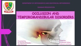
- 1. OCCLUSION AND TEMPOROMANDIBULAR DISORDERS Presented by: Dr. Deeksha Bhanotia MDS third year Guided by: Dr. Mridula Trehan Professor and Head DEPARTMENT OF ORTHODONTICS AND DENTOFACIAL ORTHOPAEDICS SEMINAR PRESENTATION
- 2. CONTENTS Introduction Anatomy Movements of TMJ TMJ examination TMJ disorders Occlusion and TMDs Orthodontic treatment and TMDs Conclusion References 2
- 3. INTRODUCTION The area where mandible articulates with the temporal bone is called the temporomandibular joint (TMJ). One of the most complex joints in the body. 3 Okeson J.: Management of Temporomandibular Disorders and Occlusion, (8th edition);5.
- 4. It provides hinging movements in one plane and therefore can be considered a Ginglymoid joint. However, at the same the time it also provides for gliding movements. Which classifies it as an arthrodial joint. Thus, it has been technically considered a Ginglymoarthrodial joint. Okeson J.: Management of Temporomandibular Disorders and Occlusion, (8th edition);5 4
- 5. Classified as compound joint as articular as articular disc acts as a non-ossified bone that permits complex movements of the joint. It is synovial type of joint. 5 Okeson J.: Management of Temporomandibular Disorders and Occlusion, (8th edition);5
- 6. Anatomy Articular surfaces: 1. Anterior Tubercle 2. Anterior part of mandibular fossa/Glenoid fossa/ Articular fossa 3. Posterior non-articular part of tympanic plate 4. The inferior articular surface formed by the head of the mandible 6 “Nothing is more fundamental to treating the patients than knowing the anatomy” - Jeffrey P. Okeson Okeson J: Management of Temporomandibular Disorders and Occlusion, (8th edition);2 Chaurasia B.D: Human anatomy volume 3, (edition 6th);121
- 7. 7
- 8. Ligaments Collateral ligament Capsular ligament Temporomandibular ligament Sphenomandibular ligament Stylomandibular ligament 8 Functional ligaments Accessory ligaments
- 9. Collateral ligaments Also called discal ligaments. Types – medial discal ligament - lateral discal ligament Attach the medial and lateral edge of the articular disc to the poles of the condyle. Divides the joint mediolaterally into the superior and inferior joint cavities. Okeson J: Management of Temporomandibular Disorders and Occlusion, (8th edition);10 9
- 10. Function: • Restricts the movement of the disc away from the condyle. • Permits the disc to be rotated anteriorly and posteriorly on the articular surface of the condyle. Vascular supply and innervation present. 10 Okeson J: Management of Temporomandibular Disorders and Occlusion, (8th edition);10
- 11. Fibrous Capsule Attached above to temporal bone along the border of articular surface of the mandibular fossa and articular eminence . Below to neck of the condyle. The lower part of the joint is surrounded by tight fibres while the upper part is surrounded by loose fibres. 11 Okeson J: Management of Temporomandibular Disorders and Occlusion, (8th edition);10 Chaurasia B.D: Human anatomy volume 3, (edition 6th);121
- 12. Function: • Resists medial, lateral, or inferior forces that to separate or dislocate articular surfaces. • Encompasses the joint, thus retaining the synovial fluid. Well innervate and provides proprioceptive feedback regarding position and movement of the joint 12 Okeson J: Management of Temporomandibular Disorders and Occlusion, (8th edition);10
- 13. Temporomandibular ligament Attached above to articular tubercle on the root of the zygomatic process of the temporal bone. It extends downwards and backwards at an angle of 45° to the horizontal, to attach to the lateral surface and posterior border of the neck of the condyle, deep to the parotid gland. 13 Chaurasia B.D: Human anatomy volume 3, (edition 6th);121
- 14. Composed of two parts: outer oblique portion and inner horizontal portion. Function: • Oblique portion- limits opening of the mouth. • Horizontal portion- limits posterior movement of the condyle and the disc. Okeson J: Management of Temporomandibular Disorders and Occlusion, (8th edition);10 14
- 15. Sphenomandibular ligament Medial to, and normally separate from, the capsule. Flat, thin band that descends from spine of sphenoid and widens as it reaches lingula of mandibular foramen. 15 Chaurasia B.D: Human anatomy volume 3, (edition 6th);121
- 16. Stylomandibular ligament Thickened band of deep cervical fascia From the apex and adjacent anterior aspect of the styloid process To the angle and posterior border of the mandible. Function- limits excessive protrusive movements of the mandible 16 Chaurasia B.D: Human anatomy volume 3, (edition 6th);122
- 17. Muscle attachments Masseter Temporalis Medial pterygoid Lateral pterygoid Digastric 17 Muscles of Mastication
- 18. Masseter Origin: Zygomatic arch and maxillary process of zygomatic bone Insertion: lateral surface of ramus of mandible 18 Drake R.L, Vogl A.W, Mitchell A.W.M :Gray’s Anatomy, (1st south Asian edition);876
- 19. Temporalis Origin: bone of temporal fossa and temporal fascia Insertion: coronoid process of mandible and anterior margin of ramus of mandible almost to last molar tooth 19 Drake R.L, Vogl A.W, Mitchell A.W.M :Gray’s Anatomy, (1st south Asian edition);876
- 20. Medial pterygoid Muscle Origin: Deep head- medial surface lateral plate of pterygoid process and pyramidal process of palatine bone Superficial head- tuberosity and pyramidal process maxilla Insertion: medial surface of mandible near angle 20 Drake R.L, Vogl A.W, Mitchell A.W.M :Gray’s Anatomy, (1st south Asian edition);876
- 21. Lateral Pterygoid Muscle Origin: Upper head- roof of infratemporal fossa Lower head-lateral surface of lateral plate of the pterygoid plate Insertion: Capsule of the TMJ in the region of attachment to the articular disc and to the pterygoid fovea on the neck of the mandible 21 Drake R.L, Vogl A.W, Mitchell A.W.M :Gray’s Anatomy, (1st south Asian edition);876
- 22. Digastric muscle 22 Origin Posterior belly: mastoid notch, just medial to the mastoid process Anterior belly: fossa on the lingual surface of the mandible, just above the lower border and close to the midline Insertion Posterior belly: fibers run forward, downward, and inward to the intermediate tendon attached to the hyoid bone. Anterior belly: fibers extend downward and backward to insert at the same intermediate tendon as does the posterior belly. Drake R.L, Vogl A.W, Mitchell A.W.M :Gray’s Anatomy, (1st south Asian edition);876
- 23. Articular Disc Shape: oval Upper surface is concavo- convex. Composed of dense connective tissue. Divides the joint into upper and lower compartments. Upper compartment – gliding movements Lower compartment – gliding+ rotatory movements 23 Chaurasia B.D: Human anatomy volume 3, (edition 6th);122
- 24. 24
- 25. In sagittal section, the disc appears to have: Anterior and posterior band A thin intermediate zone Bilaminar region 25 Chaurasia B.D: Human anatomy volume 3, (edition 6th);122
- 26. Functions of articular disc Stabilize the joint Aids in lubrication of the joint Reduces the wear 26 Drake R.L, Vogl A.W, Mitchell A.W.M :Gray’s Anatomy, (1st south Asian edition);527
- 27. Relations Lateral: skin and fascia Parotid gland Temporal branches of the facial nerve Medial: The tympanic plate separates the joint from internal carotid artery Spine of sphenoid, with the upper end of sphenomandibular ligament attached to it The auriculotemporal nerve and chorda tympani nerves Middle meningeal artery Chaurasia B.D: Human anatomy volume 3, (edition 6th);122 27
- 28. Anterior: lateral pterygoid Masseteric nerves and vessels Posterior: The parotid gland separates the gland from external auditory meatus Superficial temporal vessels Auriculotemporal nerve Chaurasia B.D: Human anatomy volume 3, (edition 6th);122 28
- 29. Inferior: Maxillary artery and vein Superior: Middle cranial fossa Middle meningeal vessels Chaurasia B.D: Human anatomy volume 3, (edition 6th);122 29
- 30. Blood supply and Innervation Blood supply: Superficial temporal artery Maxillary artery Nerve supply: Auricotemporal nerve Masseteric nerve 30 Chaurasia B.D: Human anatomy volume 3, (edition 6th);123
- 31. Movements of TMJ 31 Drake R.L, Vogl A.W, Mitchell A.W.M :Gray’s Anatomy, (1st south Asian edition);875
- 32. Lateral movements of TMJ Chewing from left side: Right lateral pterygoid Right medial pterygoid Left temporalis Left masseter Chewing from right side: Left lateral pterygoid Left medial pterygoid Right temporalis Right masseter 32 Chaurasia B.D: Human anatomy volume 3, (edition 6th;)124
- 33. 33
- 34. 34
- 35. Range of movement • The common clinical symptom associated with masticatory muscle disorders is dysfunction. • It is usually seen as a decrease in the range of mandibular movement and can be objectively measured. • Quantifying the mandibular movement is important as a record of the severity of symptoms and to show the degree of improvement. Examination of TMJ Gray et al: anatomy of TMJ- literature review (1995) 35
- 36. 36
- 37. In addition, the pathway of opening should be observed from the front for any: 1. Transient deviation -The opening pathway is straight in the beginning and then deviated to one side with maximum opening. 2. Lasting deviation - The opening pathway is straight in the beginning and followed by deviation to one side in the middle of opening but then returns to a normal midline relationship. 37 Gray et al: anatomy of TMJ- literature review (1995)
- 38. TMJ palpation 38 The TMJ can be palpated at the pre-auricular area by pressing gently over the lateral aspect of the joint. The TMJ can also be examined by intra-auricular palpation through placement of little finger in the external auditory meatus with gentle forward pressure applied. In this way both the lateral and posterior aspects of the joint can be palpated Gray et al: anatomy of TMJ- literature review (1995)
- 39. TMJ sound Pathological displacement of the disc would result in production of sound during movement. Displacement may occur due to injury to the bilaminar zone, to the disc or its attachments, or to hypertonicity in the superior head of the lateral pterygoid 39 Gray et al: anatomy of TMJ- literature review (1995)
- 40. Radiographs Radiographs are indicated only if clinical examination suggests existence of bone pathology such as erosion of condyle or fossa surfaces or presence of crepitus joint sound. All the plain TMJ radiographs have their own limitation and the information provided cannot be regarded as being conclusive. 40 Gray et al: anatomy of TMJ- literature review (1995)
- 41. Better image can be obtained by Advance imaging technique such as: 1. magnetic resonance imaging 2. computed tomographic scan 41 Gray et al: anatomy of TMJ- literature review (1995)
- 42. TMJ disorders Term suggested by Bell. Later, the term was adopted by the American Dental Association This term does not suggest merely problems that are isolated to theTMJs but includes all disturbances associated with the functional masticatory system. 42 Okeson J.: Management of Temporomandibular Disorders and Occlusion, (8th edition).
- 43. Etiology Parafunctional habits (eg, nocturnal bruxing, teeth clenching) Emotional distress Acute trauma to the jaw Trauma from hyperextension (eg, dental procedures oral intubation for general anaesthesia, yawning) Instability of maxillomandibular relationships Laxity of the joint Rheumatic or musculoskeletal disorders Poor general health and an unhealthy lifestyle 43 Greenberg, Glick, Ship: Burket’s oral medicine, (11th edition);232
- 44. 44
- 45. Hyperplasia of condyle Clinical features: Unilateral: Mandibular enlargement Facial asymmetry Shift in midline Malocclusion Bilateral: Anterior cross bite Relative microdontia Obtuse mandibular angle Ghom G.A:Textbook of oral medicine, (second edition);608 45
- 46. Radiological Features • Ramus—the vertical ramus is increased in vertical depth as well as in its anteroposterior diameter. It will result in prevention of occlusion of the posterior teeth. • Body of mandible—body of the affected side of mandible are larger as compared on unaffected side. • Condyle—the condylar enlargement is sometimes symmetrically distributed throughout the whole process. It may retain its normal shape or it may assume a conical, spherical, pear shaped or an uneven and lobulated shape. The neck of the condyle may retain its integrity, be enlarged or absorbed into the enlarged head of the condyle. • Articular eminence—the articular eminence is shallower than the opposite normal side, with the distal surface slightly evacuated. • Displacement of condyle—hyperplasia of condyle may result in displacement of condyle from the mandibular fossa. 46 Ghom G.A:Textbook of oral medicine, (second edition)
- 47. 47
- 48. Condylectomy- to improve function and esthetics Maxillary osteotomy- in cases of compensatory maxillary growth Orthodontic therapy- to treat cross bites Ghom G.A:Textbook of oral medicine, (second edition);609 48 Management
- 49. Hypoplasia of condyle Clinical features: Unilateral Appearance of face- affected side: body of mandible is short unaffected side: elongation of body of mandible Shifting on affected side Malocclusion Delayed eruption of teeth. In some cases it will cause impaction and uneruption. The external ear maybe small, deformed, partially or completely absent. Ghom G.A:Textbook of oral medicine, (second edition); 606-7 49
- 50. Bilateral Micrognathia Delayed eruption of teeth Class II malocclusion Management Surgical – directed towards increasing the length and bulk of the bone. Orthodontic treatment to correct the malocclusion. Ghom G.A:Textbook of oral medicine, (second edition);607 50
- 51. Radiological Features Condyle—condylar process is short and it tends to assume a more posterior position in the glenoid fossa. Ramus and body of mandible—there may be proportionate shortening of the ramus and body on the affected side and the bone tends to be smaller than the opposite side. A shallow sigmoid and antegonial notch is also present. • Coronoid process—coronoid process is relatively large, heavier and posteriorly directed. • Teeth—teeth may be impacted. • Ear—in some cases, there is congenital absence of the auditory canal and middle ear and the tympanic plate is poorly developed, so when the condyle is present, the articular fossa gives an appearance of an increased size. • Bilateral involvement—in cases of bilateral underdevelopment, all the above features plus bilateral antegonial notching is seen. 51 Ghom G.A:Textbook of oral medicine, (second edition)
- 52. 52
- 53. Agenesis of the condyle Clinical features: Asymmetry of face Anterior open bite Eccentric movement Altered occlusion Shift of mandible towards affected side during opening in unilateral type. Associated anomalies like under-developed ramus absent external ear. Management: Maintenance of dental health Osteoplasty Ghom G.A:Textbook of oral medicine, (second edition);607-8 53
- 54. Osteoarthritis Primary: due to wear and tear and is more common with increasing age Secondary: occurs in response to recognizable local and systemic factors Clinical features: Unilateral pain over the joint, maybe sensitive to palpation pain on movements or biting. Worsens in the evening. Deviation of jaw on the affected side Affected joint is swollen and warm and touch Stiffness of the joint Crepitations Spasm of masticatory muscles Ghom G.A:Textbook of oral medicine, (second edition);611-2 54
- 55. Radiographic Features • Location—degenerative changes located on the lateral and anterolateral wall of the fossa. • Erosion of condyle—first evidence of erosion of condyle on a radiograph occurs on an average, 6 months after the onset of TMJ pain. This will result in enlargement and shallowing of mandibular fossa. • Sclerosis—density is increased as a result of sclerosis. Small crescent-like excavation appears at the superior aspect of the condyle just behind the point of articular contact. Saucer shaped lesion—fully developed lesions are saucer shaped on PA view. This is also called as the destructive phase. • Flattened articular eminence—eminence is flattened or almost removed and anterior half of the superior convex surface of the condyle is converted into a flat plane. • Eburnation—subchondral sclerosis of the condylar head becomes more dense and more radiopaque, is sometimes referred as eburnation. • Lipping—development of lipping (shell like extension) on the anterior borders. 55 Ghom G.A:Textbook of oral medicine, (second edition)
- 56. • Osteophyte formation—little shreds of the tissue at the margins of the articular cartilage surface may undergo ossification, so that small bony outgrowths or spurs develop which are called as osteophyte (Fig. 25-12). • Beaking—extensive osteophytic formation is referred as beaking. These usually appear on the anterosuperior aspect of the condyle and lateral aspect of temporal component. In some cases, there is formation of sharp angle, either at the margins or actually on the surface of the articular process. • Joint mice—osteophytes may break off and lie within the joint space, these fragments are called as ‘joint mice’. • Loose body—osteophytes may be separated from its attachment and lie loose in the joint as a type of ‘loose body’. • Ely’s cyst (subchondral cyst) (Figs 25-13 and 25-14)— minute areas of degeneration filled with fibrous tissue are seen just below the bony surface of the condyle. The small radiolucent areas are usually less sharply defined and may have slightly irregular borders. Some are surrounded by an area of increased density, which maybe thin and well defined or relatively wide and not so sharply defined. These areas are regarded as cystic and are given the name ‘Ely’s cyst’. 56 Ghom G.A:Textbook of oral medicine, (second edition)
- 57. 57
- 58. Management Elimination of the cause Relieving the pressure on the joint Analgesic and anti-inflammatory drugs Physiotherapy Myotherapy Doxycycline Arthroscopic lavage Ghom G.A:Textbook of oral medicine, (second edition);613 58
- 59. Ankylosis Clinical features: Pain and trismus Unilateral Reduced mouth opening Asymmetry – fullness on the affected side and flattening of unaffected side Deviation of face towards affected side Shifting of the midline Cross bite Ghom G.A:Textbook of oral medicine, (second edition);621 59
- 60. Bilateral Bird face appearance- symmetrical + micrognathia No gliding movement so no protrusive or lateral movements possible Atrophy or fibrosis of muscle of mastication Class II malocclusion, protrusive incisors, anterior open bite Ghom G.A:Textbook of oral medicine, (second edition);621-4 60
- 61. Radiographic Features Fibrous ankylosis • Appearance—in some cases, there is transverse or oblique dark line, irregular in outline, crossing the mass of dense bone (Fig. 25-32). When a dark line is present, the possibility of fibrous ankylosis is more. Ramus and body of mandible—the ramus is vertical and the angle is reduced in size. The body of the jaw is short. • Condyle—the condyle may retain its normal shape, but it can be replaced by shapeless mass of bone, which finds attachment to the base of skull above and to the base of neck of condyle below. 61 Ghom G.A:Textbook of oral medicine, (second edition)
- 62. Bony ankylosis • Joint space—joint space is completely or partially obliterated with dense sclerotic bone (Fig. 25-33). Sometimes, large mass of new bone may be seen, radiographically obscuring the condyle and joint space. Antegonial notch—prominent antegonial notch on the affected side of mandible (Fig. 25-34), along with inferior arching of the mandibular body (secondary to isotonic contraction of the depressor muscles). • Condyle—bone may form around the neck of the condyle and becomes continuous with the base of the skull. Considerable destruction of bone may precede bony ankylosis. The greater part of the condyle may have been destroyed so that the sigmoid or mandibular notch is approximated to the base of the skull. The neck of the condyle, if not completely hidden by the mass of new bone, appears to be shortened; so that the mandibular notch is nearer the zygomatic process than normal. No translation of condyle head during opening. Dark linear shadow—in some cases, there is horizontal, slightly irregular, dark linear shadow in the middle of the new bone, which represents the cartilage and meniscus. • Coronoid process—there may be deepening of the notch which has escaped involvement in the new bone formation and the appearance may be accentuated by the elongation of the coronoid process, which at the same time is narrow and slender. 62 Ghom G.A:Textbook of oral medicine, (second edition)
- 63. 63
- 64. Management Brisement force- forceful opening of jaws under GA Condylectomy Gap arthroplasty 64
- 65. Disc displacement Anterior displacement with reduction Anterior displacement without reduction Ghom G.A:Textbook of oral medicine, (second edition);626 65
- 66. Anterior disc displacement with reduction Clinical features: Pain as condyle may articulate with the retrodiscal tissue Joint is tender Reciprocal click Jaw deviates towards the side of the click till click occurs and then returns to the midline Ghom G.A:Textbook of oral medicine, (second edition);628 66
- 67. Anterior disc displacement without reduction Pain Limited mouth opening Clicking with intermittent locking (locks during opening) Closed lock Limitation of lateral movement of the joint Joint tenderness Deviation towards affected side Ghom G.A:Textbook of oral medicine, (second edition);628 67
- 68. 68
- 69. 69
- 70. Radiographic Features • Plane radiography—they are not diagnostic; except in cases of perforation of the disc, where there is evidence of degenerative changes in the joint. • Arthrogram—arthrogram is useful in studying the changes. • MRI—in anterior disc displacement, posterior band of the disc located anterior to the superior portion of the condyle is seen in closed mouth sagittal image. In some cases bone marrow edema can be seen. 70 Ghom G.A:Textbook of oral medicine, (second edition)
- 71. 71
- 72. Management Bite plane appliance- positions the mandible so that mouth remains slightly open vertically and is placed anteriorly to maintain the disc normal relationship to the condyle Occlusal adjustment, restorative treatment and orthodontic treatment Surgical treatment- disc repositioning procedure, menisectomy Postoperative physiotherapy Ghom G.A:Textbook of oral medicine, (second edition);628-9 72
- 73. Condylar fracture Most common cause is blow to the chin. Unilateral Pain the affected side; it is increased when movement is attempted Paresthesia of lower lip Small localized tender swelling over the injured TMJ Bleeding from the ear on the affected side as result of laceration to external auditory meatus Ecchymosis- hematoma surrounding the fractured condyle Limited jaw movement Gagging Ghom G.A:Textbook of oral medicine, (second edition);618 73
- 74. Bilateral fracture Gagging Restricted mandibular movement (more than unilateral type) Forward displacement of the mandible Fracture dislocation Absence of condyle Unilateral Bilateral Central dislocation Ghom G.A:Textbook of oral medicine, (second edition);619 74
- 75. Radiographic Features • View taken—displaced condylar fracture is well demonstrated on AP and lateral projection. Non-displaced fracture is well seen on AP view (Fig. 25- 23). • Increased width of fracture condyle—when fracture occurs, contraction of the lateral pterygoid muscle rotates the transverse axis of the condylar head, so that the medial end moves anteriorly, thus increasing the apparent width of the fractured condyle. • Complete fracture dislocation—in complete fracture dislocation, the condylar head may be inclined medially at an approximate 45° angle to the vertical axis of the ramus. • Anterior displacement of the condyle—if the condylar head has been displaced anteriorly and turned at 90° angle to the vertical axis of the ramus as viewed from the lateral aspect, then only the articular surface of the condylar head will be seen; this will appear as a narrow radiopaque bar situated in the infratemporal fossa. • Condyle split—the condyle may split with little or no displacement of the fragments, or some portion may be separated from head of the bone. Rarely, the whole of the articular portion is crushed and flattened. 75 Ghom G.A:Textbook of oral medicine, (second edition)
- 76. CT scan—in difficult cases, CT scan has been demonstrated to show changes in the relationship of the condyle to the mandibular fossa more precisely than the conventional radiographic examination. It is also claimed that it can demonstrate fine bony alterations at the fractured site. Fracture line—the fracture line is often transverse but usually oblique, starting in the base of the mandibular notch and passing slightly or even markedly downwards. In absence of any displacement, it is difficult to visualize such fractures. In the lateral projection, there is often no evidence of any fracture line; but when the posterior margin of the ramus is followed, a sudden step is seen. In some cases there is displacement of the adjacent margins of the fragments, so that the inferior borders of the condylar fragment are superimposed over the adjacent ramus. • Articular surface—the articular surface of the condyle is usually rotated inwards with fracture dislocation. 76 Ghom G.A:Textbook of oral medicine, (second edition)
- 77. Management Simple crack fracture- heavy masticatory forces to be avoided Fracture with slight displacement- reduction and immobilization for 4 weeks Unilateral fractures with dislocation- immobilized in normal occlusion for 10 days Bilateral fractures with dislocation- arch bar is fitted and immobilization for 4 weeks in normal occlusion Ghom G.A:Textbook of oral medicine, (second edition);620 77
- 78. Neoplastic disorders Benign tumors stiffness of the joint Facial asymmetry Deviation of the mandible to the affected side Disordered occlusion Restricted movements Management Surgical excision of the tumor Orthodontic treatment to re-establish occlusion Ghom G.A:Textbook of oral medicine, (second edition);630-1 78
- 79. Malignant tumors Types – intrinsic and extrinsic Pain on opening the mouth Diminished hearing Swelling – facial asymmetry Deviation of mandible to the unaffected side Management Chemotherapy Radiotherapy Surgery combination Ghom G.A:Textbook of oral medicine, (second edition); 630-1 79
- 80. Myofacial pain disorder syndrome or TMJ dysfunction syndrome Muscle spasm, dysfunction and pain – condition is called MPDS More common in middle age Predilection for women Occurs in episodes several times a day Masticatory pain due to myalgia or arthralgia or both Ghom G.A:Textbook of oral medicine, (second edition);632-3 80
- 81. Pain in pre-auricular region but can be radiated to temporal frontal and occipital region Patient complains of tinnitus and otalgia Noise on mandibular movements Hearing loss Restricted mouth opening Restricted protrusion Soreness of muscle when palpated Myospasm In some cases myositis occurs Ghom G.A:Textbook of oral medicine, (second edition);633 81
- 82. Laskin's diagnostic criteria Four cardinal signs Unilateral pain- dull aching type which is mild in the morning worsens as the day progresses Muscle tenderness Clicking Limitation of jaw function Negative characteristics No radiographic evidence No tenderness in TMJ area Ghom G.A:Textbook of oral medicine, (second edition);633 82
- 83. Management Muscle relaxation techniques and muscle exercise Pharmacotherapy • Analgesics • Tranquilizer • Anti depressant • Sedatives and hypnotics Psychotherapy Physical medication • Hot packs • Massage • Diathermy • Electrical stimulation (TENS) Ghom G.A:Textbook of oral medicine, (second edition);634-5 83
- 84. Anesthesia is administered to the • Muscle and fascia • TMJ area (intra-capsular or extra-capsular) • Refrigerated spray – vapocoolant spray such as ethyl chloride is used reduce muscle spams by counter irritation Ghom G.A:Textbook of oral medicine, (second edition);635 84
- 85. Other therapies • Acupuncture • Hypnotherapy • Endodontic and prosthodontic therapy • Surgery- zygomectomy, eminectomy, condylectomy • Orthodontic therapy- indicated in cross bite and traumatic bite and in patients with parafunctional habits such as bruxism • Orthognathic surgery Ghom G.A:Textbook of oral medicine, (second edition);635-6,jaypee 85
- 86. OCCLUSION AND TMDs Clinicians and researchers have considered for many years occlusion as one of the major direct and/or indirect etiological factors causing temporomandibular disorders (TMDs). Dr. James Costen , an otolaryngologist in 1934, associated the development of TMJ pain and noises, as well as headache, limited mandibular opening, myofascial tenderness, and otological symptoms (summarized in the so-called “Costen's Syndrome”) to occlusal alterations and to the increase of the overbite. Thirty years later, Thompson assumed that malocclusion might cause the posterior and superior displacement of the condyle, suggesting the need to correct dental malocclusion in order to alleviate TMD symptoms. 86 Michelotti A, Rongo R, D'Antò V, Bucci R. Occlusion, orthodontics, and temporomandibular disorders: Cutting edge of the current evidence. Journal of the World Federation of Orthodontists. 2020 Oct 1;9(3):S15-8.
- 87. Transverse malocclusions Among the transverse malocclusions, unilateral posterior crossbite (UPCB) is the one most assessed in relation to TMD, and in particularly to TMJ clicking and myofascial pain. Indeed, it has been assumed that the abnormal occlusal contacts observed in patients with UPCB might affect the relationship between condyle and fossa. Moreover, the altered dental contacts between the right and the left side might create an asymmetric activation of the masticatory muscles, overloading one side over the other. 87 Michelotti A, Rongo R, D'Antò V, Bucci R. Occlusion, orthodontics, and temporomandibular disorders: Cutting edge of the current evidence. Journal of the World Federation of Orthodontists. 2020 Oct 1;9(3):S15-8.
- 88. Sagittal and vertical malocclusions Although the evidence is still scarce, it can be speculated that the relation between hyperdivergent growth pattern and TMJ disorders might be due to the early onset of the latter conditions that might bring to an abnormal development of the condyle 88 Michelotti A, Rongo R, D'Antò V, Bucci R. Occlusion, orthodontics, and temporomandibular disorders: Cutting edge of the current evidence. Journal of the World Federation of Orthodontists. 2020 Oct 1;9(3):S15-8.
- 89. Briefly, it must be considered that TMD has a multifactorial etiology, and several factors such as comorbidities, oral parafunctions, psychosocial distress, muscle overload, somatic symptoms, and genetic markers, are supported through high-quality evidence as causal factors. The role of occlusion in the etiology of TMD has not been clearly addressed and therefore should not be overstated, considering that in some cases occlusal changes could be the consequence rather than a cause for TMDs 89 Michelotti A, Rongo R, D'Antò V, Bucci R. Occlusion, orthodontics, and temporomandibular disorders: Cutting edge of the current evidence. Journal of the World Federation of Orthodontists. 2020 Oct 1;9(3):S15-8.
- 90. Orthodontic treatment and TMD It is widely believed that certain types of malocclusion may predispose patients to the development of mandibular dysfunction; Orthodontic treatment is frequently recommended to avoid such problems. However, orthodontic treatment also has been implicated as contributing to mandibular dysfunction with the possibility of the more serious sequelae of temporomandibular joint (TMJ) degeneration many years after treatment. 90 Sadowsky C and BeGole EA. Long-term status of temporomandibular joint function and functional occlusion after orthodontic treatment. Am J Orthod. 1980; 78 : 201-212.
- 91. Reynders reviewed the literature on orthodontics and temporomandibular disorders between 1966-1988 and came out with the following conclusions: (i) Unlike sample studies, viewpoint publications and case reports have little or no value in the assessment of the relationship between orthodontics and temporomandibular disorders; (ii) Sample studies demonstrated that orthodontic treatment mechanics with fixed appliances used during adolescence does not influence the risk of development of temporomandibular disorders in later life; 91 Reynders RN. Orthodontics and temporomamdibular disorders : A review of the literature (1966-1988). Am J Orthod Dentofacial Orthop. 1990; 97 : 463-71.
- 92. (iii) Longitudinal sample research showed no differences in the incidence of temporomandibular joint dysfunction among the patients treated with functional appliances (activators) without extractions as compared to patients treated with fixed orthodontic appliances and four premolar extractions; and (iv) The above findings indicate that orthodontic treatment should not be considered responsible for creating temporomandibular disorders, regardless of the orthodontic technique. They also reject the assumption that orthodontic treatment is specific or necessary to cure signs and symptoms of temporomandibular dysfunction. 92 Reynders RN. Orthodontics and temporomamdibular disorders : A review of the literature (1966-1988). Am J Orthod Dentofacial Orthop. 1990; 97 : 463-71.
- 93. Orthodontics and temporomandibular disorder: A meta-analysis In this meta-analysis, the relationship between traditional orthodontic treatment, including the specific type of appliance used and whether extractions were performed, and the prevalence of temporomandibular disorders (TMD) was investigated. After an exhaustive literature search of 960 articles, we found 31 that met the inclusion criteria (18 cross-sectional studies or surveys and 13 longitudinal studies). We divided and extracted data from the 31 articles according to study designs, symptoms, signs, or indexes. The data included in this comprehensive meta-analysis do not indicate that traditional orthodontic treatment increased the prevalence of TMD. 93 Myung-Rip Kim,Thomas M. Graber,b and Marlos A. Viana, c Kim MR, Graber TM, Viana MA. Orthodontics and temporomandibular disorder: a meta-analysis. American journal of orthodontics and dentofacial orthopedics. 2002 May 1;121(5):438-46.
- 94. Temporomandibular disorders and dental occlusion. A systematic review of association studies: end of an era? To answer a clinical research question: ‘is there any association between features of dental occlusion and temporomandibular disorders (TMD)?’ A systematic literature review was performed. Inclusion was based on: (i) the type of study, viz., clinical studies on adults assessing the association between TMD (e.g., signs, symptoms specific diagnoses) and features of dental occlusion by means of single or multiple variable analysis, and (ii) their internal validity, viz., use of clinical assessment approaches to TMD diagnosis. 94 Manfredini D, Lombardo L, Siciliani G. Temporomandibular disorders and dental occlusion. A systematic review of association studies: end of an era?. Journal of oral rehabilitation. 2017 Nov;44(11):908-23.
- 95. The search accounted for 25 papers included in the review, 10 of which with multiple variable analysis Quality assessment showed some possible shortcomings, mainly related with the unspecified representativeness of study populations. Seventeen (N = 17) articles compared TMD patients with non-TMD individuals, whilst eight papers compared the features of dental occlusion in individuals with TMD signs/symptoms and healthy subjects in non-patient populations. 95 Manfredini D, Lombardo L, Siciliani G. Temporomandibular disorders and dental occlusion. A systematic review of association studies: end of an era?. Journal of oral rehabilitation. 2017 Nov;44(11):908-23.
- 96. Findings are quite consistent towards a lack of clinically relevant association between TMD and dental occlusion. Only two (i.e., centric relation [CR]-maximum intercuspation [MI] slide and mediotrusive interferences) of the almost fort occlusion features evaluated in the various studies were associated with TMD in the majority (e.g., at least 50%) of single variable analyses in patient populations. Only mediotrusive interferences are associated with TMD in the majority of multiple variable analyses. Such association does not imply a causal relationship and may even have opposite implications than commonly believed (i.e., interferences being the result, and not the cause, of TMD). 96 Manfredini D, Lombardo L, Siciliani G. Temporomandibular disorders and dental occlusion. A systematic review of association studies: end of an era?. Journal of oral rehabilitation. 2017 Nov;44(11):908-23.
- 97. Findings support the absence of a disease specific association. Based on that, there seems to lack ground to further hypothesise a role for dental occlusion in the pathophysiology of TMD. Clinicians are encouraged to abandon the old gnathological paradigm in TMD practice. 97 Manfredini D, Lombardo L, Siciliani G. Temporomandibular disorders and dental occlusion. A systematic review of association studies: end of an era?. Journal of oral rehabilitation. 2017 Nov;44(11):908-23.
- 98. Conclusion The harmony of the interface between the teeth, muscles, nerves, supporting tissue, and temporomandibular joint all must be in balance to provide health, functional efficiency, esthetics and stability to entire stomatognathic system. 98
- 99. References Drake R.L, Vogl A.W, Mitchell A.W.M :Gray’s Anatomy, (1st south Asian edition) Michelotti A, Rongo R, D'Antò V, Bucci R. Occlusion, orthodontics, and temporomandibular disorders: Cutting edge of the current evidence. Journal of the World Federation of Orthodontists. 2020 Oct 1;9(3):S15-8. Greenberg, Glick, Ship: Burket’s oral medicine, (11th edition) Okeson J.: Management of Temporomandibular Disorders and Occlusion, (8th edition). Chaurasia B.D: Human anatomy volume 3, (edition 6th) Gray et al: anatomy of TMJ- literature review (1995) Ghom G.A:Textbook of oral medicine, (second edition);606-636 99
- 100. Sadowsky C and BeGole EA. Long-term status of temporomandibular joint function and functional occlusion after orthodontic treatment. Am J Orthod. 1980; 78 : 201-212. Reynders RN. Orthodontics and temporomamdibular disorders : A review of the literature (1966-1988). Am J Orthod Dentofacial Orthop. 1990; 97 : 463- 71. Kim MR, Graber TM, Viana MA. Orthodontics and temporomandibular disorder: a meta-analysis. American journal of orthodontics and dentofacial orthopedics. 2002 May 1;121(5):438-46. Manfredini D, Lombardo L, Siciliani G. Temporomandibular disorders and dental occlusion. A systematic review of association studies: end of an era?. Journal of oral rehabilitation. 2017 Nov;44(11):908-23. 100