1. Defining Mechanisms of Reovirus Specific IRF3 Activation
Chidinma Onyia1,2,3, Johnasha Stuart2,3, and Karl W. Boehme2,3
1Department of Biochemistry, Colorado College, Colorado Springs, CO, 80946, 2Department of Microbiology and Immunology, and
3Center for Microbial Pathogenesis and Host Inflammatory Response, University of Arkansas for Medical Sciences, Little Rock, AR 72205
Abstract
Mammalian orthoreovirus (reoviruses) are models for studying viral replication
and pathogenesis. Reoviruses are nonenveloped icosahedral viruses with a
viral genome consisting of 10 segments of double-stranded RNA. Upon entry
into a host cell, viral RNAs are detected by cellular receptors that initiate
innate immune responses, including the type 1-interferon (IFN) pathway.
Serotype 1 (T1) and serotype 3 (T3) reoviruses differ in their capacity to
activate IFN responses. T3 viruses potently induce IFN responses, whereas
T1 viruses are poor initiators of the IFN pathway. Activation of transcription
factor interferon regulatory factory 3 (IRF3) by phosphorylation is a key step
for production of interferons. Our laboratory previously showed that a T3
strain (rsT3D) induced high levels of IRF3 phosphorylation, but a T1 strain
(rsT1L) failed to elicit IRF3 phosphorylation. The mechanisms underlying the
serotype-specific differences in IFN induction are not known. In this study, we
engineered a panel of single gene rsT1L × rsT3D reassortant viruses to
identify gene segments responsible for differential IRF3 phosphorylation.
Background
Type 1-interferon host responses are critical for limiting viral replication and
spread.
Model of studies for viral replication and pathogenesis
Three serotypes: Type 1 (T1), T2, and T3
Acknowledgements
Results
I would like to thank Boehme Laboratory members Matthew Phillips and Emily
Simon. This research was supported by Center for Microbial Pathogenesis and
Host Inflammatory Response (P20 GM103625), NIAID K22 A194079, and
Summer Undergraduate Research Program (SURP) at UAMS.
Conclusions
rsT3D induces higher levels of phosphorylated IRF3 than rsT1L.
Infectivity of rsT3D and rsT1L is comparable in SVECs.
IRF3 phosphorylation correlates with rsT3D L3, S1, and S3 gene
segments.
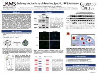
Figure 3. rsT3D induces higher
levels of phosphorylated IRF3
compared to rsT1L. SV-40
immortalized endothelial cells
(SVECs) were mock-infected (M) or
infected with rsT1L (T1) or rsT3D
(T3) at an MOI of 100 PFU/cell. At 6
hours post-Infection (HPI) western
blot analysis was done for
phosphorylated-IRF3, total IRF3,
reovirus antigen, and β-actin.
Figure 6. IRF3 phosphorylation associate with rsT3D L3, S1, and S3 gene
segments. SVECs were mock-infected (M), or infected with rsT1L (T1) or rsT3D
(T3) at an MOI of 100 PFU/cell. At 6 hours post-infection western blot analysis was
performed for phosphorylated-IRF3, total IRF3, β-actin, and reovirus antigen.
Figure 2. The reovirus virion and
activation of type-1 interferon induced
antiviral state. Upon infection viral dsRNA
is recognized by RIG-I-like receptors
(RLRs) or Toll-like-receptors (TLRs) which
induce a signaling pathway that
phosphorylates and activates interferon
regulatory factor 3 (IRF3). Phosphorylated
IRF3 translocates to the nucleus where it
induces the expression of interferons
(IFNs). IFNs binds to interferon alpha
receptor (IFNAR) to initiate a signaling
pathway that activates Signal Transducer
and Activator of Transcription (STATs)
proteins to induce the expression of
interferon stimulating genes (ISGs), which
encode proteins containing antiviral activity.
Figure 4. rsT3D and rsT1L infectivity is comparable in SVECs. SVECs were
infected with rsT1L or rsT3D at an MOI of 100 PFU/cell. At 24 hours post-infection
the cells were fixed, stained with reovirus polyclonal antiserum and visualized by
fluorescence microscopy.
Figure 7. Model for reovirus interferon induction. Reovirus attaches to and
enters the cell through endocytosis. Within the endosomes, the virus particle
dissembles and then the viral core is released into the cytosol where viral RNA is
detected by sensors of the cell. rsT3D induces higher levels of IRF3
phosphorylation compared to rsT1L. Induction of IRF3 phosphorylation correlates
with rsT3D gene segments L3, S1 and S3.
mNS
sNS
s1s
Nonstructural
Figure 1. The reovirus virion. Schematic of reovirus virion (left panel). Reovirus
particles are formed from two concentric protein shells, the outer capsid and inner
core. The core contains the viral genome, which consists of ten segments of
double-stranded RNA. Reovirus nonstructural proteins are listed to the right of the
virion schematic. Cryo-electron micrograph image reconstruction of a reovirus
virion (right panel).
6 HPI
M T1 T3
P-IRF3
IRF3
Reovirus
β-actin
N=8
T1 T3 T1 Reassortment
Figure 5. Identification of
reovirus genes responsible
for IRF3 phosphorylation. To
identify reovirus genes that
mediate serotype-specific
differences in IRF3
phosphorylation, we will use a
panel of single gene
reassortment viruses. We can
engineer viruses to contain 9
gene segments from rsT1L in
combination with a single
gene segment from rsT3D.