1. Review article
Luteinizing hormone is a primary culprit in the endometrial carcinoma
development in elderly women
C.V. Rao *
Departments of Cellular Biology and Pharmacology, Molecular and Human Genetics and Obstetrics and Gynecology, Reproduction and Development Program,
Herbert Wertheim College of Medicine, Florida International University, Miami, FL 33199, USA
1. Introduction
ECs are the most common gynecologic malignancies with a
higher incidence than ovarian and cervical cancers in Western
countries.1–3
Greater than 95% of endometrial carcinomas are
adenocarcinomas.1–3
The incidence increases with age, thus, 80% of
ECs are seen among post-menopausal women.1–3
Caucasian
women are at a greater risk than black, Hispanic, Asian and Pacific
Islanders, but black women are most likely to die from the
disease.4,5
The incidence of EC is on the rise without an increase in
survival rates during the last four decades.4–6
According to some
estimates, there were about 55,000 new cases and 10,000 died
from EC in 2015.4,5
The estimated economic impact of this
malignancy is about $2.6 billion per year.6
EC is a story of two diseases.7,8
While type 1 disease is
diagnosed in pre-menopausal women, type 2 disease primarily
occurs among post-menopausal women.7,8
The tumors from Type
1 disease are of endometriod histology, usually stages 1 or 2 and
have a favorable prognosis. The tumors from type 2 diseases, on the
other hand, have non-endometrial histology, including serous,
clear cell, mucinous and other high-grade tumors. Type 1 disease is
not usually aggressive, well differentiated, estrogen dependent,
contain estrogen, and progesterone receptors (ER and PR), slow to
spread and can be successfully treated with surgery or with
progestins.7,8
Type 2 disease, on the other hand, is aggressive,
poorly differentiated, estrogen independent, do not contain ER or
PR, vascular, spreads outside the uterus and has a poor prognosis
that requires aggressive treatment.7,8
Type 2 ECs show aneuploidy,
p53
mutations, alterations in several genes, including those
involved in cell cycle progression.9–15
Age, obesity, diabetes, reproductive and family history are some
of the risk factors for type 2 EC development.4,5,16–20
The risk is
modulated by the degree of obesity, thus body mass index has a
strong association with an increased risk.4,5,16–20
Type 2 ECs are associated with bleeding and also pelvic pain and
pressure.4,5
Definitive diagnosis is made by endometrial biopsy or
may be suspected by transvaginal ultrasound and then confirmed
by biopsy.4,5
ECs are surgically staged tumors.21
The early stages
(stages I/II) are usually curable with an excellent 5 year survival
rates.21
Stage IV disease, on the other hand, has less than 10%
survival rates at 5 years.21
Based on the scientific data, we suggest
that LH is a culprit in the type 2 EC development in elderly women.
Journal of Reproductive Health and Medicine xxx (2016) xxx–xxx
A R T I C L E I N F O
Article history:
Received 2 May 2016
Accepted 21 June 2016
Available online xxx
Keywords:
Endometrial carcinoma (EC)
type 1 and type 2 EC
Elderly women
Luteinizing hormone (LH)
LH/human chorionic gonadotropin ( hCG)
receptors
Nongonadal LH/hCG receptors
Estrogens
Gonadotropin releasing hormone analogs
A B S T R A C T
Endometrial carcinomas (ECs) are the most common gynecologic malignancies, exceeding the incidence of
ovarian and cervical cancers in elderly women (post-menopausal) in Western countries. Evidence suggests
that it is a luteinizing hormone (LH) dependent disease. ECs overexpress LH/human chorionic gonadotropin
(hCG) receptors as compared with pre and post-menopausal endometria. Activation of the LH/hCG
receptors in primary and immortalized EC cells results in an increased cell proliferation and invasion,
which are mediated by cyclic AMP(cAMP)/protein kinase A (PKA) signaling, require the presence of LH/hCG
receptors, activation of b1 integrin receptors and an increase in the secretion of metalloproteinase-2
(MMP-2) in its active form. In addition to the endometrium, LH actions in the ovaries and adrenal glands
results in an increased secretion of androgens, which are aromatized into estrogens in the adipose and EC
tissues. LH also has direct effects in the pancreas, which results in an increase in insulin secretion, which in
turn can also stimulate ovarian stromal cell proliferation, luteinization, androgens secretion and
aromatization in adipose and EC tissues. LH is further elevated in post-menopausal women who develop EC
as compared with post-menopausal women who do not develop the disease. These findings support
complex network of LH actions that promote EC development in elderly women.
ß 2016 Published by Elsevier, a division of Reed Elsevier India, Pvt. Ltd.
* Tel.: +1 3053480659.
E-mail address: crao@fiu.edu
G Model
JRHM-33; No. of Pages 7
Please cite this article in press as: Rao CV. Luteinizing hormone is a primary culprit in the endometrial carcinoma development in
elderly women, J Reprod Health Med. (2016), http://dx.doi.org/10.1016/j.jrhm.2016.06.001
Contents lists available at ScienceDirect
Journal of Reproductive Health and Medicine
journal homepage: www.elsevier.com/locate/jrhm
http://dx.doi.org/10.1016/j.jrhm.2016.06.001
2214-420X/ß 2016 Published by Elsevier, a division of Reed Elsevier India, Pvt. Ltd.
2. It works through a complex network of actions in several organs
including the EC as well as ovaries, adrenals, adipose tissue and
pancreas.
2. Evidence linking LH to EC
Several earlier studies have suggested that LH might be
involved in the development of EC in post-menopausal women,
based on the findings that circulatory LH levels were further
elevated in women who developed EC as compared to those that do
not develop the disease.22–25
This suggestion has been validated by
a study which demonstrated that ECs overexpress LH/human
chorionic gonadotropin (hCG) receptors as compared with pre- and
post-menopausal endometria.26
This study also demonstrated that
the receptor overexpression increased with the stage of the
disease.26
Subsequent studies have confirmed the receptor
presence not only in ECs, but also in primary and immortalized
EC cells.27–35
The receptor expression was higher in carcinomas
than in the surrounding microscopically normal endometrium and
closely linked to aggressive tumor behavior.30
LH was found to be
mitogenic as well as to enhance invasion in primary and
immortalized EC cells.29,31,33
The EC cells that have higher LH/
hCG receptor levels, showed a greater invasive potential when
exposed to exogenous recombinant LH.33
These actions are cAMP/
PKA mediated and require the presence of the LH receptors.31
The
invasion, which is a prerequisite for metastasis, is promoted by an
activation of b1 integrin receptors, with a subsequent increase in
metalloproteinase (MMP-2) secretion in its active form.31
The
mitogenic, invasion enhancing potential and other effects of LH/
hCG have previously been demonstrated in normal cells.36–63
These normal processes may have been amplified in EC, which is a
hallmark feature of all carcinomas.
Circulatory LH appears to drive EC pathogenesis in elderly
women. These levels (total/bioactive) are elevated up to seven fold
in the women who have developed EC than the cohorts who do not
develop EC.22,64
Therefore, LH seems to be an important factor. But
it alone may not be sufficient, as most elderly women with
elevated LH levels do not develop the disease. Therefore, genetic or
epigenetic and other inherent risk factors may also be required.
3. LH actions in EC
LH has several effects in the normal human endometrium
that are relevant to implantation of blastocyst and its
limited invasion into endometrium and pregnancy continua-
tion.39,42–44,46,49–52,54–62,65,66
When some of the normal actions are
dysregulated, the potential exists for LH to initiate malignant
changes that might lead to the development of EC. The LH actions
in normal endometrial cells fall into proliferation, invasion,
angiogenesis, and apoptosis categories.39,42–44,46,49–52,54–62,65,66
Even though, the latter two have not been demonstrated in the
context of EC development, there is a reason to believe that they
may occur. For example, type 2 ECs are highly vascular and the
increased vascularity could come from the LH, which is a
vasoactive hormone in its own right. For example, uterine
vasculature contains LH/hCG receptors and their activation results
in the formation of new blood vessels as well as dilation of the
existing ones.54,67–71
4. LH actions in ovaries, adrenals, adipose tissue, and pancreas
may contribute to the EC development
The mechanism of LH action to induce EC may also involve its
actions in the ovaries, adrenals, adipose tissue, and pancreas,
through functional LH/hCG receptors in these tissues.72–78
The ovaries of post-menopausal women actively secrete
androgens from the stromal cell compartment and LH can
stimulate this secretion.79–95
The ovaries of women with EC are
even more active in androgens secretion than cohorts without
EC.85,89,92–94,96
The increased secretion comes from hyperplasia
and luteinization of stromal cells and greater elevation of LH
levels.22,63,64,96–99
The role of the adrenal glands in EC development is related to
an increase in LH levels, which can stimulate zona fasciculate to
secrete androgens. In fact, (a) age associated increase in LH levels
correlate with an increased adrenal function in post-menopausal
women,100–103
(b) hCG challenge increases adrenal androgens
secretion in older female macaques104
and finally (c) hCG can
stimulate androgens secretion from human adrenal cortical
cells.75
Adipose tissue involvement in EC pathogenesis is related to an
increased aromatization of androgens from adrenals and ovaries,
which is perhaps under LH and/or insulin control. However, there
is no evidence yet for the LH control, but this may not be a far-
fetched possibility, considering that it contributes to the
aromatase regulation in ovaries. Insulin, on the other hand,
seems to be able to regulate aromatase in fat tissue.105
Activation
of LH/hCG receptors has been shown to increase cell proliferation,
differentiation, and leptin secretion from preadipocytes.76
These
actions are mediated by cAMP/PKA independent mitogen
activated protein kinase yet (MAPK)/c-fos signaling.76
Whether
or how these LH actions could contribute to EC pathogenesis is not
known.
The involvement of the pancreas in EC development in post-
menopausal women is related to the hyperinsulinemia, a known
risk factor in type 2 ECs.4,5,106
In fact, EC patients often have
elevated insulin levels and an increased insulin resistance.106
The
higher insulin levels could come from LH stimulation of b-cells of
pancreas.77
However, it is not known whether LH can also
contribute to increased insulin resistance. Nevertheless, the
increased insulin levels can stimulate ovarian stromal cells
proliferation, luteinization, secretion of androgens, and their
aromatization in EC tissue.106–111
Post-menopausal women regardless of EC have elevated
androgen levels.84,92–95,100–102
These elevated levels come from
a secretion from adrenals as well as ovaries, both of which are
stimulated by LH.72,103,104
The androgens are then converted to
estrogens in adipose and endometrial tissues.111–114
This
conversion is increased in post-menopausal women who
develop EC, as compared to those who do not develop the
disease.109,112
These increases likely come from LH and insulin
stimulation because their levels are elevated during EC
pathogenesis64,106
and the tissues themselves contain their
receptors.64,105–107,110,115,116
In fact, insulin can regulate
aromatase in endometrial and adipose tissues.105,111
Whether
LH is also involved is not known. But it is not a far-fetched
possibility. Estrogens formed from androgens result in only a
small increase in circulation, perhaps due to differences in
metabolic clearance rates, conversions to other steroids and the
level and binding capacity of sex steroid binding globulin
(SHBG).92
The increased aromatization in EC tissue and potential
further stimulation by LH and/or insulin could result in a
high local estrogen concentration in the tumor microenviron-
ment. The role of these estrogens is not known, but they are not
likely to induce type 2 EC because the tumors do not contain
ER.1–3
What roles do these estrogens play, remains to be
investigated.
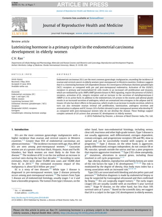
Fig. 1 presents the proposed model on how LH can induce type
2 EC in elderly women. Future research will undoubtedly bring
several modifications to this model.
C.V. Rao / Journal of Reproductive Health and Medicine xxx (2016) xxx–xxx2
G Model
JRHM-33; No. of Pages 7
Please cite this article in press as: Rao CV. Luteinizing hormone is a primary culprit in the endometrial carcinoma development in
elderly women, J Reprod Health Med. (2016), http://dx.doi.org/10.1016/j.jrhm.2016.06.001
3. 5. Estrogens versus LH
Estrogens are believed to be culprits in type I EC development
through the mitogenic actions.117,118
These actions are antago-
nized by progesterone, which promotes the differentiation of
endometrial epithelial cells.119
Thus, women who regularly
ovulate and produce progesterone rarely get EC.119
When the
cyclicity ceases and the balance tips in favor of estrogens, then
endometrial cells can continue to proliferate unabated, leading to
EC.119
This tipping can also happen in women who take only
estrogen containing birth control pills.120
The antagonism can also
explain why progesterone therapy works for type 1 ECs.119
Estrogens are not likely to be the culprits in type 2 EC
development because their circulating levels are very low and
there are no ER in the tumor. Conversely, LH are not likely to be the
culprits in type I EC development, because its levels are low, except
during a brief periovulatory period. However, LH/hCG receptors,
which are functionally coupled to physiological responses that are
required for pregnancy initiation and maintenance, are present in
endometrium.39,42–44,46,49–52,54–62,65,66
It is only when LH levels
are chronically elevated, followed by some type of dysregulation
which results in the receptor overexpression and perhaps post-
receptor changes, then the LH actions become relevant in type 2 EC
development. The molecular details of this dysregulation remain to
be investigated.
Pre-menopausal women with polycystic ovarian syndrome
(PCOS) have a 3-fold increase in EC risk, which is impacted by
the degree of obesity.121–128
These women have a higher total/
bioactive LH and androgen levels, increased insulin resistance
and low circulating estrogens, especially if they are not
ovulating.129–132
ECs arise from complex or atypical endometrial
hyperplasias in these women.28,123
Both simple and complex
hyperplasias contain higher LH/hCG receptors levels than
normal endometrium and the levels further increase from
simple to atypical hyperplasias.28
It is entirely possible,
therefore, that LH could also be a culprit in the EC development
in PCOS women.
There are two other hyperandrogenic conditions in which EC
risk also increases. One is hyperthecosis and the other is androgen
producing ovarian tumors.133,134
Insulin levels are elevated and
insulin resistance increases in these women.135
Even though ECs of
these women have not been investigated for the presence of LH/
hCG receptors, it is within the realm of possibility that LH could
also be a culprit in EC development in these pre-menopausal
women.
Even though hCG levels, surrogate for LH, are elevated during
pregnancy, they are not likely to cause EC. Quite to the contrary,
the life-time EC risk decreases with each pregnancy.136
This
pregnancy induced protection comes from progesterone, which
induces cell differentiation. Its levels are rather high to begin with
and they keep increasing to even higher levels during the second
half of pregnancy.
Setiwan et al. have suggested that the classification of ECs
requires a change due to a considerable overlap in risk factors for
type 1 and type 2 diseases.137
Moreover, not every women will
have exactly the same time course of hormonal changes as they
approach menopause and beyond. Therefore, we recommend
reclassification based on LH dependency. In the reclassification,
type 1 ECs are LH independent and type 2 ECs are LH dependent.
Estrogens will have different roles in both the diseases. In LH
independent disease, estrogens can initiate the disease through
their mitogenic effects. In LH dependent disease, estrogens have a
secondary role of increasing LH release from anterior pituitary
gland. In addition, elevation of free estrogens, due to a decrease in
SHBG in post-menopausal obese woman, can serve as a further
powerful stimulus for bioactive LH release.138
The same scenario
applies to pre-menopausal obese women with polycystic ovarian
disease.129
In both cases, obesity will be the primary trigger in
inducing the cascade of hormonal changes that are ultimately
responsible for EC development. Obesity is an important health
concern that costs the U.S. economy approximately $69 billion a
year.139
It increases an individual’s risk for many diseases,
including EC and several other forms of cancer.15,139
However,
not all post-menopausal obese women are likely to develop EC, due
Hypothalamus
Anterior
OvariesAdipose
Ɵssue
PancreasAdrenals
Endometrial
carcinoma
GnRH
LH
LH
LH
LH
Androgens Androgens
AndrogensAndrogens Estrogens Insulin
Insulin
LH
Fig. 1. Proposed model of LH induced type 2 endometrial carcinoma in elderly women. In this model, LH is a primary instigator. Besides direct actions in the endometrial
carcinoma tissue, its works through a number of other organ systems and all of which bear down on EC to drive the disease process. The molecular details of many of these
steps are unknown.
C.V. Rao / Journal of Reproductive Health and Medicine xxx (2016) xxx–xxx 3
G Model
JRHM-33; No. of Pages 7
Please cite this article in press as: Rao CV. Luteinizing hormone is a primary culprit in the endometrial carcinoma development in
elderly women, J Reprod Health Med. (2016), http://dx.doi.org/10.1016/j.jrhm.2016.06.001
4. to a lack of strong co-existing genetic predisposition and other
inherent risk factors. On the other hand, some post-menopausal
lean women may develop EC, because of the presence of a strong
genetic predisposition and/or the presence of other inherent risk
factors. Therefore, obesity and consequent elevation of LH levels
are important and can only predispose women to develop EC, when
genetic, environmental, life style and reproductive risk factors co-
exist. This reasoning should neither come as a surprise nor solely
applicable to EC. It is also important to point out that non-
hysterectomized post-menopausal women, regardless of obesity,
who take estrogen replacement therapy for the control of their
menopausal symptoms, are at an increased risk to develop EC, due
to incessant stimulation.118,120
6. Mechanism of LH actions in EC
The following mechanisms of action can be envisioned from the
known LH actions in EC, normal endometrial epithelial and in other
cells.36–63
1. LH binds to its cell surface receptors to activate them.
2. The activation results in the generation of second messengers
such as, cAMP/PKA, PKC, MAPK, b catenin/Wnt, and a cross talk
between them.
3. The second messengers can then regulate cyclins, cyclin
dependent protein kinases, b1 integrin receptors, active
metalloproteinase-2 secretion, pro and anti-inflammatory,
and apoptotic molecules, etc.
4. The receptor activation may also lead to other changes such as
the secretion of cytokines, growth factors, and eicosanoids.
5. LH may also modulate the immunity by regulating the immune
cells trafficking and their cytokines secretion in ECs.
All LH actions can be classified into non-genomic as well as
genomic. In both cases, initial cell surface receptor binding of LH is
necessary. After the binding, the non-genomic actions such as,
second messengers’ generation, activation/inactivation of kinases,
phosphorylation/dephosphorylation of proteins, ion flux changes,
etc. will commence. The non-genomic actions will be rapid and
may be required for sustaining the slow genomic actions. Non-
genomic changes can help in the genomic actions of LH, through
phosphorylation/dephosphorylation of transcription factors, their
nuclear import, subsequent binding to cis-acting elements, etc. The
genomic actions can involve up or down regulation of many genes
in the families of cell cycle, cell invasion, growth factors,
oncogenes, tumor suppressors, apoptosis inhibitory, and multitude
of others, whose identity remains unknown.
7. Need for further research
There is an obvious need for a great deal of further research for a
better understanding of the LH actions in type 2 EC development.
This research could focus on answering the following interrelated
questions.
1. What are the triggering factors for the LH/hCG receptor
overexpression in ECs?
2. What are the cellular, genetic and biochemical mechanisms that
LH uses to increase the cell proliferation, invasion, etc. in ECs?
3. Can LH induce EC pathogenesis in the absence of aromatizable
androgens from ovaries and adrenals or their aromatization in
fat and EC tissues?
4. Does LH upregulate aromatase and/or its catalytic activity in
adipose and EC tissues?
5. Can LH also increase the insulin resistance in obese EC patients?
6. How important are the insulin actions in ovarian stroma and in
EC for the disease development?
7. Can LH regulate immune cells migration and their secretion of
cytokines, chemokines, etc. in ECs?
The answers will enrich our understanding of complex basic
cellular, molecular, biochemical and genetic mechanisms that LH
employs to induce EC development. Such an understanding could
provide discovery path for novel therapeutic targets in ECs.
8. Therapeutic possibilities
When an elevated circulatory LH levels are the culprits in type
2 EC development, then the obvious treatment approach will be to
reduce the levels, which can be accomplished by treatment with
gonadotropin releasing hormone analogs (GnRHa). There are a
number of conflicting reports, however, on the success of GnRHa
treatment.140–148
The reported treatment failures could come from
the advanced disease stage, incomplete EC dependence on LH, low
LH/hCG receptor expression, etc. Complicating the interpretation
of the results are the findings that ECs contain GnRH receptors,
which could mediate the direct inhibitory effect of GnRH in
ECs.149–152
However, there is a report showing that GnRH induced
growth inhibition of EC cells does not require its receptors.146
ECs
seem to produce small amounts of hCG and how this production
impacts the EC development and/or its response to GnRHa
treatment remains unknown.34,153
Future therapies worth explor-
ing include, local delivery of pharmacologic LH/hCG receptor
inhibitors, receptor gene silencers, etc.
Conflicts of interest
The author has none to declare.
References
1. Rose PG. Endometrial carcinoma. N Engl J Med. 1996;335:640–649.
2. Wingo PA, Ries LAG, giovino GA, et al. Annual report to the nation on the status of
cancer, 1973–1996. J Natl Cancer Inst (Bethesda). 1999;91:675–690.
3. Amant F, Moerman P, Neven P, Timmerman D, Van Limbergen E, Vergote I.
Endometrial cancer. Lancet. 2005;366:491–505.
4. Endometrial Cancer. What are the Key Statistics About Endometrial Cancer?Amer-
ican Cancer Society; 2015. Date published 12.01.15, www.cancer.org/cancer/
endometrialcancer/detailedguide/endometrial-uterine-cancer-keystatistics
[accessed 01.07.15, date revised: 17.03.15]
5. Basic Information About Uterine Cancer, Uterine Cancer Fact Sheet. Center for
Disease Control and Prevention; 2015. http://www.cdc.gov/cancer/uterine/
basic_info/index.htm [accessed October 7]
6. Society of Gynecologic Oncology. Creating a New Parradigm in Gynecologic Cancer
Care: Policy Proposals for Delivery, Quality and Reimbursement. Society of Gyneco-
logic Oncology White, paper published year: 2013; 2015. https://www.sgo.org/
wp-content/uploads/2012/09/Practice_Summit_Report_FINAL.pdf [accessed Oc-
tober]
7. Bokhman JV. Two pathogenetic types of endometrial carcinoma. Gynecol Oncol.
1983;15:10–17.
8. Deligdisch L, Holinka CF. Endometrial carcinoma: two diseases? Cancer Detect
Prev. 1987;10:237–246.
9. Kacinski BM, Carter D, Mittal K, et al. High level expression of fms proto-oncogene
mRNA is observed in clinically aggressive human endometrial adenocarcinomas.
In J Radiat Oncol Biol Physiol. 1988;15:823–829.
10. Kacinski BM, Chambers SK, Stanley ER, et al. The cytokine CSF-1 (M-CSF)
expressed by endometrial carcinomas, in vivo and in vitro may also be a
circulating tumor marker of neoplastic disease activity in endometrial carcinoma
patients. In J Radiat Oncol Biol Physiol. 1990;19:619–626.
11. Borst MP, baker VV, Dixon D, Hatch KD, Shingleton HM, Miller DM. Oncogene
alterations in endometrial carcinoma. Gynecol Oncol. 1990;38:364–366.
12. Gurpide E. Endometrial cancer: biochemical and clinical correlates. J Natl Cancer
Inst. 1991;83:405–416.
13. Konishi I, Koshiyama M, Mandai M, et al. Sex steroid receptors, LH/hCG receptor
and oncogene expression in endometrial carcinomas. In: Genazzani AR, Petraglia
F, D’Ambrogio G, Genazzani AD, Artini PG, eds. In: Recent Developments in
Gynecology and Obstetrics. London: The Pathenon Publishing Group; 1995:
715–719.
C.V. Rao / Journal of Reproductive Health and Medicine xxx (2016) xxx–xxx4
G Model
JRHM-33; No. of Pages 7
Please cite this article in press as: Rao CV. Luteinizing hormone is a primary culprit in the endometrial carcinoma development in
elderly women, J Reprod Health Med. (2016), http://dx.doi.org/10.1016/j.jrhm.2016.06.001
5. 14. Abal M, Planaguma J, Gil-Moreno A, et al. Molecular pathology of endometrial
carcinoma: transcriptional signature in endometrioid tumors. Histol Histopathol.
2006;21:197–204.
15. Hecht JL, Mutter GL. Molecular and pathologic aspects of endometrial carcino-
genesis. J Clin Oncol. 2006;24:4783–4791.
16. Goodman MT, Hankin JH, Wilkens LR, et al. Diet, body size, physical activity, and
the risk of endometrial cancer. Cancer Res. 1997;57:5077–5085.
17. Zamboni M, Mazzali G, Zoico E, et al. Health consequences of obesity in
the elderly: a review of four unresolved questions. Int J Obesity. 2005;29:
1011–1029.
18. Conroy MB, Sattelmair JR, Cook NR, Manson JE, Buring JE, Lee IM. Physical
activity, adiposity, and risk of endometrial cancer. Cancer Causes Control.
2009;20:1107–1115.
19. Dal Maso L, Tavani A, Zucchetto A, et al. Anthropometric measures at different
ages and endometrial cancer risk. Br J Cancer. 2011;104:1207–1213.
20. Jenabi E, Poorolajal J. The effect of body mass index on endometrial cancer: a
meta-analysis. Public Health. 2015;1–9. http://dx.doi.org/10.1016/j.puhe.2015.
04.017.
21. Endometrial Cancer. How is Endometrial Cancer Staged?American Cancer Society;
2015. Date published 12.01.15, www.cancer.org/cancer/endometrialcancer/
detailedguide/endometrial-uterine-cancer-staging [accessed 01.07.15, revised
17.03.15]
22. Sherman AI, Wolf RB. An endocrine basis for endometrial carcinoma. Am J Obstet
Gynecol. 1959;77:233–242.
23. Varga A, Henriksen E. Urinary excretion assays of pituitary luteinizing hormone
(LH) related to endometrial carcinoma. Obstet Gynecol. 1963;22:129–136.
24. Dilman VM, Bernstein LM, Bobrov YF, Bohman YU, Kovaleva IG, Krylova NV.
Hypothalamopituitary hyperactivity and endometrial carcinoma. Am J Obstet
Gynecol. 1968;102:880–889.
25. Dilman VM, Goluber VN, Krylova NV. Dissociation of hormonal, antigenic acitivity
of luteinizing hormone excreted in endometrial carcinoma patients. Am J Obstet
Gynecol. 1973;115:966–971.
26. Lin J, Lei ZM, Lojun S, Rao Ch.., Satyaswaroop PG, Day Jr TG. Increased expression
of luteinizing hormone/human chorionic gonadotropin receptor gene in human
endometrial carcinomas. J Clin Endocrinol Metab. 1994;79:1483–1491.
27. Bax CMR, Chatzaki E, Davies S, Gallagher CJ. Elucidating the role of gonadotropins
in endometrial cancer cell growth. Biochem Soc Trans. 1996;24:443S.
28. Konishi I, Koshiyama M, Mandai M, et al. Increased expression of LH/hCG
receptors in endometrial hyperplasia and carcinoma in anovulatory women.
Gynecol Oncol. 1997;65:273–280.
29. Davies S, Bax CMR, Chatzaki E, Chard T, Iles RK. Regulation of endometrial cancer
cell growth by luteinizing hormone (LH) and follicle stimulating hormone (FSH).
Br J Cancer. 2000;83:1730–1734.
30. Ji Q, Chen P, Aoyoma C, Lui P. Increased expression of human luteinizing hormone/
human chorionic gonadotropin receptor mRNA in human endometrial cancer.
Mol Cell Probes. 2002;16:269–275.
31. Dabizzi S, Noci I, Borri P, et al. Luteinizing hormone increases human endometrial
cancer cells invasiveness through activation of protein kinase A. Cancer Res.
2003;63:4281–4286.
32. Viswanath G, Chatterjee S, Roy P. Assessment of luteinizing hormone receptor
function in an endometrial cancer cell line. Ishikawa cells in response to human
chorionic gonadotrophin (hCG). Mol Cell Endocrinol. 2007;272:14–21.
33. Noci I, Pillozzi S, Lastraioli E, et al. hLH/hCG-receptor expression correlates with in
vitro invasiveness in human primary endometrial cancer. Gynecol Oncol. 2008;
111:496–501.
34. Jankowska AG, Andrusiewicz M, Fischer N, Warchol JB. Expression of hCG and
GnRHs and their receptors in endometrial carcinoma and hyperplasia. Int J
Gynecol Cancer. 2010;20:92–101.
35. Arcangeli A, Noci I, Fortunato A, Scarselli GF. The LH/hCG axis in endometrial
cancer: a new target in the treatment of recurrent or metastatic disease. Obstet
Gynecol Int. 2010;1–5.
36. Teodorczyk-Injeyan JA, Kellen JA. Chorionic gonadotropin-induced cell prolifera-
tion and polyclonal immunoglobulin synthesis in human mononuclear cells.
Biomed Pharmacother. 1988;42(1):49–53.
37. Lei ZM, Rao Ch.., Lincoln S, Ackermann DM. Increased expression of human
chorionic gonadotropin/human luteinizing hormone receptors in adenomyosis.
J Clin Endocrinol Metab. 1993;76:763–768.
38. Kornyei JL, Lei ZM, Rao CV. Human myometrial smooth muscle cells are novel
targets of direct regulation by human chorionic gonadotropin. Biol Reprod.
1993;49(6):1149–1157.
39. Tang B, Gurpide E. Direct effect of gonadotropin on decidualization of human
endometrial stroma cells. J Steroid Biochem Mol Biol. 1993;47:115–121.
40. Eta E, Ambrus G, Rao CV. Direct regulation of human myometrial contractions by
human chorionic gonadotropin. J Clin Endocrinol Metab. 1994;79:1582–1586.
41. Ambrus G, Rao CV. Novel regulation of pregnant human myometrial smooth
muscle cell gap junctions by human chorionic gonadotropin. Endocrinology.
1994;135:2772–2779.
42. Han SW, Lei ZM, Rao CV. Up-regulation of cyclooxygenase-2 gene expression by
chorionic gonadotropin during the differentiation of human endometrial stromal
cells into decidua. Endocrinology. 1996;137:1791–1797.
43. Han SW, Lei ZM, Rao CV. Treatment of human endometrial stromal cells with
chorionic gonadotropin induces their mosphological and functional differentia-
tion into decidua. Mol Cell Endocrinol. 1999;147:7–16.
44. Zhou XL, Lei ZM, Rao CV. Treatment of human endometrial gland epithelial cells
with chorionic gonadotropin/luteinizing hormone increases the expression of
cyclooxygenase-2 gene. J Clin Endocrinol Metab. 1999;84:3364–3377.
45. Lei ZM, Taylor DD, Gercel-Taylor C, Rao CV. Human chorionic gonadotropin
promotes tumorigenesis of choriocarcinoma Jar cells. Placenta. 1999;20:147–159.
46. Uzumcu M, Coskun S, Jaroudi K, Hollanders JMG. Effect of human chorionic
gonadotropin on cytokine production from human endometrial cells in vitro.
Am J Reprod Immunol. 2000;40:83–88.
47. Horiuchi A, Nikaido T, Yoshizawa T, et al. HCG promotes proliferation of uterine
leiomyomal cells more strongly than that of myometrial smooth muscle cells in
vitro. Mol Human Reprod. 2000;6:523–528.
48. Salvador LM, Maizels E, Hales DB, Miyamoto E, Yamamoto H, Hunzicker-Dunn M.
Acute signaling by the LH receptor is independent of protein kinase C activation.
Endocrinology. 2002;143:2986–2994.
49. Srisuparap S, Strakova Z, Brudney A, et al. Signal transduction pathway activated
by chorionic gonadotropin in the primate endometrial epithelial cells. Biol Reprod.
2003;65:457–464.
50. Kayisli UA, Selam B, Guzeloglu-Kayisli O, Demir R, Arici A. Human chorionic
gonadotropin contributes to maternal immunotolerance and endometrial apo-
ptosis by regulating Fas-Fas ligand system. J Immunol. 2003;171:2305–2313.
51. Perrier d’Hauterive S, Charlet-Renard C, berndt S, et al. Human chorionic gonado-
tropin and growth factors at the embryonic endometrial interface control lue-
kemia inhibitory factor (LIF) and interleukin 6 (IL-6) secretion by human
endometrial epithelium. Hum Reprod. 2004;19:2633–2643.
52. Akoum A, Metz CN, Morin M. Marked increase in marcophage migration inhibi-
tory factor syntheisis and secretion in human endometrial cells in response
to human chorionic gonadotropin hormone. J Clin Endocrinol Metab. 2005;90:
2904–2910.
53. Hamada AL, Nakabayashi K, Sato A, et al. Transfection of antisense chorionic
gonadotropin b gene into choriocarcinoma cells suppresses the cell proliferation
and induces apoptosis. J Clin Endocrinol Metab. 2005;90:4873–4879.
54. Berndt S, d’Hauterive SP, Blacher S, et al. Angiogenic activity of human chorionic
gonadotropin through LH receptor activation on endothelial and epithelial cells of
the endometrium. FASEB J. 2006;20:E2189–E2198.
55. Herrmann-Lavoie C, Rao CV, Akoum A. Chorionic gonadotropin down-regulates
the expression of the decoy inhibitory interleukin 1 receptor type II in human
endometrial epithelial cells. Endocrinology. 2007;148:5377–5384.
56. Fluhr H, Carli S, Deperschmidt M, Wallwiener D, Zygmunt M, Licht P. Differential
effects of human chorionic goandotropin and decidualization on insulin-like
growth factors-I and II in human endometrial stromal cells. Fertil Steril. 2008;
90:1384–1389.
57. Sengupta S, Sengupta J, Mittal S, Kumar S, Ghosh D. Effect of human chorionic
gonadotropin (hCG) on expression of vascular endothelial growth factor A (VEGF-
A) in human mid-secretory endometrial cells in three-dimensional primary
culture. Indian J Physiol Pharmacol. 2008;52:19–30.
58. Berndt S, Blacher S, d’Hauterive SP, et al. Chorionic gonadotropin stimulation
of angiogenesis and pericyte recruitment. J Clin Endocrinol Metab. 2009;94:
4567–4574.
59. Paiva P, Hannan NJ, Hincks C, et al. Human chorionic gonadotropin regulates FGF2
and other cytokines produced by human endometrial epithelial cells, providing
a mechanism for enhancing endometrial receptivity. Hum Reprod. 2011;26:
1153–1162.
60. Kajihara T, Uchino S, Suzuki M, Itakura A, Brosens JJ, Ishihara O. Human chorionic
gonadotropin confers resistance to oxidative stress-induced apoptosis in decid-
ualizing human endometrial stromal cells. Fertil Steril. 2011;95:1302–1307.
61. Bourdiec A, Shao R, Rao CV, Akoum A. Human chorionic gonadotropin triggers
angiogenesis via the modulation of endometrial stromal cell responsiveness to
interleukin 1: a new possible mechanism underlying embryo implantation. Biol
Reprod. 2012;87:64829.
62. Bourdiec A, Calvo E, Rao CV, Akoum A. Transcriptome analysis reveals new
insights into the modulation of endometrial stromal cell receptive phenotype
by embryo-derived signals interleukin-1 and human chorionic gonadotrop pos-
sible involvement in early embryo implantation. PLOS ONE. 2013;8:e64829.
63. So KH, Kodithywakky SP, Kottawatta KS, et al. Human chorionic gonadotropin
stimulates spheroid attachment of fallopian tube epithelial cells through the
mitogen-activated protein kinase pathway and down-regulation of olfactome-
din-1. Fertil Steril. 2015;104:474–482.
64. Nagamani M, Doherty MG, Smith ER, Yallampalli C. Increased bioactive luteiniz-
ing hormone levels in postmenopausal women with endometrial cancer. Am J
Obstet Gynecol. 1992;167:1825–1830.
65. Rao CV. Novel concepts in neuroendocrine regulation of reproductive tract
functions. In: Bazer FW, ed. In: Endocrinology of Pregnancy. Totowa, NJ: The Human
Press Inc.; 1998:125–144.
66. Licht P, Fluhr H, Neuwinger J, Wallwiener D, Wildt L. Is human chorionic
gonadotropin directly involved in the regulation of human implantation? Mol
Cell Endocrinol. 2007;269:85–92.
67. Lei ZM, Reshef E, Rao Ch.. The expression of human chorionic gonadotropin/
luteinizing hormone receptors in human endometrial and myometrial blood
vessels. J Clin Endocrinol Metab. 1992;75:651–659.
68. Toth P, Li X, Rao Ch.., et al. Expression of functional human chorionic gonadotro-
pin/human luteinizing hormone receptor gene in human uterine arteries. J Clin
Endocrinol Metab. 1994;79:307–315.
69. Toth P, Lukacs H, Gimes G, et al. Clinical importance of vascular LH/hCG receptors
– a review. Reprod Biol. 2001;1:5–11.
70. Zygmunt M, Herr F, Keller-Schoenwetter S, et al. Charactization of human
chorionic gonadotropin as a novel angiogenic factor. J Clin Endocrinolo Metab.
2002;87:5290–5296.
71. Bourdiec A, Bedard D, Rao CV, Akoum A. Human chorionic gonadotropin
regulates endothelial cell responsiveness to interleukin 1 and amplifies the
C.V. Rao / Journal of Reproductive Health and Medicine xxx (2016) xxx–xxx 5
G Model
JRHM-33; No. of Pages 7
Please cite this article in press as: Rao CV. Luteinizing hormone is a primary culprit in the endometrial carcinoma development in
elderly women, J Reprod Health Med. (2016), http://dx.doi.org/10.1016/j.jrhm.2016.06.001
6. cytokine-mediated effect on cell proliferation, migration and the release of
angiogenic factors. Am J Reprod Immunol. 2013;70(2):127–138.
72. Peluso JJ, Steger RW, Lasyczak S, Hafez ESE. Gonadotropin binding sites in human
postmenopausal ovaries. Fertil Steril. 1976;27:789–795.
73. Nakano R, Shina K, Yarnoto M, Kobayashi M, Nishimori K, Hiraoka JI. Binding sites
for gonadotropins in human postmenopausal ovaries. Obstet Gynecol. 1989;73:
196–200.
74. Pabon J, Li X, Lei Z, Safilippo J, Yussman M, Rao CV. Novel presence of luteinizing
hormone/chorionic gonadotropin receptors in human adrenal glands. J Clin
Endocrinol Metab. 1996;81:2397–2400.
75. Rao CV, Zhou XL, Lei ZM. Functional luteinizing hormone/chorionic gonadotropin
receptors in human adrenal cortical H295R cells. Biol Reprod. 2004;71:579–587.
76. Dos Santo E, Dieudonne M-N, Leneveu M-C, Pacquery R, Serazin V, Giudicelli Y. In
vitro effects of chorionic gonadotropin hormone on human adipose development.
J Endocrinol. 2007;194:313–325.
77. Parkash J, Lei ZM, Rao CV. The presence of human chorionic gonadotropin/
luteinizing hormone receptors in pacreatic beta-cells. Reprod Sci. 2015;22:
1000–1007.
78. Lasley BL, Conley AJ, Morrison JH, Gee NA, Rao CV. Identification of immunoreac-
tive luteinizing hormone receptors in the adrenal cortex of the female rhesus
macaque. Reprod Sci. 2016;23:524–530.
79. Woll E, Hertig AT, Smoth GV, Johnson LC. The ovary in endometrial carcinoma
with notes on morphological histology of the aging ovary. Am J Obstet Gynecol.
1948;56:617–633.
80. Novak ER, Mohler DE. Ovarian changes in endometrial cancer. Am J Obstet Gynecol.
1953;65:1099–1110.
81. Novak ER, Goldberg B, Jones SG, O’Toole RV. Enzyme histochemistry of the
menopausal ovary associated with normal and abnormal endometrium. Am J
Obstet Gynecol. 1965;93:669–682.
82. Poliak A, Jones GES, Goldberg B, Soloman D, Woodruff ID. Effect of human
chorionic gonadotropin on postmenopausal women. Am J Obstet Gynecol. 1968;
101:731–739.
83. Judd HL, Judd GE, Lucas WE, Yen SSC. Endocrine function of the postmenopausal
ovary. Concentration of androgens and estrogens in ovarian and peripheral vein
blood. J Clin Endocrinol Metab. 1974;39:1020–1024.
84. Judd HL, Judd GE, Lucas WE, Yen SSC. Endocrine function of the postmenopausal
ovary: concentration of androgens and estrogens in ovarian and peripheral vein
blood. J Clin Endocrinol Metab. 1974;39:1020–1024.
85. Rizkallah TH, Tovell HMM, Kelly WG. Production of estrone and fractional
conversion of circulating androstenedione to estrone in women with endometrial
carcinoma. J Clin Endocrinol Metab. 1975;40:1045–1056.
86. Judd HL, Lucas WE, Yen SSC. Serum 17b-estradiol and estrone levels in postmen-
opausal women with and without endometrial cancer. J Clin Endocrinol Metab.
1976;43:272–278.
87. Vermeulen A. The hormonal activity of the postmenopausal ovary. J Clin Endo-
crinol Metab. 1976;42:247–253.
88. Greenblatt RB, Colle ML, Mahesh VB. Ovarian and adrenal steroid production in
postmenopausal women. Obstet Gynecol. 1976;47:383–387.
89. Judd HL, Davidson BJ, Freeman AM, Shamonki MI, Lagasse LD, Ballon SC. Serum
androgens and estrogens in postmenopausal women with and without endome-
trial cancer. Am J Obstet Gynecol. 1980;136:859–871.
90. Longcope C, Hunter R, Franz C. Steroid secretion by the postmenopausal ovary. Am
J Obstet Gynecol. 1980;138:564–568.
91. Dennefors BL, Janson PO, Knutson F, Hamberger L. Steroid production and
responsiveness to gonadotropin in isolated stromal tissue of human postmeno-
pausal ovaries. Am J Obstet Gynecol. 1980;136:997–1002.
92. Nagamani M, Hannigan EV, Dillard Jr EA, Dinh TV. Ovarian steroid secretion in
postmenopausal women with and without endometrial cancer. J Clin Endocrinol
Metab. 1986;62:508–512.
93. Nagamani M, Stuart CA, Doherty MG. Increased steroid production by the ovarian
stromal tissue of postmenopausal women with endometrial cancer. J Clin Endo-
crinol Metab. 1992;74:172–176.
94. Adashi EY. The climacteric ovary as a functional gonadotropin-driven androgen-
producing gland. Fertil Steril. 1994;62:20–27.
95. Jongen VHWM, Sluijmer AV, Heineman MJ. The post-menopausal ovary as an
androgen-producing gland: hypothesis on the etiology of endometrial cancer.
Maturitas. 2002;43:77–85.
96. Sommers SC, Meissner WA. Endocrine abnormalities accompanying human
endometrial cancer. Cancer. 1957;10:516–521.
97. Marcus CC. Ovarian cortical stromal hyperplasia and cancer of the endometrium.
Obstet Gynecol. 1963;21:175–186.
98. Fienberg R. The stromal theca cell and postmenopausal endometrial carcinoma.
Cancer. 1969;24:32–38.
99. Jongen VHWM, Thijssen JHH, Santena JG, Van der Zee AGJ, Heineman MJ.
Endometrioid endometrial cancer, ovarian stromal hyperplasia and steroid pro-
duction. Br J Obstet Gynaecol. 2003;110(2):690–695.
100. Lasley BL, Santoro N, Rnadolph J, et al. The relationship of circulating dehydro-
epiandrosterone, testosterone, and estradiol to stages of the menopausal transi-
tion and ethnicity. J Clin Endocrinol Metab. 2002;87:3760–3767.
101. Crawford S, Santoro N, Laughlin GA, et al. Circulating dehydroepiandrosterone
sulfate concentrations during the menopausal transition. J Clin Endocrinol Metab.
2009;94:2945–2951.
102. Lasley BL, Stanczyk FX, Gee NA, et al. Androstenediol complements estradiol
during the menopausal transition. Menopause. 2012;19:650–657.
103. Saxena AR, Seely EW. Luteinizing hormone correlates with adrenal function in
postmenopausal women. Menopause. 2012;19:1280–1283.
104. Moran F, Chen J, Lohstroh PN, Gee NA, Lasley BL. Dehydroepiandrosterone sulfate
(DHEAS) levels reflect endogenous LH production and response to human chori-
onic gonadotropin (hCG) challenge in the older female macaque (Macaca fasci-
cularis). Menopause. 2013;20:329–335.
105. McTernan PG, Anderson LA, Anwar AJ, et al. Glucocorticoid regulation of P450
aromatase activity in human adipose tissue: gender and site differences. J Clin
Endocrinol Metab. 2002;87:1327–1336.
106. Nagamani M, Hannigan EV, Dinh VT, Stuart CA. Hyperinsulinemia and stromal
luteinizing of the ovaries in postmenopausal women with endometrial cancer. J
Clin Endocrinol Metab. 1988;67:144–148.
107. Poretsky L, Grigorescuf F, Seibel M, Moses AC, Flier JS. Distribution and charac-
terization of insulin and insulin-like growth factor I receptors in normal human
ovary. J Clin Endocrinol Metab. 1985;61:728–734.
108. Tseng L, Mazella J, Funt MI, Mann WJ, Stone ML. Preliminary studies of aromatase
in human neoplastic endometrium. Obstet Gynecol. 1984;63:150–154.
109. Tseng L, Mozella J, Mann WT, Chumas J. Estrogen synthesis in normal and
malignant human endometrium. J Clin Endocrinol Metab. 1982;55:1029–1031.
110. Barbieri RL, makris A, Randall WR, Daniels G, Kistner RW, Ryan KJ. Insulin
stimulates androgen accumulation in incubation of ovarian stroma from women
with hyperandrogenism. J Clin Endocrinol Metab. 1986;62:904–910.
111. Randolph JF, Kipersztok S, Ayers JWT, Ansbacker R, Peegal H, Menon KMJ. The
effect of insulin on aromatase activity in isolated human endometrial glands and
stroma. Am J Obstet Gynecol. 1987;157:1534–1539.
112. Schindler AE, Ebert A, Friedrich E. Conversion of androstenedione to estrone by
human fat tissue. J Clin Endocr Metab. 1972;35:627–630.
113. MacDonald PC, Edman CD, Hernsell DI, Porter JC, Siiteri PK. Effect of obesity on
plasma androstenedione to estrone in postmenopausal women with and without
endometrial cancer. Am J Obstet Gynecol. 1978;130:448–455.
114. Ackerman GE, Smoth ME, mendelson CR, MacDonald PC, Simpson ER. Aromati-
zation of androstenedione by human adipose tissue stromal cells in monolayer
culture. J Clin Endocrinol Metab. 1981;53:412–417.
115. Cuatrecasas P. Insulin-receptor interactions in adipose tissue cells: direct mea-
surement and properties. Proc Natl Acad Sci USA. 1971;68:1264–1268.
116. Sheets EE, Tsibris JCM, Cook NI, Virgin ST, DeMay RM, Spellacy WN. In vitro
binding of insulin and epidermal growth factor to human endometrium and
endocervix. Am J Obstet Gynecol. 1985;153:60–65.
117. Lucas WE. Casual relationships between endocrine-metabolic variables in
patients with endometrial carcinomas. Obstet Gynecol Surv. 1974;29:507–528.
118. Pike MC, Peters RK, Cozen W, et al. Estrogen-progestin replacement therapy and
endometrial cancer. J Natl Cancer Inst. 1997;89:1110–1111.
119. Yang S, Thiel KW, Leslie KK. Progesterone: the ultimate endometrial tumor
suppressor. Trends Endocrinol Metab. 2011;22:145–152.
120. Hammond CB, Jelovsek FR, Lee KL, Creasman WT, Parker RT. Effects of long
term estrogen replacement therapy: neoplasia. Am J Obstet Gynecol. 1979;133:
537–547.
121. Jackson RL, Dockerty MD. The Stein Leventhal Syndrome: analysis of 43 cases
with special reference to association with endometrial cancer. Am J Obstet
Gynecol. 1957;73:161–173.
122. Fechner RE, Kaufman R. Endometrial adenocarcinoma in Stein-Leventhal Syn-
drome. Cancer. 1974;34:444–452.
123. Wood GP, Boronow RC. Endometrial adenocarcinoma and the polycystic ovary
syndrome. Am J Obstet Gynecol. 1976;124:140–142.
124. Chittenden BG, Fullerton G, Maheshwari A, Bhattacharya S. Polycystic ovary
syndrome and the risk of gynaecological cancer: a systematic review. Reprod
Biomed Online. 2009;19:398–405.
125. Fauser BC, Tarlatzis BC, Rabar RW, et al. Consensus on women’s health aspects of
polycystic ovary syndrome (PCOS): the Amsterdam ESHRE/ASRM-Sponsored 3rd
PCOS Consensus Workshop Group. Fertil Steril. 2012;97:28–38.
126. Haoula Z, Salman M, Atiomo W. Evaluating the association between endometrial
cancer and polycystic ovary syndrome. Hum Reprod. 2012;27:1327–1331.
127. Dumesic DA, Lobo RA. Cancer risk and PCOS. Steroids. 2013;78:782–785.
128. Hart R, Doherty DA. The potential implications of a PCOS diagnosis on a woman’s
long-term health using data linkage. J Clin Endocrinol Metab. 2015;100:911–919.
129. Lobo RA, Granger L, Gobelsmann U, Mishell DR. Elevation of unbound serum
estradiol as a possible mechanism for inappropriate gonadotropin secretion in
women with PCO. J Clin Endocrinol Metab. 1981;52:156–158.
130. Lobo RA, Kletzky OA, Campeau J, diZerega G. Elevated bioactive luteinizing
hormone in women with polycystic ovary syndrome. Fertil Steril. 1983;39:674.
131. Lobo RA, Shoupe D, Chang PS, Campeau J. The control of bioactive luteinizing
hormone secretion in women with polycystic ovary syndrome. Am J Obstet
Gynecol. 1984;148:423–428.
132. Shoupe D, Kumar DD, Lobo RA. Insulin resistance in polycystic ovarian syndrome.
Am J Obstet Gynecol. 1983;147:588–592.
133. Aiman J, Edman CD, Worley RJ, Vellios F, MacDonald PC. Andorgen and estrogen
formation in women with ovarian hyperthecosis. Obstet Gynecol. 1978;51:1–9.
134. Mohammed CN, Cardena A, Villasanta U, Toker C, Ances IF. Hilus cell tumor of the
ovary and endometrial carcinoma. Obstet Gynecol. 1978;52:486–490.
135. Nagamani M, Dinh TV, Kelver ME. Hyperinsulinemia in hyperthecosis of the
ovaries. Am J Obstet Gynecol. 1986;154:384–389.
136. Jongen VHWM, Hollema H, Van der Zee AGJ, Heineman MJ. Aromatase in the
context of breast and endometrial cancer. Min Endocrinol. 2006;31:47–60.
137. Setiawan VW, Yang HP, Pike MC, et al. Type I and II endometrial cancers: have
they different risk factors? J Clin Oncol. 2013;31:2607–2618.
138. Urban RJ, Veldhuis JD, Dufau LM. Estrogen regulates the gonadotropin-releasing
hormone-stimulated secretion of biologically active luteinizing hormone. J Clin
Endocrinol Metab. 1991;72:660–668.
C.V. Rao / Journal of Reproductive Health and Medicine xxx (2016) xxx–xxx6
G Model
JRHM-33; No. of Pages 7
Please cite this article in press as: Rao CV. Luteinizing hormone is a primary culprit in the endometrial carcinoma development in
elderly women, J Reprod Health Med. (2016), http://dx.doi.org/10.1016/j.jrhm.2016.06.001
7. 139. Wang YC, Pamplin J, Long MW, Ward ZJ, Gortmaker SL, Andreyeva T. Severe
obesity in adults cost state medicaid programs nearly $8 Billion in 2013. Health Aff
(Millwood). 2015;34:1923–1931.
140. Gallagher CJ, Oliver RT, Oram DH, et al. A new treatment for endometrial cancer
with gonadotrophin releasing-hormone analogue. Br J Obstet Gynaecol. 1991;98:
1037–1041.
141. Kleinman D, Douvdevani A, Schally AV, Levy J, Sharoni Y. Direct growth inhibition
of human endometrial cancer cells by the gonadotropin-releasing hormone
antagonist SB-75: role of apoptosis. Am J Obstet Gynecol. 1994;170:96–102.
142. Jeyarajah AR, Gallagher CJ, Blake PR, et al. Long-term follow-up of gonadotrophin-
releasing hormone analog treatment for recurrent endometrial cancer. Gynecol
Oncol. 1996;63:47–52.
143. Covens A, Thomas G, Shaw P, et al. A phase II study of leuprolide in advanced/
recurrent endometrial cancer. Gynecol Oncol. 1997;64:126–129.
144. Markman M, Kennedy A, Webster K, Peterson G, Kulp B, Belinson J. Leuprolide in
the treatment of endometrial cancer. Gynecol Oncol. 1997;66:542.
145. Lhomme C, Vennin P, Callet N, et al. A multicenter phase II study with
triptorelin (sustained-release LHRH agonist) in advanced or recurrent endo-
metrial carcinoma: a French anticancer federation study. Gynecol Oncol. 1999;
75:187–193.
146. Noci I, Coronnello M, Borri P, et al. Inhibitory effect of luteinizing hormone-
releasing hormone analogues on human endometrial cancer in vitro. Cancer Lett.
2000;150:71–78.
147. Noci I, Borri P, Bonfirraro G, et al. Long standing survival without cancer
progression in a patient affected by endometrial carcinoma treated primarily
with leuprolide. Br J Cancer. 2001;85:333–336.
148. Asbury RF, Brunetto VL, Lee RB, Reid G, Rovereto TF, Gynecologic Oncology Group.
Goserelin acetate as treatment for recurrent endometrial carcinoma: a gyneco-
logic oncology group study. Am J Clin Oncol. 2002;25:557–560.
149. Srkalovic G, Wittliff JL, Schally AV. Detection and partial characterization of
receptors for [D-trp6]-luteinizing hormone-releasing hormone and epidermal
growth factor in human endometrial carcinoma. Cancer Res. 1990;50:1841–1846.
150. Pahwa GS, Kullander S, Volimer G, Oberheuser F, Knuppen R, Emons G. Specific
low affinity binding sites for gonadotropin-releasing hormone in human endo-
metrial carcinoma. Eur J Obstet Gynecol Reprod Biol. 1991;41:135–142.
151. Emons G, Schroder B, Ortmann O, Westphalen S, Schulz K-D, Schally AV. High
affinity binding and direct ant proliferative effects of luteinizing hormone-
releasing hormone analogs in human endometrial cancer cell lines. J Clin Endo-
crinol Metab. 1993;77:1454–1464.
152. Chatzaki E, Bax CMR, Eidne KA, Anderson L, Grudzinskas JG, Gallagher CJ. The
expression of gonadotropin releasing hormone and its receptor in endometrial
cancer and its relevance as an autocrine growth factor. Cancer Res. 1996;
56:2059–2065.
153. Nowak-Markwitz E, Jankowska A, Szcerba A, Andrusiewicz M. Human chorionic
gonadotropin-beta in endometrium cancer tissue. Eur J Gynaecol Oncol. 2004;25:
251–254.
C.V. Rao / Journal of Reproductive Health and Medicine xxx (2016) xxx–xxx 7
G Model
JRHM-33; No. of Pages 7
Please cite this article in press as: Rao CV. Luteinizing hormone is a primary culprit in the endometrial carcinoma development in
elderly women, J Reprod Health Med. (2016), http://dx.doi.org/10.1016/j.jrhm.2016.06.001