SlideShare a Scribd company logo
1 of 68
Download to read offline
AUTOStroke solution
As part of our Automation Platform offering, we’ve created an innovative solution that helps
optimize treatment outcomes for stroke patients when speed and accuracy are everything.
Non-contrast CT
Intracranial Hemorrhage
ASPECTS CT Large Vessel Occlusion CT Brain Perfusion maps
Automation Platform is an AI-based, zero-click solution that uses deep learning technology to
streamline your workflow for fast, actionable results every time. From scanner to clinical decision,
you’ll be supported by leading-edge deep learning technologies that process and deliver images
for accurate triage, worklist prioritization and treatment decisions.
For more information, contact your local sales representative or visit our website:
https://eu.medical.canon/products/healthcare_it/automation-platform
The right insights. Accelerated by AI.
Automation Platform
https://eu.medical.canon
MAGAZINE
FOR
HEALTH
PROFESSIONALS
//
European
Edition
//
No
37
//
September
2021
MAGAZINE FOR HEALTH PROFESSIONALS
European Edition //No 37 //September 2021
Diving into the
Future of CT with
Deap Learning
Spectral Imaging
18 //CT
Providing a Hub
to Advance
Interventional
Radiology
12 //HIT
Supporting
Advances in
Gynecology
Oncology
23 //UL
A Seamless
Transition
that Boosts
Productivity
38 //MR
Cover Image:
Adobe Stockphoto, edited
with Canon Medical’s
clinical images.
VISIONS magazine is a publication of Canon Medical Europe and is offered free of charge to health professionals.
The magazine is published twice a year. Registration to access full, previously published, digital editions can be
done via the website: https://eu.medical.canon/visions-magazine. Canon Medical stores and uses personal data of
the registration to send out the magazine and inform members about new developments. Members can customize
preferences or opt-out, after registration.
VISIONS magazine is covering Canon Medical’s European region and as such reflects products, technologies and
services for this particular area. The mentioned products may not be available in other geographic regions.
Please consult your Canon Medical representative sales office in case of any questions.
No part of this publication may be reproduced in whole or in part, stored in an automated storage and retrieval
system or transmitted in any manner whatsoever without written permission of the publisher. The opinions
expressed in this publication are solely those of the authors and not necessarily those of Canon Medical.
Canon Medical does not guarantee the accuracy or reliability of the information provided herein.
News, articles and the full edition of VISIONS magazine are announced firstly, as pre-publication, via the dedicated
VISIONS LinkedIn Group: https://www.linkedin.com/groups/3698045. In this group you can actively participate in
discussions about the content and future direction of the magazine.
Alphenix, Aquilion, Aquilion ONE, Aquilion ONE GENESIS, Aquilion ONE PRISM, Aquilion Precision, Astelion,
Aquilion Lightning, Vantage Galan, Vantage Elan, Vantage Orian,Vantage Titan, Pianissimo, Aplio, Xario and
Made for Life are trademarks of Canon Medical Systems Corporation.
Xephilio is a trademark of Canon Inc.
Vitrea is a trademark of Vital Images, Inc.
Vital Images, Inc., will change its company name to Canon Medical Informatics, Inc., in October 2021. This document was
created prior to the name change and therefore uses the former company name.
Publisher
Canon Medical Systems Europe B.V.
Zilverstraat 1, 2718 RP Zoetermeer
The Netherlands
+31 79 368 92 22
W: https://eu.medical.canon/
E: visions.eu@eu.medical.canon
Editor-in-chief
Jacqueline de Graaf
jacqueline.degraaf@eu.medical.canon
Design & Layout
Boerma Reclame
boermareclame.com
Printmanagement
Printweb Media B.V.
printweb.nl
Photography
Cojan van Toor
www.cojanvantoor.nl
Text contributions and editing
Mélisande Rouger - Independent Journalist
Sara Sharp - The Creative Practice
Follow us:
© 2021 by Canon. All rights reserved. ISSN 1617-2876
Works are generally classified into two categories: Full length articles (e.g. clinical added value of new/special
applications & technologies) and Short contributions (e.g. system testimonials, case reports, technical notes).
General guidelines for authors
C
o
p
y
ri
g
h
t
©
2
0
1
5
S
o
c
ia
l
M
e
d
ia
E
x
a
m
in
e
r®
All articles should be double-spaced.
Full length articles
Full length articles should generally include the following:
- Author’s full name and highest academic degree, employer
medical institution
- Author’s biography (150 words)
- Author’s passport-size photograph (suitable for publication);
(image of 300 dpi)
- 200-word abstract
- Text including headline, sub-title, introduction and sections like:
materials  methods (which should include a full description of
equipment used), results, discussion and references
- Text approx 4 to 5 pages or 12.000 to 14.000 characters (not
including figures, tables and photographs)
- Correspondence address
- Literature (no more than 10 references)
- Separate, continuous numbered image- and table captions
Short contributions
Short contributions should generally include the following:
- Author’s full name and highest academic degree
- Author’s employer medical institution
- Author’s biography (150 words)
- Author’s passport-size photograph (suitable for publication);
(image of 300 dpi)
- Text including headline, sub-title, introduction and full
description of methods  materials/equipment used
- Case Report or description of system improvements (Technical
Notes)
- Correspondence address
- Literature (no more than 10 references)
- Separate, continuous numbered image- and table captions
Text
The article should be saved in Microsoft Word (PC format) if
possible, and, if not, in text only.
Please indicate the software program and version used (Microsoft
Word 2007, etc.) and whether it is a PC or Macintosh formatted
document. If e-mailing, make sure to send it as an attachment,
rather than embedded in the e-mail message.
Symbols, formulas and abbreviations
Symbols, Greek letters superscripts/Subscripts must to be
identified clearly. Furthermore, the figure 1 (one) and the letter l (el)
as well as the capital letter 0 and the figure 0 (zero) should be easy
to differentiate.
All abbreviations including units of measure, chemical names,
technical or medical acronyms, names of organisations or
institutions should be defined when they first appear in the text
(e.g. congestive heart failure (CHF). Please refrain from using
unfamiliar abbreviations, clinical slang or jargon.
Images, art and tables
Cite all figures and tables in text, preferably in consecutive order.
Please include a caption for each figure. All captions for each figure,
should be separate from the text, at the end of the manuscript on a
separate page. Captions should avoid duplication of text material.
Credit lines for artwork can appear at the end of the corresponding
caption by stating: (Provided by first initial, last name). Black out (or
give clear instructions which parts should be blackened out) of the
images to not violate any data protection regulations (e.g. patient
data)
Do not embed figures, charts, or graphs into your document file.
Please provide them as a separate file, as well as hard copy/correct
.pdf file. Please use one the following formats: EPS , TIFF or JPEG.
Arrows stuck onto images for purposes of delineation should be
clearly visible and reproducible.
Authors should indicate if they would like to have artwork returned.
Each table should have a title, and all abbreviations should be
spelled out or explained in a footnote.
Style
Title page should include full names, degrees and titles of authors,
and affiliations (name of institution, city and state) for use in a by-
line, as well as phone and fax numbers to facilitate sending edited
copy back to author for approval.
Define all symbols, abbreviations and acronyms on first reference.
All manuscripts should be written in a third-person style, unless the
article is specifically an editorial or first-hand review.
References
A maximum of 10 references is suggested. Complete references
should be listed in order of citation in text, NOT alphabetically.
Up to four authors will be listed; if there are five or more authors,
only the first three will be listed, followed by et al. Within the text,
reference numbers should appear as footnotes in parentheses or
in superscript text at the end of each appropriate citation. Please
do not use Microsoft Words endnote feature, as this causes major
problems in the editing phase.
In addition, if the reference is not in English, please indicate the
language of publication.
Journal example
Oberhaensli RD, Galloway GJ, Hilton-Jones D, et al. The study of
human organs by phosphorus-31 topical magnetic resonance
spectroscopy. Br J Radiol 1987;60:367-373
Book example
Welch KMA, Barkley GL. Biochemistry and pharmacology of
cerebral ischemia. In: Barnett JHM, Stein BM, Mohr JP, Yatsu FM,
eds. Stroke: pathophysiology, diagnosis and management. New
York: Churchill Livingstone, 1986;75-90.
VISIONS 37 // 3
JOS RUIS
Chief Operating Officer
CANON Medical Systems Europe
With kind regards,
//EDITORIAL
© 2021 CANON MEDICAL SYSTEMS
Dear Reader,
With COVID-19 measures increasingly being eased in many European countries, we are now
entering a ‘back to a new normal’ era.
Over the past year, our lives changed suddenly and drastically. Virtual interactions largely
replaced face-to-face contact, and words, like eConsultation, Zoom and Microsoft Teams
became prominent in our vocabulary.
Some of these changes made us realize that we can work (more) efficiently in these new ways;
we must keep those changes as part of our ‘new normal’ way of working.
Some others, we will gladly say “goodbye” to. Social isolation, for example, does not work well
for us, as a herd animal species.
‘Back to a new normal’ also comes with plenty of catching up. Catching up on overdue visits to
those close to us, catching up on missed holidays, catching up on intentions, tasks, appoint-
ments, interventions, diagnosis, treatments etc., all of which have been delayed due to COVID-
19 priorities, and, of course, catching up on the latest and greatest in our professional fields.
Our new professional life will likely be one of less travelling, a balanced mix of face-to-face and
electronic business- and social interactions, an improved and continuous preparedness for the
unexpected, to name just a few elements.
From our side, we are looking forward to meeting you again in person, in your professional
environments, and during congresses and exhibitions. We have many things to share, of which
some are covered in this new edition of our VISIONS magazine.
In the meantime I wish you well. Remain vigilant and stay safe.
4 // VISIONS 37
23
Supporting
Advances in
Gynecologic
Oncology
ULTRASOUND
26
 Taking a Qualitative
Leap Forwards
MULTI-MODALITY
//CONTENTS
03	Editorial
06	News
11	 President's Message
12	
Providing a Hub to Advance
Interventional Radiology
	 HEALTHCARE IT
18	
Diving into the Future of
CT with Deep Learning
Spectral Imaging
	 COMPUTED TOMOGRAPHY
23	
Supporting Advances in
Gynecologic Oncology
	 ULTRASOUND
26	
Taking a Qualitative
Leap Forwards
	 MULTI-MODALITY
30	
A Significant Investment
in Canon Medical CT
	 COMPUTED TOMOGRAPHY
38	
A Seamless Transition that
Boosts Productivity
	 MAGNETIC RESONANCE
42	
How Deep Learning
Reconstruction Became
the Standard
	 COMPUTED TOMOGRAPHY
06
NEWS
(Canon
Webinars
2021)
12
Providing a
Hub to Advance
Interventional
Radiology
HEALTHCARE IT
VISIONS 37 // 5
56
Offering a Wealth
of Unexplored
Possibilities
EYE CARE
62
How Hygienic Positioning
Aids are Simplifying
Work Processes during
the Pandemic
COMPUTED TOMOGRAPHY
42
How Deep Learning
Reconstruction
Became the Standard
COMPUTED TOMOGRAPHY
© 2021 CANON MEDICAL SYSTEMS
48	
Digital Efficiencies in
the COVID-19 Pandemic
	 DIAGNOSTIC X-RAY
53	Maximum Efficiency,
Prioritized Patient Safety
and Increased Revenue
with Canon Medical’s
4D CT Technology
	 INTERVENTIONAL X-RAY
56	
Offering a Wealth of
Unexplored Possibilities
	 EYE CARE
60	Deep Learning
Reconstruction –
a Game Changer in
CT Imaging
	 COMPUTED TOMOGRAPHY
62	
How Hygienic Positioning
Aids are Simplifying
Work Processes during
the Pandemic
	 COMPUTED TOMOGRAPHY
6 // VISIONS 37
//NEWS
Canon Medical Systems Corporation has recently announced a major operational plan to
strengthen its healthcare information technology (HIT) division.
In a world, in which data is a business’s most valuable asset,
there simply isn’t room for silos of any kind. Cohesion, flow,
and integration are the watchwords of our time, which is
why it should be no surprise to anyone familiar with Canon
Medical that they have recently announced a major opera-
tional plan to strengthen their HIT division.
The entire reform, which is expected to take some time
to complete, will see Vital Images, Inc. adopt the Canon
Medical brand to support a more unified business approach.
The consolidated business unit will leverage Canon Medical’s
global infrastructure to accelerate the delivery of multiple
Enterprise, AI, and Collaborative imaging solutions.
“This development represents a bold new future for our
company, customers and partners,” says Toshio Takiguchi,
CEO and President of Canon Medical Systems Corporation.
“Enhancing our HIT solution and service provision is the
Canon Medical Solidifies HIT as a Key Growth Pillar
natural next step in a world that demands accurate and
immediate results beyond our traditional modality-centered
business model.”
Jim Litterer, President and CEO of Vital Images, Inc. echoed
these sentiments adding: “Canon Medical has a well-es-
tablished reputation for delivering leading solutions that
support the end-to-end needs of the evolving healthcare
systems. I am confident that customers around the globe can
look forward to enhanced service delivery, uncompromised
quality, and an innovative approach to healthcare – which is
why I am excited to strengthen the HIT Division, leveraging
the global reach of Canon Medical.”
Vital Images, Inc. will be fully integrated into the global Canon
Medical brand from October 1, 2021. To find out more about
our HIT solutions, visit https://eu.medical.canon/products/
healthcare_it or contact your local sales representative. //
VISIONS 37 // 7
© 2021 CANON MEDICAL SYSTEMS
Visit our website or scan
the QR code:
https://eu.medical.canon/
➔ Events Calender ➔ Webinars
➔ Past Webinars ➔ WHC - Ultrasound
Last May 2021, Canon Medical hosted
the webinar ‘Fetal Neurosonography
– three essential talks to boost your
confidence.’
Fetal Neurosonography is very
important as malformations of central
nervous system (CNS) are common
and can often have a poor neurological
outcome. As the fetal brain undergoes
tremendous development during
pregnancy it can be challenging in
differential diagnosis of pathologies
and often depend on examiner skills.
The main technique for the diagnosis
of fetal CNS anomalies is ultrasound.
In specific cases, the use of MRI plays a
role as well.
In 2020, it became obvious that
holding face-to-face fetal cardiology
courses would be impossible due to the
COVID-19 pandemic. Therefore, the
collaboration between Canon Medical
and the Evelina London Children’s
Hospital plus King’s College Hospital,
London, UK, was translated into the
comprehensive online course “State
of the Heart in Fetal Cardiology”. The
course comprised of eight two-hour
sessions, run every two weeks from
January to May 2021.
The course covered all major facets
of fetal cardiology, including normal
cardiac patterns, features of major
groups of structural abnormalities,
and fetal arrhythmias. Some areas of
practice have undergone significant
advances, including first trimester
scanning, diagnosis of fetal vascular
Webinar Fetal Neurosonography
Worldwide Online Course ‘State of the Heart in Fetal Cardiology’ was a Success!
Our excellent well-known speakers
Prof Laurent Guibaud (Hospital
Femme Mère Enfant - University
Claude Bernard Lyon/France), Prof Luc
de Catte (University Hospital Leuven/
Belgium) and Dr Miguel Branco
(Bissaya Barreto Coimbra Hospital and
University Center/Portugal) explained
a detailed multiplanar neurosonogra-
phy examination, how to differentiate
cystic brain lesions and the fetal
cortex development in normal and
abnormal cases.
This successful webinar was intended
to boost confidence and with attendees
from 86 countries all over the world
joining we saw the high demand for
rings and the introduction of motion-
corrected fetal cardiac MRI, which
has been pioneered at the Hospital.
Novel ultrasound imaging modalities,
including Superb Micro-vascular
Imaging have had a significant impact
in the first trimester and in diagnosis
of vascular rings.
With numbers capped at 200 to permit
meaningful interaction with the audi-
ence, delegates registered from all over
the world and could interact live with
the panel for each session.
“Prenatal detection and accurate
diagnosis of congenital heart disease
is essential, because of its potential
profound impact,” said Professor John
Simpson, Professor of Pediatric and
Fetal Cardiology at Evelina London
Children’s Hospital. “We have been
running foundation and advanced
courses in fetal cardiology together
with Canon Medical for over 10 years.
The online course has enabled us to
continue this vital education despite
the COVID-19 pandemic.”
Feedback from the course has been
extremely positive, with respect to
content, image quality and the faculty-
audience discussions. The demand for
the course exceeded all expectations
and we believe this approach will con-
tinue to be an effective complemen-
tary means of delivering education,
even when things return to a more
‘normal’ situation. //
supporting our customers with high-
level education not only performing
daily routine ultrasound, but detailed
neurosonography scan to improve
scanning skills and confidence in the
final diagnosis. //
Dr. Miguel
Branco
Prof. John
Simpson
Dr. Owen
Miller
Dr. Vita
Zidere
Dr. Kuberan
Pushparajah
Dr. Thomas
Day
Prof. Gurleen
Sharland
Dr. Marietta
Charakida
Dr. Trisha
Vigneswaran
Dr. David F.A.
Lloyd
Dr. Ioana
Dumitrascu-Biris
Prof. Laurent
Guibaud
Prof. Luc
de Catte
8 // VISIONS 37
//NEWS
July
Eye Care Webinar (Italian)
Dr. Barbara
Parolini
Eye Care Webinar
Presentato dai seguenti Relatori
Dr. Barbara Parolini Prof. Enzo M. Vingolo
Interpretazione Clinica
su OCT e OCTA nella
Pratica Quotidiana
15 Luglio, 2021 | 19:00-20:00 CET
August 24, 25, 26 -
September 1,2
Online
Neurology
Days
Dr. Mahtab
Zamani
Prof. Neil D.
Pugh
Dr. Grant
Mair
Dr. Anton
Meijer
Dr. Joseph
Puig
Dr. Nevia
Caputo
Prof. Thomas
Tourdias
Dr. Alexandra
Borchert
Prof. Nens
van Alfen
Dr. Benoît Doche
de Laquintane
Prof. Bart van
Wijmeersch
Prof. Adnan
Siddiqui
Dr. Andrés
González Mandly
Canon Webinars 2021
Learn from the experts and gain new insights
In online educational webinars, leading experts share their expertise and exchange
knowledge. Register to join one of our upcoming webinars or visit our website to
watch the past recorded webinars.
Visit our website
https://eu.medical.canon/
events_calendar/webinars
Or scan the
QR code for the
complete online
overview.
November 9	 Ultrasound (MSK)
November 18	 Eye Care OCT (Dutch)
September 30	 Eye Care OCT
The clinical benefits of scanning wider and deeper in the retina.
October 26, 27, 28 - 	 Online Cardiology Days
November 3, 4
	
Day 1 - Oct 26	 | Cardiovascular disease - New Imaging Approaches | Eye Care/MR/CT
Day 2 - Oct 27	 | Cardiovascular disease - Complex Imaging ​Procedures | MR/CT/HIT/VL
Day 3 - Oct 28	 | Structural Heart Disease | UL/CT/VL
Day 4 - Nov 3	 | Pediatric Cardiology | UL/CT/VL
Day 5 - Nov 4	 | Sports Cardiology | UL/VL
VISIONS 37 // 9
© 2021 CANON MEDICAL SYSTEMS
Lars
Lindgren
May
Seminario web Ecografía (Spanish)
Dr. Pedro Mora
Sanz
Dra. Gloria Ruiz
Fernández
Dr. Antonio
Olveira Martín
Dra. Marta
Abadía Barnó
Seminario web Ecografía
Presentado por los siguientes ponentes
Dra.GloriaRuizFernández
Dra.MartaAbadíaBarnó
Ecografía Avanzada en la
Valoración del Daño Hepático
13 de Mayo, 2021 | 19:00-20:00 CET
Dr. Pedro Mora Sanz Dr.AntonioOlveiraMartín
June
Echografie / MRI Webinar (Dutch)
Dr. Bas
Maresch
Dr. Jan
Veryser
Dr. Kurt
Vanderdood
10 juni 2021 | 19:00 CEST
De Complementariteit van
MRI en Echografie;
Een Enkel Onderzoek
Gepresenteerd door de volgende sprekers
Registeer hier of bezoek onze website: eu.medical.canon
Dr. Kurt Vanderdood
Dr. Bas Maresch Dr. Jan Veryser
Echografie / MRI Webinar
June
X-Ray Webinar
X-Ray Webinar
Presented by the following speaker
Register here or go to our website: eu.medical.canon
Lars Lindgren
Managing Challenges
in Orthopaedic Implant
Imaging
June 17, 2021 | 19:00 CET
July
Webinar
Prof. Joao
Lima
Dr. Yoko
Kato
Prof. Leopoldo
Pérez de Isla
Prof. Laurent
Feldman
Prof. Mickaël
Ohana
Webinar
Presented by the following speakers
Post-COVID Conditions
July 13, 2021 | 19:00 (CET) / 13:00 (EST)
Dr. Yoko Kato
Prof. Mickaël Ohana Prof. Laurent Feldman
Prof.LeopoldoPérezdeIsla
Prof. Joao Lima
May
MRI Webinar
Prof. Dr. Alberto
Aliprandi
Prof. Dr. Reinhard
Tomczak
MRI Webinar
Presented by the following speakers
Redefining MRI:
Unleash Smart AI Solutions
to Enhance Your Diagnostic
Confidence and Increase
Throughput
May 11, 2021 | 19:00 – 20:00 CET
Prof.Dr.ReinhardTomczak
Prof.Dr.AlbertoAliprandi
10 // VISIONS 37
Visit our website to view the
complete overview online
and to stay up-to-date
about changes or newly
added webinars!
January
MSK Webinar
Dr. Jan
Veryser
Dr. Ramon
Balius
MSK Webinar
Presented by the following speakers
Dr. Jan Veryser Dr. Ramon Balius
January 14, 2021 | 19:00 CET
Ultrasound  Guided interventions:
High-resolution ultrasound
of shoulder
March
Eye Care Webinar
Prof. Tariq
Aslam
Dr. Sal
Rassam
Dr. Barbara
Parolini
Dr. Orlaith Mc
Grath
Eye Care Webinar
Presented by the following speakers
Dr. Orlaith Mc Grath
Prof. Tariq Aslam Dr. Barbara Parolini
Dr. Sal Rassam
Advanced application 
Interpretation of OCT and OCT
Angiography in clinical practice
March 25, 2021 | 19:00-20:00 CET
April
CT Webinar
Prof. Antoine
Khalil
Dr. Monique
Brink
Prof. Ernesto
Di Cesare
Dr. Russell
Bull
CT Webinar
Presented by the following speakers
Dr. Russell Bull
Prof. Antoine Khalil Prof. Ernesto Di Cesare
Dr. Monique Brink
CT for COVID-19 Patients:
From EmergencyTriaging to
Follow-up Assessment
April 29, 2021 | 19:00-20:00 CET
April
MSK Ultrasound Webinar
Dr. Jan
Veryser
Prof. Dr. Karsten
Knobloch
MSK Ultrasound Webinar
Presented by the following speakers
Prof. Dr. Karsten
Knobloch
Dr. Jan Veryser
High resolution imaging
in challenging
MSK examinations
April 22, 2021 | 19:00-20:00 CET
MSK Ultrasound Webinar Series
Prof. Laurent
Guibaud
May
Ultrasound WHC Webinar
Presented by the following speakers
Fetal Neurosonography
Three Essential Talks to
Boost Your Confidence
May 6, 2021 | 19:30 – 21:00 CET
Ultrasound WHC Webinar
Prof. Laurent Guibaud Prof. Luc de Catte
Dr. Miguel Branco
Dr. Miguel
Branco
Prof. Luc de
Catte
VISIONS 36 // 11
© 2021 CANON MEDICAL SYSTEMS
L
et me start by expressing my
deepest thanks for your continued
patronage and the work of our
valued health care practitioners as they
fight against COVID-19.
If I had to sum up the last few months in
two words, they would be gratitude and
success. It was recently announced that
former employee, Mitsue Miyazaki, had
been awarded the Medal with Purple
Ribbon at the Medal with the Spring of
the Third Year of Reiwa in recognition of
his pioneering work in medical imaging.
Mitsue Miyazaki, who now holds an
esteemed position at the University of
California, San Diego, was instrumental
to the development of an MRI device that
can visualize blood vessels without the
use of contrast media – making MRI sig-
nificantly safer for patients with kidney
and liver dysfunction.
In addition, Canon Medical has been
awarded the National Invention Award
for the Third Year of Reiwa, the Gift
TOSHIO TAKIGUCHI
President and Chief Executive Officer
Canon Medical Systems Corporation
Invention Award, and the Invention
Implementation Achievement Award for
its creation of a data reading method for
large-field CT detectors. This technol-
ogy, which was key to the development
of our high-end CT systems, enables
clinicians to image the entire heart and
brain in just one scan – thereby reducing
exposure dose and pushing the potential
of CT beyond its limits.
I am extremely proud of all those who
played a part in this great achievement,
and I remain ever grateful to the work
you do in service of our Made for Life
philosophy. Thank you once again and
I wish you every success moving forward.
President’s message
12 // VISIONS 37
VISIONS 37 // 13
INTERVIEW //HEALTHCARE IT //Aquilion PRIME, Astelion,VantageTitan,Vantage Orian, Aplio a450, Aplio 300,Vitrea
Rome in Italy has emerged as one of the world’s centers of
Interventional Radiology expertise, and the San Pietro Hospital,
located in the north of the city, makes a significant contribution.
Focusing on several specialisms including Oncology, Pediatrics,
Women’s Health, Obstetrics, and Musculoskeletal (MSK) sur-
gery, it has a strong team of Interventional Radiologists led by
Professor Alberto Bellelli. He explained to VISIONS how a long
collaboration with Canon Medical that has spanned two decades
has enabled the Hospital to develop as a center of excellence in
Interventional Radiology.
Providing a
Hub to Advance
Interventional
Radiology
© 2021 CANON MEDICAL SYSTEMS //MMEU2105
VISIONS spoke with Prof. Bellelli,
Head of the Radiology Department at
the San Pietro Hospital, Rome, Italy.
S
an Pietro’s Radiology Department
has opted for several different
modalities from Canon Medical,
including CT, MR and Ultrasound
- all supported by Vitrea Advanced
Visualization (AV) post-processing
software). The Aquilion PRIME SP CT
is used for all advanced diagnostic
examinations, including Cardiac and
Oncology. The Astelion Advance CT is
primarily used for MSK Interventional
Radiology procedures, often in
combination with Smart Fusion. The
MR systems (Vantage Titan and Orian)
are used for all specialties. Vitrea
connects all the modalities, collects all
the information that is obtained, and
consolidates this into a single report.
14 // VISIONS 37
Quality and integration
Prof. Bellelli has found that the
quality of the cardiac examinations
obtained from the Aquilion PRIME
SP and Vantage Orian is very high,
and Vitrea AV maximizes and
consolidates the information that
the specialists can obtain from the
tomographic images.
“Vitrea AV streamlines the post pro-
cessing of the cardiac examinations
and the radiologists and the cardiol-
ogists using the system are pleased
by the quality and presentation of the
results, which is fundamental for a
highly specialized department,” he
remarked.
Surgical MSK procedures, another core
activity of the hospital, are required on
a very large number of patients.
VISIONS 37 // 15
© 2021 CANON MEDICAL SYSTEMS //MMEU2105
with the SEMAR algorithm of the CT
system and the post-surgical control
examination, the images provided are
incredible in case of metal implants.
Global Illumination provides an out-
standing rendering of the anatomical
structures, and the easy segmentation
enables a quick method to accurately
map all the fracture fragments.”
“Global Illumination is a feature that
has surprised the hospital’s surgeons.
It is the first time they have had the
opportunity to interact with such
realistic rendering of the displacement
of all the bone structures,” said Prof.
Bellelli. “3D images are really suitable
for the depth of anatomical structure
that we need to see. In combination
“Global Illumination provides an
outstandingrenderingoftheanatomical
structures and easy segmentation tools
enable a quick method to accurately
map all the fracture fragments.”
Prof. Bellelli.
“The possibility to combine images
from different modalities is very
well-designed,” he continued. “In
a department, such as ours, where
we use a full range of information
sources and modalities in the plan-
ning of interventional procedures,
this feature is mandatory. Vitrea gives
many benefits in modality integra-
tion. Ever since my first experience
with Vitrea, I could appreciate the
smooth workflow that it provides
for complex post-processing, in
comparison to other products that I
have used before. After the selection
of the images to be processed, Vitrea
automatically extracts the qualitative
and the quantitative information
needed, according to the specific
examination and clinical application.
The Radiologist can focus their efforts
just on formulating the diagnosis, and
not on extracting the information.”
Best independent post-processing
Prof. Bellelli leads a team of 64 col-
laborators focused on Interventional
Radiology procedures. In a broader
context, Vitrea also provides post-pro-
cessing capability that is multi-vendor,
which enables the experts to integrate
their work more easily.
“I prefer to have all the images directly
acquired in my department, on the
Canon Medical equipment, but this
is not always possible. Sometimes,
I need to process examinations that
are scanned with the systems of other
vendors. Vitrea doesn’t put any limits
in the processing of images from
other vendors and this provides the
possibility to always have the best
post-processing, independent of the
vendor,” he explained.
“One of the biggest benefits that
the Vitrea provides is that it can
support full collaboration with
other specialists, who are not always
certain of images. Vitrea presents
information in a very user-friendly
and easy-to-understand way, also for
specialties that are not confident
with radiologic images.”
16 // VISIONS 37
San Pietro Hospital, Rome, Italy.
Vitrea AV has also helped facilitate com-
munication across Prof. Bellelli’s clinical
team, as the representation of the
relevant clinical structures is extremely
clear. There are currently 16 radiolo-
gists in the Department. All of them
support their reports with the Vitrea
AV. In particular, the generic vascular
MSK interventional procedures.
and cardiac examinations require the
Vessel Analysis tools. Communication
among team members is also facilitated
for MSK surgical planning, in which the
Radiologist is required to provide a clear
representation of the anatomical struc-
ture to enable the surgeon to choose the
best surgical approach.
“Of course, the most important benefit
is in the improvement of selecting the
most suitable Interventional procedure
in order to avoid, or limit treatment
complications,” added Prof. Bellelli.
Key role in continuous
development
Prof Bellelli started working with
Canon Medical twenty years ago.
During this time, the relationship has
been consolidated through continuous
support of Canon Medical’s team in
training, advanced clinical sessions
and problem solving. Prof. Bellelli has
been able to improve and develop his
clinical protocols with the support of
Canon Medical’s experts.
“Canon Medical plays a key role in the
planning and ongoing development
of our Radiology Department. And
continuous dialogue and collaboration
between Canon Medical and our hos-
pital in terms of new technologies and
deployments is essential for the realiza-
tion of our vision,” said Prof. Bellelli.
VISIONS 37 // 17
Biography
Prof. Bellelli is the Head of
the Interventional Radiology
department of the San Pietro
Hospital, in Rome, Italy, and has
been working for the past 30
years in the fields of diagnostic
radiology, emergency and later
specializing in pain therapy,
diagnostic and therapeutic
treatment of lumbar and
joint pain. He has joined San
Pietro Hospital in 2018, where
he is now leading a team of
64 collaborators focused on
interventional procedures.
“In combination with the SEMAR
algorithm of the CT system and the
post-surgical control examination,
the images provided are incredible
in case of metal implants.”
Prof. Bellelli.
“Our aim is to provide the best patient
care, but at the same time there is a
necessity to optimize the efforts of
the internal resources to guarantee
the best possible experience to the
patient. To achieve this result, it is
mandatory to implement a fast and
reliable modality integrated solution
across all the imaging devices in the
department. The department of today
must also aspire to the department of
the future. This means that all modal-
ities installed must be open to further
implementation and upgrade to ensure
that the clinical offer to patients is
always ‘state-of-the-art’. Efficient
workflow means that our department
is ready for the introduction of the
latest technologies,” Prof. Bellelli
concluded. //
© 2021 CANON MEDICAL SYSTEMS //MMEU2105
18 // VISIONS 37
Spectral Imaging
“Featuring a single X-Ray tube with an automatic adap-
tive exposure, the field of view is up to a 50cm centi-
meters, and scans be obtained by helical or sequential
acquisition. Spectral reconstruction is performed by deep
learning. With one click on the Vitrea workstation you
can obtain a great deal of information with the spectral
analysis application. It's entirely automatic and doesn't
take too much time. The Vitrea workstation automati-
cally provides you with a visceral and/or a vascular study.
In addition, you can use spectral imaging in multiple
body regions,” explained Prof. Roy.
CLINICAL PRESENTATION //COMPUTED TOMOGRAPHY //Aquilion ONE / PRISM Edition, SURE
Subtraction, AiCE
T
he Aquilion ONE / PRISM Edition complements the
Hospital’s existing Aquilion ONE / GENESIS Edition
system, which was installed in 2018.
“With our vast and growing demand for high-quality imaging,
our CT systems are at the heart of the hospital. They serve the
needs of our numerous radiologists, clinicians and technolo-
gists,” said Prof. Roy. “Aquilion ONE / PRISM Edition has become
invaluable for us, due to its reliability, fast workflow and high
versatility. In particular, it has impressed us through key technol-
ogies that deliver spectral imaging. The system is so useful that
my co-workers are almost fighting to work on the new machine!”
Diving into the Future of CT with
Deep Learning Spectral Imaging
Strasbourg University Hospital is one of the top five research hospitals in France. It is renowned for
the quality of its healthcare, clinical research and leadership in training. As a center of excellence
in a wide range of specialties including cardiac surgery, transplantation, rheumatology, microsur-
gery, cryotherapy, immunology and surgical robotics, it contributes to advancing medicine globally.
The hospital acquired an Aquilion ONE / PRISM Edition CT system from Canon Medical in 2019.
Prof. Catherine Roy, Head of the Radiology department at the hospital, explains how its deep learn-
ing spectral imaging delivers new diagnostic efficiencies.
Prof. Catherine Roy
Deep learning spectral scanning, reconstruction and analysis.
VISIONS 37 // 19
© 2021 CANON MEDICAL SYSTEMS //CTEU210158
Radiology Team Strasbourg University Hospital, France.
Monochromatic imaging from 35 KeV to 135 KeV showing improved contrast enhancement in the lower KeV images.
Despite the low iodine load of 100 ml of 270 mgI/ml and a flow of 1.3 ml/sec, Spectral CT helps to enhance the contrast at low keV free of noise. It
provided an excellent contrast for the evaluation of the aortic dissection (type B).
20 // VISIONS 37
“Supplementary information available with the software
is to have the quantification of the iodine load in mg per
ml. You can obtain the spectral curve by placing a region of
interest inside the tissue.”
“Additionally, iodine maps improve visibility of nodules with
iodine uptake. For examinations, like scanning for kidney
stones, the software is very easy to use. In the future, I guess
that we will have a better evaluation of tissue perfusion, for
instance: for the follow-up of oncologic patients and to prove
that there is an efficiency of antiangiogenic treatment. Or
perhaps for immunotherapy. With just only one injected
phase, you can characterize tissue.”
Extraordinary Images
The Aquilion ONE / PRISM Edition has been able to meet the
needs of the whole team at the Hospital.
Monochromatic images to see better
One type of image that is available with spectral imaging are
high quality monochromatic images.
“We use the monochromatic images to better visualize to
contrast on any abnormality. The monochromatic images
produce excellent image quality and to visualize iodine
uptake even better, you can use a slidebar to adjust the keV
level. Using the best contrast-to-nose ratio, you can optimize
the contrast inside the suspicious region by placing a region
of interest,” she added.
“It's excellent for detecting abnormalities, even the small
ones, and you can decrease the iodine loads, which is also
very important. We use the lowest volume or concentration
of contrast agent possible. Concerning the radiation dose, it's
quite similar to that obtained with iterative reconstruction.”
Multiple tools to characterize and quantify
The Aquilion ONE / PRISM edition delivers a great deal
of additional information that enables Prof. Roy and her
team to better characterize and to quantify what they
are scanning.
“You obtain an iodine map, which is the information on
the iodine uptake; the virtual non-contrast (VNC), with no
iodine at all; and you can select an evaluation of the spec-
tral curve by placing a region of interest and evaluation of
the effective Z image or electron density image. With these
three elements, you can differentiate contrast enhance-
ment from non-contrast enhancement. With the VNC, you
can avoid the non contrast acquisition that was previously
done,” she continued.
Vitrea Advanced Visualization Spectral analysis software.
Monochromatic Image (80keV) Iodine map Virtual Non-Contrast (VNC)
Monochromatic Image (60 keV) Effective Z Spectral curve
“The best descriptive
classification of the
Aquilion ONE / PRISM
Edition CT is - Versatility
for all diagnostic
purposes.”
Prof. Catherine Roy, Head of the Radiology department
at the Strasbourg University Hospital, France.
VISIONS 37 // 21
© 2021 CANON MEDICAL SYSTEMS //CTEU210158
Deep Learning Spectral CT used to evaluate liver and renal masses. Spectral DLR delivers excellent energy separation, enabling assessment of Iodine
uptake. Additionally, the Iodine map clearly outlines the regions of Iodine uptake.
Deep Learning Spectral CT of the lung to evaluate nodules. Spectral DLR
delivers excellent energy separation and low-noise properties, enabling
assessments of Iodine uptake within the lesions, which can help facili-
tate assessment of benign vs. malignant lesions.
Deep Learning Spectral CT for visualization and quantification of kidney
stones. 2 kidney stones are analyzed as calcium oxalate.
22 // VISIONS 37
Myocardium Iodine Maps helps to evaluate the attenuation of the
hypo-enhancing area compared to normal myocardium.
Deep Learning Spectral CT of the foot for visualization and quantifica-
tion of Monosodium Urate (MSU) in the metatarsophalangeal joint of
the hallux. Spectral DLR enables volume calculations of the MSU, and
color overlay on 3D/2D images for improved visualization of MSU.
Deep Learning Spectral CT for Pulmonary Embolism assessment. Spectral
DLR delivers excellent energy separation and low-noise properties,
enabling assessment of perfusion defect within the lung parenchyma,
associated with the pulmonary emboli. Courtesy: Kyorin University, Japan.
Prof. Catherine Roy
Head of the Radiology
department at the
Strasbourg University
Hospital, France.
“Our clinicians love, and they are very interested by innova-
tive things, especially with spectral imaging. Their feeling
is that with this new scanners spectral imaging capabilities,
they are more confident with their diagnoses,” said Prof. Roy.
“Our technologists need easy-to-use hardware and software
and this is available with the Aquilion ONE / PRISM Edition.
They need a reliable scanner, and this is especially true for
the Aquilion ONE / PRISM Edition. There have been no
problems with it at all. The best descriptive classification of
the Aquilion ONE / PRISM Edition CT is - Versatility for all
diagnosis purposes.” //
VISIONS 37 // 23
IOTA-ADNEX model in case of advanced stage Ovarian Cancer.
Ultrasound in research and daily practice
“My story changed significantly in the year 2000, when I met
Professor Lil Valentin (Lund University, Sweden), Professor
Tom Bourne (Imperial College, London, UK) and Professor Dirk
Timmerman(KULeuven,Belgium).Togetherwiththem,Istarted
an adventure that is the International Ovarian Tumor Analysis
group (IOTA)” she explained. “We started to prospectively collect
data of ultrasound and histology, of patients with ovarian masses
and developed mathematical models, like simple rules and
ADNEX, to discriminate between benign and malignant ovarian
masses. So far, our database includes more than 25.000 cases.”
Prof. Testa has been working with ultrasound engineers to
develop new softwares and algorithms to improve the clinical
management of patients with gynecological disease. In partic-
ular, she has contributed for the integration of IOTA predictive
INTERVIEW //ULTRASOUND //Aplio i900, Aplio i700, Xario 200G
T
hroughout her career, Prof. Testa has completed
pioneering research and advanced gynecology with
the help of ultrasound. She graduated in Medicine at
the Catholic University in Rome (Italy) and is now Associate
Professor of the Institute of Obstetric and Gynecological
Clinics at the Catholic University of the Sacred Heart
in Rome, Italy and Scientific Director of the Center for
Ultrasound in Gynecological Oncology “Class Ultrasound” of
the Fondazione Policlinico Agostino Gemelli IRCCS, Rome.
She has been a Board member of the International Society of
Ultrasound in Obstetrics and Gynecology (ISUOG) for eight
years, and Vice-President of the Italian Society of Gynecology
and Obstetrics (SIGO Federation) for two years. Prof. Testa is
member of the Steering Committee of International Ovarian
Tumor Analysis (IOTA) group.
Supporting Advances in
Gynecologic Oncology
Professor Antonia Testa is one of Europe’s leading experts in gynecologic oncology and has
conducted more than fifty research projects on gynecological cancer and chemotherapy proto-
cols. She also founded a world-class school for ultrasound in gynecology in Rome, Italy, that has
trained thousands of specialists since its inception. Professor Testa is also Board Member of the
International Ovarian Tumor Analysis group (IOTA). Her overall work has contributed to improve
early and confident diagnosis in gynecological disease. Professor Testa explained to VISIONS how
important Canon Medical’s ultrasound systems are in daily clinical practice and research.
Prof. Antonia Testa
model (ADNEX model) into ultrasound
equipment for discrimination between
benign and malignant ovarian masses,
which is integrated into the Aplio series.
“In addition, through working as part
of the gynecology team, I realized the
great value of ultrasound in the daily
clinical management of our patients. In
particular, during follow-up, when we
can use ultrasound as a bedside tool.
For example, if we have a suspicion of
a recurrence, we can check it immedi-
ately to understand the nature of one or
more lesions and assess the possibility
to surgically remove the lesion. In
addition we can obtain a biopsy of that
lesion to follow up the situation during
treatment,” she continued.
© 2021 CANON MEDICAL SYSTEMS //ULEU210081
24 // VISIONS 37
“I wanted to share my knowledge with my colleagues, with
residents and with students, which is why I created the Center
for Ultrasound in Gynecological Oncology “Class Ultrasound”
at the Agostino Gemelli University Hospital Foundation in
Rome, Italy. Since its inception six years ago, around 2,500 col-
leagues have attended courses there.” The Center has trained
Italian and European physicians and health professionals,
provides patients with the highest professional service and
plays a key role in multi-center scientific studies.
Celebrating five years of scientific collaboration
This year marks the fifth anniversary of a collaboration with
Canon Medical that has brought great results for Prof. Testa’s
research and clinical practice, and has helped Canon to
advance its ultrasound systems.
“My story with Canon Medical started in 2016 after ISUOG World
Congress, which I had the honor to organize. Canon profession-
als offered me the opportunity to begin a scientific collaboration.
As I am always looking for something innovative I was very
happy with their proposal to explore new software and new
ultrasound equipment.”
Aplio i700 Women’s Health System
Prof. Testa started to work with Canon Medical’s Aplio i900
ultrasound system at the beginning of the collaboration, which
was later exchanged for a dedicated Aplio i700 Women’s Health
system when it became available. The School of Ultrasound
also have a smaller Canon Medical Xario 200G for evaluation in
their surgery department with intraoperative and laparoscopic
transducers. Initially, they used the Aplio for this functionality,
but instead of moving the system between departments, the
Xario 200G was made continuously available in the surgery
department. With its smaller footprint, little space was
required. Because it runs on batteries, it offers optimal mobility.
“The most important requirements relating to an ultrasound
system that I am always looking for are first - resolution, so
OvarianFibromawithflowvisualizedbySuperbMicro-vascularImaging(SMI).
Ovarian Cancer.
high-quality grayscale images. Then,
intuitive software, that allows the
examiners to use the equipment at its
best. In other words, easy-to-manage
tools that are not very complicated to
use. Then, high-quality vascularization
assessment,” she continued. “When I
discovered Aplio ultrasound machines,
I was fascinated by its very high image
quality with crystal-clear images and
enhanced penetration. Then I found
the equipment very good, able to
deliver superb images for a wide range
of clinical applications. Moreover,
regarding the vascularization, it was
amazing to find that the machine
had the possibility to detect very, very
small vessels, which is crucial for us to
discriminate papillary projections or
STANDARD LAPAROSCOPY ROBOT ASSISTED LAPARATOMY
Laparoscopic and intraoperative surgery.
VISIONS 37 // 25
amorphous material, for instance, within an ovarian cyst.
I have to say that also the Doppler Luminance is fantastic,
because it gives the impression of 3D rendering images.”
Meeting challenges
“In gynecologic oncology there are several challenges for the
management of our patients. First, pre-operative assessment: to
detect a lesion and to be able to discriminate between benign and
malignant; Furthermore to stage cancer, that means not only to
see the lesion, not only to understand that it can be malignant,
but to assess the extent of the disease. We know that the trans-
vaginal ultrasound examination is fundamental and superior to
other sophisticated imaging methods, because of the dynamic
examination. It offers the possibility to check the reciprocal
movements of other organs, to check for pain, while pushing
against the organ, and to assess the deformability of the organ….
these are of upmost importance in the examination of the pelvis.”
“Secondly, the challenge is to have a bedside tool to get a quick
assessment to solve some clinical problems, to check for pleural
effusion and presence of ascites or do simple procedures, such
as paracentesis, drainage of fluid or place intraperitoneal cath-
eters. And thirdly, we discovered that ultrasound can play an
important role in the surgical field. As our patients very often
had been already treated with radiotherapy, and/or previous
surgical procedures, sometimes, it's very challenging to detect a
small recurrent lesion located in the lower part of the pelvis, for
example. With the transvaginal transducer, we can guide the
surgeon to reach that small area. Besides that, Canon Medical
provided us with a laparoscopic ultrasound transducer, and this
is fantastic, because for some patients with borderline ovarian
tumors, for instance, we are able to provide ‘fertility-saving’
surgery. We can select the area and localize the small lesion in
order to spare the largest amount of normal tissue.”
New research possibilities
The systems have also enabled new possibilities in research. “As
far as innovation is concerned, I was very interested in the possi-
© 2021 CANON MEDICAL SYSTEMS //ULEU210081
Publications:
1
	Intraoperative ultrasound diagnosis of metastatic lymph
node in serous borderline ovarian tumor
De Blasis I, Tortorella L, Macchi C, Arciuolo D, Scambia G,
Testa AC (UOG 2019).
2
	Ultrasound Technologies in the diagnosis and treatment
of ovarian cancer
Antonia Testa, Canon Medical webinar at ISUOG World
Congress 2020.
3
	Fusion imaging of ultrasound and MRI in the assessment
of locally advanced cervical cancer: a prospective study
Moro F, Gui B, Arciuolo D, Bertoldo V, Borzi R, Romeo P,
Petta F, Cambi F, Pasciuto T, Zannoni GF, Valentini V,
Manfredi R, Scambia G, Testa AC Italy (IJGC 2019).
bility to explore Smart Fusion that fuses Ultrasound with MR or
CT images. So, we decided to run a prospective study on patients
with cervical cancer and a study in patients with advanced
ovarian cancer. We have already published the results on the
first study - cervical cancer. We have been analyzing data of a
large series of patients with ovarian cancer,” said Prof. Testa. “At
the moment, I can say that I'm very happy about this experience
and, in my opinion, Smart Fusion can play a prominent role in
education. To have the possibility to share the experience, being
the radiologist and gynecologist together the same room - It's
very important for the improvement of the experience of both
disciplines.”
Working on the future
Canon Medical continuously develops its ultrasound
systems to provide even better image quality and workflow
that enable clinicians to advance medicine. One important
aspect is integrating Artificial Intelligence (AI) enabled
technologies into the ultrasound systems in the future.
“I look confidently towards the future,” added Prof. Testa. “I
really hope that ultrasound equipment will provide us with
Artificial Intelligence analysis. With this, we will be able to store
a lot of images using a simple method plus define and recog-
nize more of what we cannot do with our eyes and brains.” //
Smart Fusion Image of Low-Grade Appendiceal Mucinous Neoplasm. Prof. Antonia Testa
26 // VISIONS 37
INTERVIEW //MULTI-MODALITY //Vantage Galan 3T, Vantage Titan 3T, Vantage Elan 1.5 T , Aquilion Lightning SP, Aplio a-series, AiCE
Taking a Qualitative
Leap Forwards
Dr. Jordi Catalá March and Dr. Jorge Salmerón Pintos.
VISIONS 37 // 27
© 2021 CANON MEDICAL SYSTEMS //MMEU2106
A
cross its seven facilities in
Barcelona, the diagnostic cen-
ter has recently acquired one
Vantage Galan 3T MRI with Advanced
intelligent Clear-IQ Engine (AiCE), one
Vantage Titan 3T MRI, three Vantage
Elan 1.5 T MRI systems, one Aquilion
Lightning SP CT with AiCE and two
Aplio a-series Ultrasound systems
from Canon Medical.
Shared medical philosophy
Instituts Guirado is a company created
and directed by doctors.
“We think like doctors, and combine
technology, research, and academic
publications, to provide the highest stan-
dards of comprehensive and innovative
care in everything we do,” remarked Dr.
Catalá March. “We were looking for a
technological partner that would share
our medical philosophy, and in partic-
ular, share our objective to get the most
out of the images we acquire, enabling us
to, for example, improve the diagnosis of
tumors by detecting them earlier, map
articular cartilage, and perform brain
volumetric studies. We can achieve these
things with tools that leverage Canon
Medical's innovative technology that
utilize AI and Deep learning.”
Technology to serve both
patients and medical team
Dr. Catalá March appreciates the
constant improvement that is made
possible through Canon technology
and ongoing collaboration with
Canon Medical.
“Just as an example, Canon Medical’s
Deep Learning has allowed us to go
further in diagnosis and patient care
through enabling examinations with
80% less radiation dose, 40% less
dose of iodinated contrast, and higher
image quality. This is what we are
looking for: technological changes
that are beneficial for patients and for
the medical team,” he explained.
Qualitative leaps forward in
CT and MRI
The Aquilion Lightning SP has
enabled the diagnostic center to
take a qualitative leap forward. With
a multi-slice detector, allowing a
minimum thickness of 0.5mm;
simultaneous acquisition of data
with 160 slices in a single rotation;
3D and MPR images with a precise
and uniform level of detail can be
obtained in routine scans.
“It is really important that patients are
scanned with a minimum exposure
time, and with a minimum radiation
dose, which is adjusted at all times
to the anatomical region studied and
the size of the patient's body. And, of
course, that the CT produces excellent
image quality, from which the patient
benefits through more accurate
diagnosis and a lower radiation dose,”
said Dr. Catalá March.
“With our new acquisition of the
latest generation, Canon Medical’s MR
system the Vantage Galan 3T, we have
taken a great qualitative leap in our
protocols, expanding new sequences
that improve diagnosis - changing
diagnostic paradigms in some of our
processes. Particularly relevant is the
integration of AI through Canon’s
AiCE technology,” he continued.
Dr. Jordi Catalá March with Canon's Vantage Galan 3T MR system.
Instituts Guirado in Barcelona, Spain, is a high quality medical imaging center at the forefront of
radiology. It is dedicated to providing rapid, accurate and trustworthy diagnosis through continuous
commitment to investment in the latest technology, an optimized center for patients, and support
from its highly skilled medical and professional team. With the latest MRI, CT and Ultrasound
technology from Canon Medical, the diagnostic center can improve its services and knowledge even
further. Dr. Jordi Catalá March, Executive Director - CEO of the Instituts Guirado, explained how the
systems have supported a qualitative leap in many diagnostic procedures.
28 // VISIONS 37
“Equally important, is the possibility
to perform comfortable and silent
examinations using new techniques,
from which the patient gains in
comfort and in much shorter scan-
ning times, all thanks to the rapid
acquisition of Compressed SPEEDER
for 3D sequences. Another aspect to
highlight is the wider gantry diameter,
which is a help for claustrophobic and
obese patients. We also have advanced
post-processing.
Continually advancing
The collaboration agreement with
Canon Medical Systems allows the
Instituts Guirado to constantly update
the latest diagnostic techniques, such
as, tractography, functional studies and
MRI-Neurography, etc.
With the Vantage Galan 3T MRI, in
particular, the diagnostic center has
been able to add more sequences that
add great value.
“One example of this is our studies of
Meniere's disease and hydrops, which
we have carried out for some years. The
high resolution and post-processing
of the acquired images, allows us to
detect changes that have not been
visible until now, related to pathologies
in the membranous labyrinth.”
Another example is in oncology
studies. The acquisitions achieved
with Compressed SPEEDER for 3D
technology, AiCE - Deep Learning,
Atlas SPEEDER coils and all MRI expert
packages, offer customization and
rigor to these very sensitive diagnoses
for the patient.
“By using the latest systems and techniques
developed by Canon Medical, we have
been able to remain at the forefront as
leaders in diagnostic imaging.”
Dr. Jordi Catalá March, Executive Director - CEO of the
Instituts Guirad, Barcelona, Spain.
VISIONS 37 // 29
© 2021 CANON MEDICAL SYSTEMS //MMEU2106
Leaders in diagnostic imaging
Over the last six years, Instituts
Guirado has strived for excellence in
imaging. “By using the latest systems
and techniques developed by Canon
Medical, we have been able to remain
at the forefront as leaders in diagnostic
imaging,” remarked Dr. Catalá March.
“Our collaboration with Canon is a
commitment to the future, in which
the teamwork that we have established
brings us great advantages, and ulti-
mately continual progression enabling
us to continue to offer an excellent
medical service and maximize diag-
nostic value.” //
“We continue, hand-in-hand with
Canon Medical. Growing and sharing
enables us to continually advance.
Canon Medical understands our philos-
ophy, listens to our needs, and observes
our working methods,” said Dr. Catalá
March. “Together, we achieve synergy
in our work. We feel very comfortable
with Canon’s response to our diagnostic
requirements, which are focused on
early and better diagnosis leading to
optimal treatment, we aim to build a
better and healthier future.”
New generation expertise
The “know how” of an institution
provides an active value that we pay
a lot of attention to and from Instituts
Guirado we promote the acquisition of
state of the art technology that gives to
the professionals and new talents the
tools and the environment we need for
our research and continuous training,
said Dr. Catalá March.
Instituts Guirado, Barcelona, Spain
The Instituts Guirado is a high quality
medical imaging center for diagnos-
tics located in Barcelona, Spain.
Established in 1977, it is renowned
as a leader in imaging, and provides
radiology services and diagnos-
tics that span MR, CT, CBCT and
Ultrasound imaging, conventional
digital radiology, dental radiology,
dental 360, sports medicine, reha-
bilitation medicine, clinical analysis
and preoperative imaging. Through
use of the latest imaging equipment
and techniques, it operates at the
forefront in diagnostic imaging.
Headed by Dr. Jordi Catalá March,
the Instituts Guirado currently
operates more than seven centers in
Barcelona. It employs a total of 25
radiologists. In addition to providing
a wide range of diagnostics, it is
involved in cutting-edge research in
radiology and radiology techniques,
and has produced the following
studies:
https://www.institutsguirado.com/
?lang=es
Labyrinthine Hydrops.
Meniere´s disease.
Dr. Jorge Salmerón.
Barcelona, 2021.
Complex odontoma.
Dra. Araceli Martínez
Mirabé
Barcelona, May 2019
Transmission and mixed
hypoacusia.
Dr. Jorge Salmerón.
Barcelona, February 2019.
Alterations of labyrinthine
signals in the MR acoustic-
facial package studies.
Dr. Jorge Salmerón.
Barcelona, November 2018.
Radiological clinical
study.
Dr. Jorge Salmerón.
Barcelona, October 2018.
“Thanks to the technology we currenty
have we are able to detect haemorrhages,
tumors and vascular problems in just
a few minutes.”
Dr. Jorge Salmerón Pintos, Medical Director at
Instituts Guirado in Barcelona, Spain.
30 // VISIONS 37
Creating a New Platform in CT
The Sainte Elisabeth Clinic in
Namur, Belgium, has acquired
an Aquilion Lightning SP, an
Aquilion ONE / PRISM Edition
and an Aquilion Precision.
VISIONS 37 // 31
INTERVIEW //COMPUTED TOMOGRAPHY //Aquilion Lightning SP, Aquilion ONE / PRISM Edition, Aquilion Precision, AiCE
The Sainte Elisabeth Clinic is one of three hospitals that comprise
the CHU UCL University Hospital in Namur, Belgium. The Clinic
specializes in oncology and radiotherapy, maxillofacial surgery,
implantology and weight-loss surgery, but is also active with
other medical and surgical disciplines, including interventional
cardiology, dialysis, maternity care and pediatrics. It has
recently acquired three CT scanners from Canon Medical,
including the first Aquilion Precision in Belgium. Dr. Frédéric
Alexis, Head of Radiology at the Clinic, explained how the
systems are not only supporting better diagnostic confidence,
but also enabling new research.
A Significant
Investment in
Canon Medical CT
© 2021 CANON MEDICAL SYSTEMS //CTEU210159
Which factors were particu-
larly important in choosing
new CT scanners?
As we perform a great number of
pain clinic and peri-rachidial infiltra-
tions, biopsies, and drainage of fluid
collections, we were looking for a
scanner for interventional imaging.
We also required scanners with the widest
anatomical coverage for perfusion, car-
diac imaging, and ‘difficult’ patients, such
as elderly people or children, or those with
dyspnea. In addition, all our CTs require
a large gantry to enable interventional
imaging, diagnosis and treatment of
bariatric and Intensive Care patients.
VISIONS spoke with Dr. Frédéric Alexis
(Head of the Radiology department at
Sainte Elisabeth Clinic, Namur, Belgium)
about the experiences with Canon
Medical's CT systems; the Aquilion
Lightning SP, the Aquilion ONE / PRISM
Edition, and the Aquilion Precision.
32 // VISIONS 37
Technically, we wanted one of the
devices to be equipped with Spectral
imaging, and we also wanted to
obtain the best spatial resolution.
Ease-of-use for technologists was a
very important factor, as well as com-
fort for patients and technologists.
Great capabilities and fast post-pro-
cessing were also key needs.
Why did you choose these
particular scanners from
Canon Medical Systems?
The Aquilion Lightning SP, the
Aquilion Precision and the Aquilion
ONE / PRISM Edition scanners utilize
The Aquilion Precision. The Aquilion ONE / PRISM Edition.
Sainte Elisabeth Clinic, Namur, Belgium. The Aquilion Lightning SP.
a similar user interface for image
acquisition, the same image pro-
duction chain, and the same quali-
tative components. All of them have
Advanced intelligent Clear-IQ Engine
(AiCE) reconstruction, a large gantry,
and are patient-friendly, providing
lateral table movement and a very
low table height, which is ideal for
patients with reduced mobility.
They also offer wide anatomical
coverage, which is especially useful
for cardiac imaging and cerebral
perfusion (as required in cases of
stroke). And high resolution for bone
and oncology imaging.
Canon Medical offers such high
professional standards in all com-
ponents of its products and services;
from technology to construction of
the products, and from installation
to support.
What did you expect the
systems to bring in terms
of improvements in daily
practice?
Firstly, we anticipated that they
would help ease and harmonize the
acquisition of examinations for the
technologists, thanks to the com-
mon user interface of the three
CT systems.
VISIONS 37 // 33
© 2021 CANON MEDICAL SYSTEMS //CTEU210159
We were expecting fast and efficient
post-processing, and user-friendly
features. It was important for us to
be able to work with reduced dose,
or improved image quality. We were
also looking forward to exploring
spectral imaging.
Have the Canon Medical
scanners met these
expectations?
Yes. Thanks to training of our
technologists that began before the
arrival of the scanners, we were able
to facilitate rapid use of the new
equipment by the majority of our
staff that need to use the systems.
Other staff were introduced later
with progressive installation of the
systems.
A COVID-19 appropriate inaugura-
tion of the first Aquilion Precision in
Belgium took place on 29 June 2021.
The representatives of Canon Medical
during this inauguration where:
Kristoff Reyntjens (General Manager
at Canon Medical Systems Belgium),
Roy Verlaan (European Modality
Director CT at Canon Medical Systems
Europe), René Degros (Vice President
Sales  Marketing at Canon Medical
Systems Europe) and Mr. Tetsuya
Kawagishi (President and CEO at
Canon Medical Systems Europe).
From left to right: Mr. Tetsuya Kawagishi
(President and CEO at Canon Medical
Systems Europe), Mrs. Patricia Winandy
(Hospital General Manager, Sainte
Elisabeth Clinic), Mrs. Yolande Hudsen
(Deputy Chief of Staff to the Regional
Minister of Health), Dr. Frédéric Alexis
(Head of the Radiology department, Sainte
Elisabeth Clinic), and Mr Jean-Marc Dieu
(President of the Board of Directors of the
CHU UCL Namur) cutting the ribbon on
the inauguration day.
34 // VISIONS 37
“Canon Medical offers such high
professional standards in all components of
its products and services; from technology
to construction of the products, and from
installation to support.”
Dr. Frédéric Alexis, Head of the Radiology department
at Sainte Elisabeth Clinic, Namur, Belgium.
and interventionnal radiologist) - it
is more precise and responsive than
previous systems, not exposes to any
radiation.
What is the most significant
difference that AiCE has made
to your practice?
It clearly improves the quality of the
images and reduces the dose of the
examinations.
There is less noise, even at low
dose, with preserved or improved
We have been able to develop common
examination protocols, but also
specific protocols for each system,
for example, for use in Ultra-High
resolution Ear Nose and Throat
(ENT), Musculoskeletal (MSK), and in
Oncology for the Aquilion Precision.
We have developed protocols
in oncology, vascular, cardiac,
perfusion and spectral imaging for
the Aquilion ONE / PRISM Edition
and Interventional imaging for the
Aquilion Lightning SP.
The availability of AiCE on each system
guarantees improved image quality
and a reduction in radiation dose.
Cardiac examinations have particu-
larly improved, in terms of dose and
image quality, even for patients with
high heart rates, or when we are not
able to use beta-blockers. The Vitrea
coronary CTA post-processing is
fast and robust. For Interventional
procedures, the joystick is easy of use
for Dr. Peter Verbeek, (future head
of departement and MSK diagnostic
Dr Sergio Pestieau, MSK Radiologist at the
Sainte Elisabeth Clinic in Namur, Belgium.
Dr. Peter Verbeek, (new) Head of Radiology
Department at the Sainte Elisabeth Clinic
in Namur, Belgium.
M. Thomas Solot, CT Lead CT Technologist at
the Sainte Elisabeth Clinic in Namur, Belgium.
VISIONS 37 // 35
© 2021 CANON MEDICAL SYSTEMS //CTEU210159
possibilities for optimization, with
an maximum dose reduction of 80%
in cardiac imaging. In ideal rythm
and patient conditions the dose can
drop under DLP under 40 mGy.cm!
How was adoption of AiCE
with your staff?
It is an ongoing learning and evalu-
ation process. They have adapted to
the new procedures and learned how
to optimize parameters and appre-
ciate the improvements in images
compared to our old scanners.
image quality. This brings us more
diagnostic confidence.
How does AiCE improve the
image quality?
As a denoiser, the algorithms used
improve contrast resolution and image
quality. Without dose optimization,
image quality is excellent. If you
optimize the radiation dose, the quality
remains very good, but allows us to scan
effectively with low dose. After a few
months of use, the average dose of the
examinations decreased by 46% with
What are the advantages of
using high resolution images
acquired with the Aquilion
Precision?
We have already been able to acquire
detailed studies of bone structures,
small vascular anomalies, particularly
neurological ones (for Arteriovenous
Malformation (AVM), aneurysms etc.),
the sinuses, and the fine structures
of the temporal bone. Stent imaging
for coronary arteries and other small
vessels is now better. With these
high-quality imaging capabilities,
Dr Marie Gheur, Thoraco abdominal
Radiologist at the Sainte Elisabeth Clinic
in Namur, Belgium.
M. Didier Vandeput, Chief Technologist of the
Medical Imaging Department at at the Sainte
Elisabeth Clinic in Namur, Belgium.
Dr Hélène Antoine, Neuro – ORL Radiologist at
the Sainte Elisabeth Clinic in Namur, Belgium.
36 // VISIONS 37
we hope to improve surgical tumor
extension assessments, especially for
pancreatic and lung cancer.
Cone Beam CT (CBCT) also
offers high-resolution imag-
ing. How do you compare the
two technologies?
Our CBCT is mainly used for high res-
olution at low dose for examinations
of maxillofacial, dental and ENT (for
sinus and temporal bone) pathology,
and bone structures of the extremities.
It allows resolution from 150 to 75
microns. The lower contrast resolution
does not allow the study of the soft
tissue.
With the Aquilion Precision we are able
to image bone structures at high resolu-
tion with a dose that begins to compete
with CBCT with soft tissue contrast.
For example, the CT Dose Index (CTDI)
for temporal bone CBCT is 21 mGy and
we have developed a protocol with the
Aquilion Precision at a dose of 30 mGy
with the same spatial resolution of
150 microns.
For what clinical indications
do you use the spectral tech-
nology from Canon Medical
Systems?
Oncologic patients, emergency
mostly ischemic and hemorrhagic
situations, characterization of renal
lesion especially atypical cystic
lesions and urinary stone charac-
terization. It also gives us better
depiction of contrast extravasation
for EVAR-TEVAR follow up.
How does spectral analysis
contribute to better diagnosis
in clinical practice?
We can avoid irradiation from
non-contrast acquisition, reduce the
amount of iodine contrast media
required and combine arterial and
portal enhancement at different kVps.
It provides better depiction of contrast
uptake, or absence of it, in cases of
pathology (for oncology or ischemia).
How would you describe
your collaboration with
Canon Medical Systems?
From start to finish, the support from
Canon Medical has always been very
professional, whether it was for the
room construction and fitting-out
teams, the device assembly team, or
the engineers for the maintenance of
the machines. The involvement and
commitment of Canon’s Managers
was also appreciated. We continue
to work closely with Canon Medical’s
Belgian and European Application
staff to improve examinations and
post-processing. //
VISIONS 37 // 37
© 2021 CANON MEDICAL SYSTEMS //CTEU210159
Clinical images from the Sainte Elisabeth Clinic, Namur, Belgium.
Comparison of Hybrid Iterative
Reconstruction and AiCE using
Aquilion ONE / PRISM Edition.
A Ultra High Resolution scan of the knee performed on Aquilion Precision.
Analysis of kidney lesions using the Spectral Curves on Aquilion ONE / PRISM Edition.
Comparison between conventional CT
and Ultra High Resolution CT.
A gated Cardiac  Aorta scanned on the Aquilion ONE / PRISM Edition.
The fine structures in the inner ear visualized with the Aquilion Precision.
38 // VISIONS 37
VISIONS 37 // 39
INTERVIEW //MAGNETIC RESONANCE //Vantage Orian 1.5T, AiCE, Compressed SPEEDER, Pianissimo
Matthieu Butavant is a Radiographer with IRM 39 Nord, a
radiology center based in the town of Dole, in France. Last
Summer, the center installed a next-generation 1.5T MRI,
Vantage Orian, which comes equipped with Canon Medical’s
Advanced intelligent Clear-IQ Engine (AiCE). We spoke to
Butavant about his experience with this new MRI and his
relationship with Canon Medical.
A Seamless Transition
that Boosts Productivity
© 2021 CANON MEDICAL SYSTEMS //MREU210048
VISIONS spoke with Matthieu Butavant
(Radiographer at IRM 39 Nord, Dole,
France) about his experiences with the
Vantage Orian 1.5T.
C
an you tell us a little about the
center and the work you do?
I am the referring radiographer at
IRM 39 Nord, which is a public/private
radiology center located in Dole, France.
It is shared between the Dole Hospital
and the private group, IM2P, which is
based in the East of France.
We carry out a broad spectrum of medical
imaging for Neurology, MSK, Cardiology,
Oncology and so on. We work with both
outpatients and inpatients at Dole
Hospital. We opened the center in 2007.
Could you describe the Vantage
Orian?
We work with the Vantage Orian. It
comes equipped with AiCE, an artificial
intelligence reconstruction algorithm, as
well as all the productivity boosting tech-
nology, including Compressed SPEEDER
as well as k-t SPEEDER for cardiology.
We have all the standard coils (neuro-
vascular, body, breast and spine), and
we chose 16ch Flex SPEEDER coils for
MSK scans.
What initiated the recent change
in supplier?
Since opening the center, we worked with
the same image equipment manufac-
turer for the 14 years. We had a close
partnership with them, and we were even
a visitor center for other specialists to
see their systems. However, we needed a
breath of fresh air. We felt that we had
to shake things up. All manufacturers
responded to the call for tenders that we
put out. Canon Medical’s response was
very impressive, and we were immediately
interested, especially technically, from an
operator’s perspective. Most impressive
was the reduced acoustic noise and the
availability of Deep learning reconstruc-
tion with AiCE which no one could offer.
How would you rate the support
that you have received?
We received pre-training during the instal-
lation works, which involved an application
specialist giving us on-site training using a
simulator. They also provided us with a lot
of training materials ahead of time, which
meant that we were familiar with the
system even before it was delivered.
40 // VISIONS 37
Afterthat,thefirstpatientwasscheduled
andwehadtwoweeksoftrainingduring
ourclinicalroutineatareducedactivityto
giveusmoretimetolearn.Wethenhada
thirdweek,followingwhichwewereback
toournormalthroughputof40-45patients
aday.WeareintouchwithourCanon
MedicalApplicationSpecialistallthetime,
andweregularlytalktoherabouthowwe
canimprovesequencesandprotocols.
How do you rate the image quality
of your system compared to the
old one?
Honestly, it’s on a completely different level!
What’s impressive is how it brings together
image quality and scan time. It can produce
very fast images but with better image
quality compared to what we had before.
We already realized this when we per-
formed a site visit, and this was the reason
why Canon Medical was at the top of our
list. But everyone knows that what you’re
shown during a demo doesn’t always reflect
how it will perform in practice. However,
this time, we were very satisfied. It’s a real
improvement across the board, particularly
in MSK scans, where the technology is
excellent. We have increased through-plane
resolution by 30% to enhance radiologists
confidence. We’re now going even thinner,
with higher resolution, which makes the
operator’s job easier.
On top of that, we’ve integrated a lot of
3D imaging, specially for brain and spine
studies. We were using 3D imaging before,
but not with this image quality, and we’ve
extended it to a range of other sequences.
Have you noticed an improvement
in your workflow?
After two weeks with the system, we
were able to get back up to our previous
throughput of 40 patients per day. Now,
we have accelerated up to 45-50 patients
a day, even taking into account the new
workflows opened up by this leap forward
in examination quality. We’re doing more
of our most complex, time-consuming
examinations, such as cardiology and
abdomen, which we used to have some
difficulty taking on. And all this despite
the fact that we’ve had to get to grips
with a whole new system.
What is your opinion on 16ch Flex
SPEEDER coils?
We thought long and hard about them,
because we had exclusively used rigid coils
up until that point. We considered it to be
a bit of a gamble, especially in terms of
workflow. We decided to go with these coils
because we had been given guarantees
about image quality, which results from
the 16-element array. We felt they would be
at least as good as the rigid coils.
Withsixmonths’hindsight,wehaveno
regretsatall.Itwasdefinitelytheright
choice.Ittookthetechnologistabitof
gettingusedto,butitturnsoutthatflex
coilsareverypractical,verylightweightand
theimagequalityisexcellent.They’reeven
fastertopositionthantherigidcoils:Where
Iusedtochangethecoilsasperbodypart.
Now, I don’t have to change them at all,
I just use the same flex coil for different
body regions. They’re superb coils. It was
the right call to get, and I would recom-
mend them to others.
Has patient experience improved?
I think it’s a very good set up. Obviously,
the opening is wider than before, but it’s
the whole experience. The system is really
bright, the bore is very short.
It also makes less acoustic noise, which
is not only a plus for the patients, but for
us as well. We don’t get any complaints
from our patients about it being too loud.
What is your opinion on AiCE?
It was one of the main things that
intrigued us with Canon Medical; we
think it is very innovative. There really is
a huge benefit to using AiCE.
We set up sequences with and without
AiCE, and there really is no comparison.
AiCE gave us the possibility to substan-
tially increase the overall image quality.
This is a major advantage, and Canon
Medical is the only one to offer it.
It’s makes a big difference and is not just
a gimmick. It is also a flexible tool that
that can be adapted to your taste.
No doubt, the best thing about it is
knowing that we’ve still not seen
everything possible with this technol-
ogy. We’ll be rolling it out for cardiac
imaging soon. As an operator, it’s
really exciting to refine the use this
technology.
What is your opinion on
Compressed SPEEDER?
We started to use Compressed SPEEDER
for different anatomies and different
imaging types. It allows us to perform a
scan that normally take three minutes in
just a minute and a half.
I’m now using it to get the most out of
the system, with improvements in both
examination time and image quality.
“The Flex coils are
super coils. It was the
right call to get them,
and I would recommend
them to others.”
Matthieu Butavant, Radiographer at IRM 39 Nord, Dole, France.
VISIONS 37 // 41
© 2021 CANON MEDICAL SYSTEMS //MREU210048
Next would be Responsiveness, because, as
I’ve said, it was a big adjustment. Not only
did we have the new system, but also a new
manufacturer, and there were significant
installation works to contend with. It
all went well, right from the start, and I
feel like you can really count on Canon
Medical, even for the tiniest problem.
People may say: “It can’t be that perfect”,
but the main thing is the ability to man-
age a range of concerns and resolve them
Another thing we can do is lower the table
between patients, so they don’t have to
leap up onto the step.
What do you think are the other
strengths of the system?
In general, it is obviously the short scan
time combined with fantastic image
quality, as well as the way it makes it easy
to set up sequences.
On top of that, picking up how to use the
console was fairly straightforward. It’s
absolutely intuitive, especially for those of
us who still do filming and printing.
What three words would you use
to describe Canon Medical?
Firstly, I’d say Family. It might seem ‘over
the top’, but I sometimes feel like Canon
Medical application specialist is my
colleague at work.
I speak to her on the phone most days,
I text her to ask for advice, and she always
gets back to me very quickly.
Canon Medical's Vantage Orian 1.5T at the IRM 39 Nord, France.
“What’s impressive is
how it brings together
image quality and
acquisition time.”
Matthieu Butavant, Radiographer at IRM 39 Nord, Dole, France.
very quickly. This is something that makes
Canon Medical who they are today.
Lastly, I would pick Novelty or
Inventiveness, because, as I see it, Canon
Medical offers something that other sup-
pliers just don’t have right now. Whether
it's AiCE or the very quiet MRI ,thanks to
Pianissimo, these are exclusive features
that aren’t just gimmicks and things that
we actually use on a daily basis. //
42 // VISIONS 37
VISIONS 37 // 43
TECHNOLOGY //COMPUTED TOMOGRAPHY //AiCE, Aquilion ONE / PRISM Edition
© 2021 CANON MEDICAL SYSTEMS //CTEU210157
A
iCE has guided us in
a new era of image
reconstruction that
we don’t want to miss
Initially, we had the option of using
DLR either for further dose reduction
or, alternatively, to increase image
quality. Regarding dose reduction,
we’ve always been very far ahead with
Canon CT equipment. In the previous
protocols, we’ve been well below the
diagnostic reference values of the
Federal Office for Radiation Protection.
We made a conscious decision to use
the new technology to achieve higher
image quality.
Choosing this new workflow has proven
extremely successful, and we now ben-
efit from significantly increased image
quality on a daily basis.
The decision to add the new software
to the Aquilion ONE / PRISM Edition
was also very much appreciated by
the staff, who now prefer to have AiCE
available on all CT equipment.
Switching from previous equipment to
the Aquilion ONE / PRISM Edition CT
with AiCE technology pleased every-
one. They didn’t have to refamiliarize
themselves with the new system or
relearn how to interpret the images.
Prof. Dr. Stefan M. Niehues, MHBA,
Senior physician in the department
of Radiology at Charité – University
Hospital in Berlin, Germany.
About Charité
Charité is one of the largest university hospitals
in Europe. Charité extends over four campuses,
and has close to 100 different Departments and
Institutes, which make up a total of 17 different
Charité Centers. Having marked its 300-year
anniversary in 2010, Charité is now one of the
largest employers in Berlin, employing 15,500
staff (or 18,700 if including its subsidiaries).
www.charite.de/en
About a year ago, we installed a wide area detector CT, the
Aquilion ONE / PRISM Edition and the Advanced intelligent
Clear-IQ Engine (AiCE) AI technology in our department. Adding
the software to high-end CT equipment offers a fundamentally
new possibility to reconstruct the images with superior image
quality, great sharpness of detail and image contrast. Deep
Learning Reconstruction (DLR) technology became standard
practice on the very first day it was installed. The department
now uses AiCE reconstruction whenever possible.
How Deep Learning
Reconstruction Became
the Standard
44 // VISIONS 37
“
In daily practice,
images are
enhanced
significantly.
Choosing the
Aquilion ONE /
PRISM Edition
was the right
decision.”
Prof. Dr. Stefan M. Niehues, Senior physician
in the department of Radiology at Charité –
University Hospital in Berlin, Germany
The images are now much
clearer and show a signifi-
cantly increased signal-to-
noise ratio (SNR), with
a much better delineation of
possible disease than with
conventional CTs.
The noise in particular has once
again significantly decreased, which
facilitates better diagnosis.
Many of the Charité specialists share
our enthusiasm. Cardiologists have
reported that our department now
delivers the best imaging results
with high-tech CT compared to other
radiological modalities, especially
in cases where small structures are
involved, e.g. revascularization of
chronic coronary obstructions.
Our imaging is now considered the
standard for planning recanalizations
of chronic total coronary occlusions
(CTO). We receive nothing but praise
and great satisfaction from the car-
diology department. In cardio diag-
nostics, volume CT is now absolutely
prioritized.
VISIONS 37 // 45
© 2021 CANON MEDICAL SYSTEMS //CTEU210157
Case 1:
39-year-old patient undergoing venous stenting for post-thrombotic syndrome. Request vascular imaging for stent revision.
Aquilion PRISM (AiCE)
DLP: 833.9 mGy.cm vs. 569.1 mGy.cm
Aquilion PRIME (AIDR 3D)
Cardiac examinations have
now reached a level that is
difficult to improve today.
The necessary dose – less than 1 mSv
for the Cardiac CTA - is consistently
very low. Thanks to the short scan
time and high temporal resolution, we
can now directly examine the heart in
volume CT, something we would have
been hesitant about in the past - for
example, in the case of a patient with
arrhythmia and/or extremely high
Agatson calcium scores (Case 2).
Reconstruction speed has also increased
with the Aquilion ONE / PRISM Edition,
but I hardly noticed the difference with
previous Canon equipment, which
already offered the possibility to com-
pletely reconstruct the images as soon
as the patient left the room.
We never had a problem with recon-
struction speed. It’s never been a
bottleneck in our workflow.
DLR technology provides extremely
clear and distinct images, although
previous imaging with AIDR 3D on
Canon CTs had quickly established
itself and was long considered the
highest technical level by the team.
AiCE shows it can be even better.
Inevitably, we would like to offer this
high image quality to all our patients,
and not only those who are examined
with the latest system. //
46 // VISIONS 37
GIR
Aquilion PRISM (AiCE)
Aquilion PRIME (AIDR 3D)
VISIONS 37 // 47
Case 2:
Cardiac CTA on a 82–year-old patient with arrhythmia and an Agatston score of 2392.
© 2021 CANON MEDICAL SYSTEMS //CTEU210157
LAD
RCA
LCx
IM
CTDIVOL
6.30 mGy. | DLP 75.40 mGy.cm
48 // VISIONS 37
Left: Adj. Professor Dr. Thomas Bayer, Head of
General Radiology (Fürth Hospital, Germany).
Right: Professor Stephan Kloska,Head of
Neuroradiology (Fürth Hospital, Germany)
VISIONS 37 // 49
INTERVIEW //DIAGNOSTIC X-RAY //Adora DRi
Since mid-2020, the Radiology Department at Fürth Hospital
(Klinikum Fürth) in Germany, has been working with two fully
digital Adora DRi systems and three mobile X-Ray units from
Canon Medical. In this interview, Adj. Professor Dr. Thomas
Bayer, Head of General Radiology, Professor Stephan Kloska, Head
of Neuroradiology, and Stefanie Mattner, Chief Radiographer,
discuss how the hospital has benefitted from the new systems,
particularly during the COVID-19 pandemic.
Digital Efficiencies
in the COVID-19
Pandemic
© 2021 CANON MEDICAL SYSTEMS //XREU210048
VISIONS spoke with Adj. Professor Dr. Thomas Bayer,
Head of General Radiology, Professor Stephan Kloska,
Head of Neuroradiology, and Stefanie Mattner, Chief
Radiographer (Fürth Hospital, Germany).
Could you tell us a little about your
hospital, specifically about the
Radiology Department?
Dr. Bayer: Our hospital has more than
800 beds and the number continues to
grow. Our X-Ray Department is used for
the entire spectrum of procedures – acute,
elective, out-patient and in-patient. Fürth
Hospital has a large AE department
that is located right next door to the
X-Ray Department. Due to this, our
conventional X-Ray units are in constant
use for emergencies, even during the
night and at weekends. We cover the
entire spectrum of acute care, both for
neuroradiological and general radiologi-
cal indications.
What demands does your department
and its X-Ray systems face?
Prof. Kloska: We have a very busy AE
Department, and a large proportion of our
patients have restricted mobility. In addi-
tion, we have staff limitations; on some
nights there is only one radiographer at
work. This means that we need a system
that enables patients to be positioned
easily by one person alone. The functions
of the Adora system – that requires
no repositioning in Level 2 - are really
helpful to us. In the first wave of the pan-
demic, about 50% of the COVID-19 cases
in the region were treated at our hospital.
During times of heaviest demand, we had
up to 90 in-patients with COVID-19.
50 // VISIONS 37
In that phase in particular, when the rush
of patients with suspected COVID-19
was especially marked, and we needed to
presort COVID-19 patients and suspected
cases quickly. We were able to respond
very fast in AE using the Adora system.
You have fully re-equipped your depart-
ment in the area of conventional X-Ray
diagnostics. Was there a specific reason
for the switch to a fully digital system?
Dr. Bayer: Switching completely to a fully
digital system was the only conceivable
option for us. Nowadays, you simply
can’t get by anymore without robot-
based X-Rays.
work processes, a well-thought-out,
menu-driven system and fully auto-
mated positioning are a great help to our
radiographers. The system can be oper-
ated remotely, without needing to get
right up close to the patient. Particularly
during the COVID-19 pandemic, with
all the isolation processes and hygiene
regulations that have to be observed,
the fact that the system can be operated
remotely is an important plus point.
Prof. Kloska: ... and we can work much
more economically than
with the old imaging plate system that
we used to use.
Prof. Kloska: Essentially, it would be true
to say that switching to a fully digital
system had been under consideration
for quite a while. However, the pandemic
was another factor that significantly
accelerated the procurement.
Your bucky rooms were replaced by
two Adora DRi systems. Are you happy
with the new systems? What are their
particular benefits?
Dr. Bayer: With the Adora system, we
can adjust the imaging angle precisely
whenever we need to, without having to
reposition the patient – for axial images
of both hip joints, for example. Effortless
“The functionality of the
Adora system, which does
not require repositioning
on the 2nd level, therefore
suits us very well.”
Prof. Stephan Kloska, Head of Neuroradiology.
“There is simply
no getting around
robot-based X-rays
these days.”
Dr. Thomas Bayer, Head of General Radiology.
VISIONS 37 // 51
© 2021 CANON MEDICAL SYSTEMS //XREU210048
inflation in the use of mobile X-Rays,
our radiographers now carry out one
‘X-Ray round’ for elective COVID-19
X-Ray cases in the morning, and one in
the afternoon.
Has this changed the workload for
the staff?
Stefanie Mattner: Fundamentally, it’s a
great benefit for us that with the Adora
system we can spontaneously switch
between beds and mobile operation. The
logistical work involved has reduced sig-
nificantly with the use of mobile devices
on the wards. Instead of the cassettes, all
we need to carry around now is a detector
– it makes the handling process much
simpler for our team. Alongside this, we
can move the patients around quickly
and under complete automation, which
makes repositioning them, especially
with obese patients, very much easier.
And, of course, it’s much more convenient
for the patients themselves.
How satisfied are you with the image
quality and the reduced exposure to
radiation?
Dr. Bayer: With a robotic system, you can
get a perfect angle from the radiation
source to the detector. That makes it a lot
easier to get the ideal image quality at a
much-reduced dosage.
Prof. Kloska: This all depends on the
settings, of course, we needed to make
some subsequent adjustments there. We
changed the positioning and adjusted the
measuring chambers, and the radiog-
raphers had to change their procedures
somewhat. In the end we got the settings
for the Canon Medical image processing
to the point where the image and the dose
reduction met our high expectations.
You work with special flat detectors. What
particular characteristics do these have?
Dr. Bayer: The flat detectors are easy to
operate and are self-explanatory.
The mobile X-Ray on the wards was
also converted. Could you tell us about
this experience?
Prof. Kloska: The mobile devices have
significantly improved handling and
workflows. The old system was working
right at its limits. In the COVID-19 areas
in particular, a lot of our activity is
mobile, because then the staff only need
to get changed once. With the old system
we needed one cassette per patient that
we had to read. Now we are spared all
the bother of switching cassettes, and
with worklists available wirelessly,
we can also assign data direct to the
individual patients.
Dr. Bayer: With the motorized units, we
can move quickly around the hospital.
And they fit into the lifts without any
trouble, that’s an important argument.
It means we can quickly pick up on the
individual jobs from the wards and
process them. To ensure that there’s no
59-year-old male patient with periprosthetic fracture after hip replacement; Fig. left: image of the thigh using the stitching technique for preoperative
planning, Fig. right: Post-OP documentation.
52 // VISIONS 37
Fürth Hospital:
•	Fürth Hospital (Klinikum Fürth), Germany, is the teaching hospital
for the Friedrich-Alexander University of Erlangen-Nürnberg.
•	It employs a staff of 2500.
•	It cares for approximately 58,000 out-patients and 42,000
in-patients a year.
•	The hospital specializes in Interventional Radiology and
Neuroradiology. It performs all of the common, minimally invasive,
radiological- and neuroradiological procedures.
65-year-old male patient; Fig. left: emergency chest x-ray lying down if COVID-19 pneumonia is suspected, Fig. right: CT confirmation of the COVID-19
pneumonia on the same day.
Prof. Kloska: We’ve also had feedback
from our radiographers that the flat
detectors are not much heavier than
the previous X-Ray cassettes. For daily
handling, that’s crucial, of course.
Stefanie Mattner: ... and with the recessed
grips on the back, transporting them
around is better. The risk of dropping
them is significantly lower. Cleaning
the flat detectors is uncomplicated, too,
because they are very resistant to water.
And let’s not forget detector-sharing: We
have a total of six Canon Medical flat
detectors of two different sizes in use
in the hospital and they can all be used
interchangeably with all Canon Medical
systems, which provides great flexibility
and increases reliability.
How was introducing the new system
under the more complicated conditions
of the COVID-19 pandemic?
Dr. Bayer: That worked out successfully
despite the exceptional situation. Canon
has behaved in an exemplary manner. //
Stefanie Mattner, Chief Radiographer (Fürth Hospital, Germany).
VISIONS 37 // 53
Maximum Efficiency, Prioritized
Patient Safety and Increased Revenue
with Canon Medical’s 4D CT Technology
Osman Ahmed, MD
Section of Vascular and Interventional Radiology
Department of Radiology University of Chicago Medical Center
Yiemeng Hoi, PhD, Dale Marek, RT(R),
Mark Hohn, Andrew Kuhls-Gilcrist, PhD, DABR
Medical Affairs, Interventional X-ray Canon Medical Systems USA, Inc.
R
eal visionary innovation creates technology that
improves efficiency, increases diagnostic and inter-
ventional revenue, expands radiology services and
simultaneously delivers high-quality care while maximizing
patient safety. Canon Medical’s hybrid 4D CT system combines
two powerful imaging modalities in the same room, allowing
CT examinations and image-guided interventions without
moving your patient to another department. This innovative
solution may help you to support the expansion of complex
interventional procedures, reduce treatment time, while
improving throughput of other diagnostic imaging suites.
Additionally, during an outbreak of communicable disease,
having a hybrid 4D CT system reduces the need to transfer
patients, thereby minimizing risk of exposure to both staff and
patients.
Integrated Modalities Boost Efficiencies
Mixing CT guided interventions in shared CT rooms perform-
ing both diagnostic and interventional procedures can create
inefficiencies associated with scheduling and patient transfers.
In a 3-year study a reduction in interventional procedures from
5% to 2% in the shared CT rooms increased the CT throughput
and resulted in 52% more diagnostic CT scans, or an addition
of 391 cases per month (Figure 1). Increased CT throughput
increases imaging revenue, creates more flexibility in patient
scheduling, and results in an overall better patient experience.
As the 4D CT suite is fully capable of supporting all image-
guided interventions, it may also help to ease staff scheduling
and increases productivity by reducing the need to have the
medical teams juggle their time between multiple rooms
Improved Operational Efficiency
Comparing the first year case volume with the 4D CT system
operational to the case volume 24-months prior, the number
of interventional procedures grew to an average of 109 cases
per month, equivalent to a 25% increase in interventional
volume, excluding the transition period (Figure 2).1
As the
4D CT suite promotes more complex procedures, it also
creates the opportunity for other conventional IR suites to
optimize their efficiency.
Figure 1 A significant increase in the number of diagnostic CT scans
resulted when more interventional procedures were able to be allocated
to a 4D CT system.
Figure 2 The 4D CT system reliably supported 25% more cases and con-
sistently exceeded the average case volumes of the previous IR suite.
Increased Radiology Revenues
Increased procedure volumes can also translate into increased
revenue. Evaluating five payor types (Medicare, Medicaid,
commercial insurance, managed care programs and out-
of-pocket), the total annual revenue increased 23% over the
first year after implementation of the 4D CT (Figure 3) (See
Appendix for analysis). The 4D CT suite expanded many new,
diverse and/or high revenue interventional procedures
supported by its innovative imaging technology.
Including the increase in case volume in both diagnostic CT (+
4692 cases/year) and 4D CT (+ 264 cases/year) suites and using
the national average Medicare rates, the additional
© 2021 CANON MEDICAL SYSTEMS //XREU210047
TECHNOLOGY //INTERVENTIONAL X-RAY //Alphenix 4D CT
54 // VISIONS 37
Patient procedures were com-
pared before and after the 4D
CT installation over the study
periods from March 2016 to
February 2018 and March 2018
to February 2019, respectively.
Several benefits were observed
with implementation of a 4D CT,
including increases in efficiencies
and revenue.
annual payments can be estimated. For example, at the low
end, an additional 4692 diagnostic chest CT scans (CPT 71250)
and 264 lung biopsies (CPT 32405) would add an additional
annual payment of approximately $900,000. At the high end,
assuming all CT scans were abdomen pelvis with contrast and
all interventional procedures were transcatheter radioemboli-
zations (CPT C 2616) would add approximately $5.1M in annual
revenue. Actual increases in revenue likely fall somewhere in
between the minimum and maximum estimates.
Figure 3 Modeling of revenue between the previous IR suite and the 4D CT IR suite during the study period estimated at least 23% increase in revenue
was gained after installing the 4D CT system.
Diversification of Services
Implementing 4D CT technology provided innovative capa-
bilities and new synergies to tackle the most complex inter-
ventions (Figure 4). In addition to increasing complexity and
diversity of procedures, there was an observable expansion
in the number of cases and services provided.
Prioritized Safety
Without the need to transfer a patient between diagnostic
CT and interventional suites, the risk of transmission of or
exposure to infectious diseases is minimized. Reducing patient
transfer may also lessen transport accidents, diminish non-
productive time between cases, and reduce the disinfection
costs of treatment in multiple locations. Utilizing a negative
pressure 4D CT suite also maintains an appropriate level of
preparedness during acute outbreak of respiratory pandemic
events.
Figure 4 When compared to published literature, Canon’s 4D CT delivers
lowest total effective dose in TACE procedure.5
AI-powered stroke solution accelerates treatment

More Related Content

What's hot

Xray views of skull1
Xray views of skull1Xray views of skull1
Xray views of skull1Sai Kumar Sai
 
Piezoelectric Devices: From Bulk to Thin-Film 2019 report by Yole Développement
Piezoelectric Devices: From Bulk to Thin-Film 2019 report by Yole DéveloppementPiezoelectric Devices: From Bulk to Thin-Film 2019 report by Yole Développement
Piezoelectric Devices: From Bulk to Thin-Film 2019 report by Yole DéveloppementYole Developpement
 
Intel 45nm high-k metal-gate press release
Intel 45nm high-k metal-gate press releaseIntel 45nm high-k metal-gate press release
Intel 45nm high-k metal-gate press releaseangsikod
 
Immobilisation in Radiotherapy
Immobilisation in RadiotherapyImmobilisation in Radiotherapy
Immobilisation in RadiotherapyYamini Baviskar
 
IMMOBILIZATION DEVICES IN RADIOTHERAPY.pptx
IMMOBILIZATION DEVICES IN RADIOTHERAPY.pptxIMMOBILIZATION DEVICES IN RADIOTHERAPY.pptx
IMMOBILIZATION DEVICES IN RADIOTHERAPY.pptxArindham Debbarma
 
Breast Thermography /Digital Infrared Analysis of the Breast
Breast Thermography /Digital Infrared Analysis of the BreastBreast Thermography /Digital Infrared Analysis of the Breast
Breast Thermography /Digital Infrared Analysis of the BreastEnrique Martín del Campo Mena
 
Radioisotopes seminar
Radioisotopes seminarRadioisotopes seminar
Radioisotopes seminarPramod Tike
 
Introduction to the parts of x ray machine
Introduction to the parts of x ray machineIntroduction to the parts of x ray machine
Introduction to the parts of x ray machineHuzaifa Oxford
 
Upper limb radiography
Upper limb radiographyUpper limb radiography
Upper limb radiographykosar kamal
 
Stereotactic Body Radiation Therapy
Stereotactic Body Radiation TherapyStereotactic Body Radiation Therapy
Stereotactic Body Radiation Therapyfondas vakalis
 
Profiler2_reference guide
Profiler2_reference guideProfiler2_reference guide
Profiler2_reference guideLuca Gastaldi
 
Interventional Radiology Powerpoint.pptx
Interventional Radiology Powerpoint.pptxInterventional Radiology Powerpoint.pptx
Interventional Radiology Powerpoint.pptxPrabakaran Palanisamy
 
Information Technology and Radiology: challenges and future perspectives
Information Technology and Radiology: challenges and future perspectivesInformation Technology and Radiology: challenges and future perspectives
Information Technology and Radiology: challenges and future perspectivesErik R. Ranschaert, MD, PhD
 
Technique skull x ray
Technique skull x ray Technique skull x ray
Technique skull x ray Double M
 

What's hot (20)

Xray views of skull1
Xray views of skull1Xray views of skull1
Xray views of skull1
 
Piezoelectric Devices: From Bulk to Thin-Film 2019 report by Yole Développement
Piezoelectric Devices: From Bulk to Thin-Film 2019 report by Yole DéveloppementPiezoelectric Devices: From Bulk to Thin-Film 2019 report by Yole Développement
Piezoelectric Devices: From Bulk to Thin-Film 2019 report by Yole Développement
 
Intel 45nm high-k metal-gate press release
Intel 45nm high-k metal-gate press releaseIntel 45nm high-k metal-gate press release
Intel 45nm high-k metal-gate press release
 
Immobilisation in Radiotherapy
Immobilisation in RadiotherapyImmobilisation in Radiotherapy
Immobilisation in Radiotherapy
 
IMMOBILIZATION DEVICES IN RADIOTHERAPY.pptx
IMMOBILIZATION DEVICES IN RADIOTHERAPY.pptxIMMOBILIZATION DEVICES IN RADIOTHERAPY.pptx
IMMOBILIZATION DEVICES IN RADIOTHERAPY.pptx
 
PION RADIOTHRAPY
PION RADIOTHRAPYPION RADIOTHRAPY
PION RADIOTHRAPY
 
Basic principles of CT scanning
Basic principles of CT scanningBasic principles of CT scanning
Basic principles of CT scanning
 
Breast Thermography /Digital Infrared Analysis of the Breast
Breast Thermography /Digital Infrared Analysis of the BreastBreast Thermography /Digital Infrared Analysis of the Breast
Breast Thermography /Digital Infrared Analysis of the Breast
 
Radioisotopes seminar
Radioisotopes seminarRadioisotopes seminar
Radioisotopes seminar
 
Radiologic Technology program powerpoint
Radiologic Technology program powerpointRadiologic Technology program powerpoint
Radiologic Technology program powerpoint
 
Introduction to the parts of x ray machine
Introduction to the parts of x ray machineIntroduction to the parts of x ray machine
Introduction to the parts of x ray machine
 
Upper limb radiography
Upper limb radiographyUpper limb radiography
Upper limb radiography
 
Radiation protection
Radiation protectionRadiation protection
Radiation protection
 
Stereotactic Body Radiation Therapy
Stereotactic Body Radiation TherapyStereotactic Body Radiation Therapy
Stereotactic Body Radiation Therapy
 
Xeroradiography
XeroradiographyXeroradiography
Xeroradiography
 
IMRT and 3DCRT
IMRT and 3DCRT IMRT and 3DCRT
IMRT and 3DCRT
 
Profiler2_reference guide
Profiler2_reference guideProfiler2_reference guide
Profiler2_reference guide
 
Interventional Radiology Powerpoint.pptx
Interventional Radiology Powerpoint.pptxInterventional Radiology Powerpoint.pptx
Interventional Radiology Powerpoint.pptx
 
Information Technology and Radiology: challenges and future perspectives
Information Technology and Radiology: challenges and future perspectivesInformation Technology and Radiology: challenges and future perspectives
Information Technology and Radiology: challenges and future perspectives
 
Technique skull x ray
Technique skull x ray Technique skull x ray
Technique skull x ray
 

Similar to AI-powered stroke solution accelerates treatment

Technical report writing
Technical report writingTechnical report writing
Technical report writingHina Honey
 
Writing Issc Cpaper J Vd S Nov06b
Writing Issc Cpaper J Vd S Nov06bWriting Issc Cpaper J Vd S Nov06b
Writing Issc Cpaper J Vd S Nov06blucamagnelli
 
Requirements Specification Document foreGrants ProjectInst.docx
Requirements Specification Document foreGrants ProjectInst.docxRequirements Specification Document foreGrants ProjectInst.docx
Requirements Specification Document foreGrants ProjectInst.docxaudeleypearl
 
EAUN Poster Presentation Tool
EAUN Poster Presentation ToolEAUN Poster Presentation Tool
EAUN Poster Presentation ToolMarc van Gurp
 
Instructions to authors
Instructions to authorsInstructions to authors
Instructions to authorsnaranbatn
 
Course project demographic site analysis the demographic site an
Course project demographic site analysis the demographic site anCourse project demographic site analysis the demographic site an
Course project demographic site analysis the demographic site anAISHA232980
 
How to Prepare a Manuscript for Scientific Journal Publication
How to Prepare a Manuscript for Scientific Journal PublicationHow to Prepare a Manuscript for Scientific Journal Publication
How to Prepare a Manuscript for Scientific Journal PublicationAkshat Tanksale
 
1 mime 1650 materials science & engineering spring 2
  1 mime 1650 materials science & engineering spring 2  1 mime 1650 materials science & engineering spring 2
1 mime 1650 materials science & engineering spring 2abhi353063
 
Report Writing.ppt
Report Writing.pptReport Writing.ppt
Report Writing.pptZenithMBA1
 

Similar to AI-powered stroke solution accelerates treatment (20)

Canon Medical Systems VISIONS Magazine - issue 31
Canon Medical Systems VISIONS Magazine - issue 31Canon Medical Systems VISIONS Magazine - issue 31
Canon Medical Systems VISIONS Magazine - issue 31
 
Canon Medical Systems VISIONS Magazine - issue 32
Canon Medical Systems VISIONS Magazine - issue 32Canon Medical Systems VISIONS Magazine - issue 32
Canon Medical Systems VISIONS Magazine - issue 32
 
Canon Medical Systems VISIONS Magazine - issue 33
Canon Medical Systems VISIONS Magazine - issue 33Canon Medical Systems VISIONS Magazine - issue 33
Canon Medical Systems VISIONS Magazine - issue 33
 
Canon Medical Systems VISIONS Magazine - issue 34
Canon Medical Systems VISIONS Magazine - issue 34Canon Medical Systems VISIONS Magazine - issue 34
Canon Medical Systems VISIONS Magazine - issue 34
 
Toshiba Medical's VISIONS Magazine - issue 29
Toshiba Medical's VISIONS Magazine - issue 29Toshiba Medical's VISIONS Magazine - issue 29
Toshiba Medical's VISIONS Magazine - issue 29
 
VISIONS Veterinary Special
VISIONS Veterinary SpecialVISIONS Veterinary Special
VISIONS Veterinary Special
 
Canon Medical Systems VISIONS Magazine - issue 35
Canon Medical Systems VISIONS Magazine - issue 35Canon Medical Systems VISIONS Magazine - issue 35
Canon Medical Systems VISIONS Magazine - issue 35
 
Toshiba's VISIONS Magazine - issue 27
Toshiba's VISIONS Magazine - issue 27Toshiba's VISIONS Magazine - issue 27
Toshiba's VISIONS Magazine - issue 27
 
Technical report writing
Technical report writingTechnical report writing
Technical report writing
 
Writing Issc Cpaper J Vd S Nov06b
Writing Issc Cpaper J Vd S Nov06bWriting Issc Cpaper J Vd S Nov06b
Writing Issc Cpaper J Vd S Nov06b
 
Requirements Specification Document foreGrants ProjectInst.docx
Requirements Specification Document foreGrants ProjectInst.docxRequirements Specification Document foreGrants ProjectInst.docx
Requirements Specification Document foreGrants ProjectInst.docx
 
EAUN Poster Presentation Tool
EAUN Poster Presentation ToolEAUN Poster Presentation Tool
EAUN Poster Presentation Tool
 
Instructions to authors
Instructions to authorsInstructions to authors
Instructions to authors
 
Course project demographic site analysis the demographic site an
Course project demographic site analysis the demographic site anCourse project demographic site analysis the demographic site an
Course project demographic site analysis the demographic site an
 
How to Prepare a Manuscript for Scientific Journal Publication
How to Prepare a Manuscript for Scientific Journal PublicationHow to Prepare a Manuscript for Scientific Journal Publication
How to Prepare a Manuscript for Scientific Journal Publication
 
1 mime 1650 materials science & engineering spring 2
  1 mime 1650 materials science & engineering spring 2  1 mime 1650 materials science & engineering spring 2
1 mime 1650 materials science & engineering spring 2
 
Sample
SampleSample
Sample
 
IEEE word template
IEEE word templateIEEE word template
IEEE word template
 
Report Writing.ppt
Report Writing.pptReport Writing.ppt
Report Writing.ppt
 
Aclguide
AclguideAclguide
Aclguide
 

More from Canon Medical Systems Europe

Exercise Myocardial Performance - Dr. Guido E. Pieles
Exercise Myocardial Performance - Dr. Guido E. PielesExercise Myocardial Performance - Dr. Guido E. Pieles
Exercise Myocardial Performance - Dr. Guido E. PielesCanon Medical Systems Europe
 
Lung SURESubtraction in Everyday Practice - Dr R. Bull
Lung SURESubtraction in Everyday Practice - Dr R. BullLung SURESubtraction in Everyday Practice - Dr R. Bull
Lung SURESubtraction in Everyday Practice - Dr R. BullCanon Medical Systems Europe
 
White paper "Advances in ultrasound - smart fusion technology"
White paper "Advances in ultrasound - smart fusion technology"White paper "Advances in ultrasound - smart fusion technology"
White paper "Advances in ultrasound - smart fusion technology"Canon Medical Systems Europe
 
White paper "coronary ct angiography with reduced radiation dose using the aq...
White paper "coronary ct angiography with reduced radiation dose using the aq...White paper "coronary ct angiography with reduced radiation dose using the aq...
White paper "coronary ct angiography with reduced radiation dose using the aq...Canon Medical Systems Europe
 
White paper "SMI – a new technique for the analysis of the microvascular tree...
White paper "SMI – a new technique for the analysis of the microvascular tree...White paper "SMI – a new technique for the analysis of the microvascular tree...
White paper "SMI – a new technique for the analysis of the microvascular tree...Canon Medical Systems Europe
 
Quantitative Elastography - A useful ultrasound tool for differentiating thyr...
Quantitative Elastography - A useful ultrasound tool for differentiating thyr...Quantitative Elastography - A useful ultrasound tool for differentiating thyr...
Quantitative Elastography - A useful ultrasound tool for differentiating thyr...Canon Medical Systems Europe
 

More from Canon Medical Systems Europe (17)

Canon Medical Systems VISIONS Magazine issue #36
Canon Medical Systems VISIONS Magazine issue #36Canon Medical Systems VISIONS Magazine issue #36
Canon Medical Systems VISIONS Magazine issue #36
 
VISIONS MR Special
VISIONS MR SpecialVISIONS MR Special
VISIONS MR Special
 
Canon Medical Systems VISIONS Magazine - issue 30
Canon Medical Systems VISIONS Magazine - issue 30Canon Medical Systems VISIONS Magazine - issue 30
Canon Medical Systems VISIONS Magazine - issue 30
 
Toshiba's VISIONS Magazine - issue 28
Toshiba's VISIONS Magazine - issue 28Toshiba's VISIONS Magazine - issue 28
Toshiba's VISIONS Magazine - issue 28
 
Exercise Myocardial Performance - Dr. Guido E. Pieles
Exercise Myocardial Performance - Dr. Guido E. PielesExercise Myocardial Performance - Dr. Guido E. Pieles
Exercise Myocardial Performance - Dr. Guido E. Pieles
 
Toshiba's VISIONS Magazine - issue 26
Toshiba's VISIONS Magazine - issue 26Toshiba's VISIONS Magazine - issue 26
Toshiba's VISIONS Magazine - issue 26
 
Lung SURESubtraction in Everyday Practice - Dr R. Bull
Lung SURESubtraction in Everyday Practice - Dr R. BullLung SURESubtraction in Everyday Practice - Dr R. Bull
Lung SURESubtraction in Everyday Practice - Dr R. Bull
 
Toshiba's VISIONS Magazine - issue 23
Toshiba's VISIONS Magazine - issue 23Toshiba's VISIONS Magazine - issue 23
Toshiba's VISIONS Magazine - issue 23
 
Toshiba's VISIONS Magazine - issue 25
Toshiba's VISIONS Magazine - issue 25Toshiba's VISIONS Magazine - issue 25
Toshiba's VISIONS Magazine - issue 25
 
Dual energy case study
Dual energy case studyDual energy case study
Dual energy case study
 
First Clinical Results of Coronary CT Subtraction
First Clinical Results of Coronary CT SubtractionFirst Clinical Results of Coronary CT Subtraction
First Clinical Results of Coronary CT Subtraction
 
White paper "Advances in ultrasound - smart fusion technology"
White paper "Advances in ultrasound - smart fusion technology"White paper "Advances in ultrasound - smart fusion technology"
White paper "Advances in ultrasound - smart fusion technology"
 
Toshiba's VISIONS Magazine - issue 24
Toshiba's VISIONS Magazine - issue 24Toshiba's VISIONS Magazine - issue 24
Toshiba's VISIONS Magazine - issue 24
 
White paper "coronary ct angiography with reduced radiation dose using the aq...
White paper "coronary ct angiography with reduced radiation dose using the aq...White paper "coronary ct angiography with reduced radiation dose using the aq...
White paper "coronary ct angiography with reduced radiation dose using the aq...
 
White paper "the clinical utility of smi"
White paper "the clinical utility of smi"White paper "the clinical utility of smi"
White paper "the clinical utility of smi"
 
White paper "SMI – a new technique for the analysis of the microvascular tree...
White paper "SMI – a new technique for the analysis of the microvascular tree...White paper "SMI – a new technique for the analysis of the microvascular tree...
White paper "SMI – a new technique for the analysis of the microvascular tree...
 
Quantitative Elastography - A useful ultrasound tool for differentiating thyr...
Quantitative Elastography - A useful ultrasound tool for differentiating thyr...Quantitative Elastography - A useful ultrasound tool for differentiating thyr...
Quantitative Elastography - A useful ultrasound tool for differentiating thyr...
 

Recently uploaded

raisen Call Girls 👙 6297143586 👙 Genuine WhatsApp Number for Real Meet
raisen Call Girls 👙 6297143586 👙 Genuine WhatsApp Number for Real Meetraisen Call Girls 👙 6297143586 👙 Genuine WhatsApp Number for Real Meet
raisen Call Girls 👙 6297143586 👙 Genuine WhatsApp Number for Real MeetCall Girls Service
 
Call Girl Gorakhpur * 8250192130 Service starts from just ₹9999 ✅
Call Girl Gorakhpur * 8250192130 Service starts from just ₹9999 ✅Call Girl Gorakhpur * 8250192130 Service starts from just ₹9999 ✅
Call Girl Gorakhpur * 8250192130 Service starts from just ₹9999 ✅gragmanisha42
 
VIP Call Girl Sector 32 Noida Just Book Me 9711199171
VIP Call Girl Sector 32 Noida Just Book Me 9711199171VIP Call Girl Sector 32 Noida Just Book Me 9711199171
VIP Call Girl Sector 32 Noida Just Book Me 9711199171Call Girls Service Gurgaon
 
Chandigarh Escorts, 😋9988299661 😋50% off at Escort Service in Chandigarh
Chandigarh Escorts, 😋9988299661 😋50% off at Escort Service in ChandigarhChandigarh Escorts, 😋9988299661 😋50% off at Escort Service in Chandigarh
Chandigarh Escorts, 😋9988299661 😋50% off at Escort Service in ChandigarhSheetaleventcompany
 
Mangalore Call Girls 👙 6297143586 👙 Genuine WhatsApp Number for Real Meet
Mangalore Call Girls 👙 6297143586 👙 Genuine WhatsApp Number for Real MeetMangalore Call Girls 👙 6297143586 👙 Genuine WhatsApp Number for Real Meet
Mangalore Call Girls 👙 6297143586 👙 Genuine WhatsApp Number for Real MeetCall Girls Service
 
Russian Call Girls in Noida Pallavi 9711199171 High Class Call Girl Near Me
Russian Call Girls in Noida Pallavi 9711199171 High Class Call Girl Near MeRussian Call Girls in Noida Pallavi 9711199171 High Class Call Girl Near Me
Russian Call Girls in Noida Pallavi 9711199171 High Class Call Girl Near Memriyagarg453
 
(Sonam Bajaj) Call Girl in Jaipur- 09257276172 Escorts Service 50% Off with C...
(Sonam Bajaj) Call Girl in Jaipur- 09257276172 Escorts Service 50% Off with C...(Sonam Bajaj) Call Girl in Jaipur- 09257276172 Escorts Service 50% Off with C...
(Sonam Bajaj) Call Girl in Jaipur- 09257276172 Escorts Service 50% Off with C...indiancallgirl4rent
 
(Ajay) Call Girls in Dehradun- 8854095900 Escorts Service 50% Off with Cash O...
(Ajay) Call Girls in Dehradun- 8854095900 Escorts Service 50% Off with Cash O...(Ajay) Call Girls in Dehradun- 8854095900 Escorts Service 50% Off with Cash O...
(Ajay) Call Girls in Dehradun- 8854095900 Escorts Service 50% Off with Cash O...indiancallgirl4rent
 
dhanbad Call Girls 👙 6297143586 👙 Genuine WhatsApp Number for Real Meet
dhanbad Call Girls 👙 6297143586 👙 Genuine WhatsApp Number for Real Meetdhanbad Call Girls 👙 6297143586 👙 Genuine WhatsApp Number for Real Meet
dhanbad Call Girls 👙 6297143586 👙 Genuine WhatsApp Number for Real MeetCall Girls Service
 
Call Girl Raipur 📲 9999965857 ヅ10k NiGhT Call Girls In Raipur
Call Girl Raipur 📲 9999965857 ヅ10k NiGhT Call Girls In RaipurCall Girl Raipur 📲 9999965857 ヅ10k NiGhT Call Girls In Raipur
Call Girl Raipur 📲 9999965857 ヅ10k NiGhT Call Girls In Raipurgragmanisha42
 
VIP Call Girl Sector 10 Noida Call Me: 9711199171
VIP Call Girl Sector 10 Noida Call Me: 9711199171VIP Call Girl Sector 10 Noida Call Me: 9711199171
VIP Call Girl Sector 10 Noida Call Me: 9711199171Call Girls Service Gurgaon
 
Call Girls Chandigarh 👙 7001035870 👙 Genuine WhatsApp Number for Real Meet
Call Girls Chandigarh 👙 7001035870 👙 Genuine WhatsApp Number for Real MeetCall Girls Chandigarh 👙 7001035870 👙 Genuine WhatsApp Number for Real Meet
Call Girls Chandigarh 👙 7001035870 👙 Genuine WhatsApp Number for Real Meetpriyashah722354
 
Call Girls Patiala Just Call 9907093804 Top Class Call Girl Service Available
Call Girls Patiala Just Call 9907093804 Top Class Call Girl Service AvailableCall Girls Patiala Just Call 9907093804 Top Class Call Girl Service Available
Call Girls Patiala Just Call 9907093804 Top Class Call Girl Service AvailableDipal Arora
 
Russian Call Girls Kota * 8250192130 Service starts from just ₹9999 ✅
Russian Call Girls Kota * 8250192130 Service starts from just ₹9999 ✅Russian Call Girls Kota * 8250192130 Service starts from just ₹9999 ✅
Russian Call Girls Kota * 8250192130 Service starts from just ₹9999 ✅gragmanisha42
 
Sambalpur Call Girls 👙 6297143586 👙 Genuine WhatsApp Number for Real Meet
Sambalpur Call Girls 👙 6297143586 👙 Genuine WhatsApp Number for Real MeetSambalpur Call Girls 👙 6297143586 👙 Genuine WhatsApp Number for Real Meet
Sambalpur Call Girls 👙 6297143586 👙 Genuine WhatsApp Number for Real MeetCall Girls Service
 
Bareilly Call Girls 👙 6297143586 👙 Genuine WhatsApp Number for Real Meet
Bareilly Call Girls 👙 6297143586 👙 Genuine WhatsApp Number for Real MeetBareilly Call Girls 👙 6297143586 👙 Genuine WhatsApp Number for Real Meet
Bareilly Call Girls 👙 6297143586 👙 Genuine WhatsApp Number for Real MeetCall Girls Service
 
nagpur Call Girls 👙 6297143586 👙 Genuine WhatsApp Number for Real Meet
nagpur Call Girls 👙 6297143586 👙 Genuine WhatsApp Number for Real Meetnagpur Call Girls 👙 6297143586 👙 Genuine WhatsApp Number for Real Meet
nagpur Call Girls 👙 6297143586 👙 Genuine WhatsApp Number for Real MeetCall Girls Service
 
Call Girls Service Faridabad 📲 9999965857 ヅ10k NiGhT Call Girls In Faridabad
Call Girls Service Faridabad 📲 9999965857 ヅ10k NiGhT Call Girls In FaridabadCall Girls Service Faridabad 📲 9999965857 ヅ10k NiGhT Call Girls In Faridabad
Call Girls Service Faridabad 📲 9999965857 ヅ10k NiGhT Call Girls In Faridabadgragmanisha42
 
Ernakulam Call Girls 👙 6297143586 👙 Genuine WhatsApp Number for Real Meet
Ernakulam Call Girls 👙 6297143586 👙 Genuine WhatsApp Number for Real MeetErnakulam Call Girls 👙 6297143586 👙 Genuine WhatsApp Number for Real Meet
Ernakulam Call Girls 👙 6297143586 👙 Genuine WhatsApp Number for Real MeetCall Girls Chandigarh
 

Recently uploaded (20)

raisen Call Girls 👙 6297143586 👙 Genuine WhatsApp Number for Real Meet
raisen Call Girls 👙 6297143586 👙 Genuine WhatsApp Number for Real Meetraisen Call Girls 👙 6297143586 👙 Genuine WhatsApp Number for Real Meet
raisen Call Girls 👙 6297143586 👙 Genuine WhatsApp Number for Real Meet
 
Call Girl Gorakhpur * 8250192130 Service starts from just ₹9999 ✅
Call Girl Gorakhpur * 8250192130 Service starts from just ₹9999 ✅Call Girl Gorakhpur * 8250192130 Service starts from just ₹9999 ✅
Call Girl Gorakhpur * 8250192130 Service starts from just ₹9999 ✅
 
9316020077📞Goa Call Girls Numbers, Call Girls Whatsapp Numbers Goa
9316020077📞Goa  Call Girls  Numbers, Call Girls  Whatsapp Numbers Goa9316020077📞Goa  Call Girls  Numbers, Call Girls  Whatsapp Numbers Goa
9316020077📞Goa Call Girls Numbers, Call Girls Whatsapp Numbers Goa
 
VIP Call Girl Sector 32 Noida Just Book Me 9711199171
VIP Call Girl Sector 32 Noida Just Book Me 9711199171VIP Call Girl Sector 32 Noida Just Book Me 9711199171
VIP Call Girl Sector 32 Noida Just Book Me 9711199171
 
Chandigarh Escorts, 😋9988299661 😋50% off at Escort Service in Chandigarh
Chandigarh Escorts, 😋9988299661 😋50% off at Escort Service in ChandigarhChandigarh Escorts, 😋9988299661 😋50% off at Escort Service in Chandigarh
Chandigarh Escorts, 😋9988299661 😋50% off at Escort Service in Chandigarh
 
Mangalore Call Girls 👙 6297143586 👙 Genuine WhatsApp Number for Real Meet
Mangalore Call Girls 👙 6297143586 👙 Genuine WhatsApp Number for Real MeetMangalore Call Girls 👙 6297143586 👙 Genuine WhatsApp Number for Real Meet
Mangalore Call Girls 👙 6297143586 👙 Genuine WhatsApp Number for Real Meet
 
Russian Call Girls in Noida Pallavi 9711199171 High Class Call Girl Near Me
Russian Call Girls in Noida Pallavi 9711199171 High Class Call Girl Near MeRussian Call Girls in Noida Pallavi 9711199171 High Class Call Girl Near Me
Russian Call Girls in Noida Pallavi 9711199171 High Class Call Girl Near Me
 
(Sonam Bajaj) Call Girl in Jaipur- 09257276172 Escorts Service 50% Off with C...
(Sonam Bajaj) Call Girl in Jaipur- 09257276172 Escorts Service 50% Off with C...(Sonam Bajaj) Call Girl in Jaipur- 09257276172 Escorts Service 50% Off with C...
(Sonam Bajaj) Call Girl in Jaipur- 09257276172 Escorts Service 50% Off with C...
 
(Ajay) Call Girls in Dehradun- 8854095900 Escorts Service 50% Off with Cash O...
(Ajay) Call Girls in Dehradun- 8854095900 Escorts Service 50% Off with Cash O...(Ajay) Call Girls in Dehradun- 8854095900 Escorts Service 50% Off with Cash O...
(Ajay) Call Girls in Dehradun- 8854095900 Escorts Service 50% Off with Cash O...
 
dhanbad Call Girls 👙 6297143586 👙 Genuine WhatsApp Number for Real Meet
dhanbad Call Girls 👙 6297143586 👙 Genuine WhatsApp Number for Real Meetdhanbad Call Girls 👙 6297143586 👙 Genuine WhatsApp Number for Real Meet
dhanbad Call Girls 👙 6297143586 👙 Genuine WhatsApp Number for Real Meet
 
Call Girl Raipur 📲 9999965857 ヅ10k NiGhT Call Girls In Raipur
Call Girl Raipur 📲 9999965857 ヅ10k NiGhT Call Girls In RaipurCall Girl Raipur 📲 9999965857 ヅ10k NiGhT Call Girls In Raipur
Call Girl Raipur 📲 9999965857 ヅ10k NiGhT Call Girls In Raipur
 
VIP Call Girl Sector 10 Noida Call Me: 9711199171
VIP Call Girl Sector 10 Noida Call Me: 9711199171VIP Call Girl Sector 10 Noida Call Me: 9711199171
VIP Call Girl Sector 10 Noida Call Me: 9711199171
 
Call Girls Chandigarh 👙 7001035870 👙 Genuine WhatsApp Number for Real Meet
Call Girls Chandigarh 👙 7001035870 👙 Genuine WhatsApp Number for Real MeetCall Girls Chandigarh 👙 7001035870 👙 Genuine WhatsApp Number for Real Meet
Call Girls Chandigarh 👙 7001035870 👙 Genuine WhatsApp Number for Real Meet
 
Call Girls Patiala Just Call 9907093804 Top Class Call Girl Service Available
Call Girls Patiala Just Call 9907093804 Top Class Call Girl Service AvailableCall Girls Patiala Just Call 9907093804 Top Class Call Girl Service Available
Call Girls Patiala Just Call 9907093804 Top Class Call Girl Service Available
 
Russian Call Girls Kota * 8250192130 Service starts from just ₹9999 ✅
Russian Call Girls Kota * 8250192130 Service starts from just ₹9999 ✅Russian Call Girls Kota * 8250192130 Service starts from just ₹9999 ✅
Russian Call Girls Kota * 8250192130 Service starts from just ₹9999 ✅
 
Sambalpur Call Girls 👙 6297143586 👙 Genuine WhatsApp Number for Real Meet
Sambalpur Call Girls 👙 6297143586 👙 Genuine WhatsApp Number for Real MeetSambalpur Call Girls 👙 6297143586 👙 Genuine WhatsApp Number for Real Meet
Sambalpur Call Girls 👙 6297143586 👙 Genuine WhatsApp Number for Real Meet
 
Bareilly Call Girls 👙 6297143586 👙 Genuine WhatsApp Number for Real Meet
Bareilly Call Girls 👙 6297143586 👙 Genuine WhatsApp Number for Real MeetBareilly Call Girls 👙 6297143586 👙 Genuine WhatsApp Number for Real Meet
Bareilly Call Girls 👙 6297143586 👙 Genuine WhatsApp Number for Real Meet
 
nagpur Call Girls 👙 6297143586 👙 Genuine WhatsApp Number for Real Meet
nagpur Call Girls 👙 6297143586 👙 Genuine WhatsApp Number for Real Meetnagpur Call Girls 👙 6297143586 👙 Genuine WhatsApp Number for Real Meet
nagpur Call Girls 👙 6297143586 👙 Genuine WhatsApp Number for Real Meet
 
Call Girls Service Faridabad 📲 9999965857 ヅ10k NiGhT Call Girls In Faridabad
Call Girls Service Faridabad 📲 9999965857 ヅ10k NiGhT Call Girls In FaridabadCall Girls Service Faridabad 📲 9999965857 ヅ10k NiGhT Call Girls In Faridabad
Call Girls Service Faridabad 📲 9999965857 ヅ10k NiGhT Call Girls In Faridabad
 
Ernakulam Call Girls 👙 6297143586 👙 Genuine WhatsApp Number for Real Meet
Ernakulam Call Girls 👙 6297143586 👙 Genuine WhatsApp Number for Real MeetErnakulam Call Girls 👙 6297143586 👙 Genuine WhatsApp Number for Real Meet
Ernakulam Call Girls 👙 6297143586 👙 Genuine WhatsApp Number for Real Meet
 

AI-powered stroke solution accelerates treatment

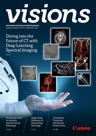
  • 1. AUTOStroke solution As part of our Automation Platform offering, we’ve created an innovative solution that helps optimize treatment outcomes for stroke patients when speed and accuracy are everything. Non-contrast CT Intracranial Hemorrhage ASPECTS CT Large Vessel Occlusion CT Brain Perfusion maps Automation Platform is an AI-based, zero-click solution that uses deep learning technology to streamline your workflow for fast, actionable results every time. From scanner to clinical decision, you’ll be supported by leading-edge deep learning technologies that process and deliver images for accurate triage, worklist prioritization and treatment decisions. For more information, contact your local sales representative or visit our website: https://eu.medical.canon/products/healthcare_it/automation-platform The right insights. Accelerated by AI. Automation Platform https://eu.medical.canon MAGAZINE FOR HEALTH PROFESSIONALS // European Edition // No 37 // September 2021 MAGAZINE FOR HEALTH PROFESSIONALS European Edition //No 37 //September 2021 Diving into the Future of CT with Deap Learning Spectral Imaging 18 //CT Providing a Hub to Advance Interventional Radiology 12 //HIT Supporting Advances in Gynecology Oncology 23 //UL A Seamless Transition that Boosts Productivity 38 //MR
  • 2. Cover Image: Adobe Stockphoto, edited with Canon Medical’s clinical images. VISIONS magazine is a publication of Canon Medical Europe and is offered free of charge to health professionals. The magazine is published twice a year. Registration to access full, previously published, digital editions can be done via the website: https://eu.medical.canon/visions-magazine. Canon Medical stores and uses personal data of the registration to send out the magazine and inform members about new developments. Members can customize preferences or opt-out, after registration. VISIONS magazine is covering Canon Medical’s European region and as such reflects products, technologies and services for this particular area. The mentioned products may not be available in other geographic regions. Please consult your Canon Medical representative sales office in case of any questions. No part of this publication may be reproduced in whole or in part, stored in an automated storage and retrieval system or transmitted in any manner whatsoever without written permission of the publisher. The opinions expressed in this publication are solely those of the authors and not necessarily those of Canon Medical. Canon Medical does not guarantee the accuracy or reliability of the information provided herein. News, articles and the full edition of VISIONS magazine are announced firstly, as pre-publication, via the dedicated VISIONS LinkedIn Group: https://www.linkedin.com/groups/3698045. In this group you can actively participate in discussions about the content and future direction of the magazine. Alphenix, Aquilion, Aquilion ONE, Aquilion ONE GENESIS, Aquilion ONE PRISM, Aquilion Precision, Astelion, Aquilion Lightning, Vantage Galan, Vantage Elan, Vantage Orian,Vantage Titan, Pianissimo, Aplio, Xario and Made for Life are trademarks of Canon Medical Systems Corporation. Xephilio is a trademark of Canon Inc. Vitrea is a trademark of Vital Images, Inc. Vital Images, Inc., will change its company name to Canon Medical Informatics, Inc., in October 2021. This document was created prior to the name change and therefore uses the former company name. Publisher Canon Medical Systems Europe B.V. Zilverstraat 1, 2718 RP Zoetermeer The Netherlands +31 79 368 92 22 W: https://eu.medical.canon/ E: visions.eu@eu.medical.canon Editor-in-chief Jacqueline de Graaf jacqueline.degraaf@eu.medical.canon Design & Layout Boerma Reclame boermareclame.com Printmanagement Printweb Media B.V. printweb.nl Photography Cojan van Toor www.cojanvantoor.nl Text contributions and editing Mélisande Rouger - Independent Journalist Sara Sharp - The Creative Practice Follow us: © 2021 by Canon. All rights reserved. ISSN 1617-2876 Works are generally classified into two categories: Full length articles (e.g. clinical added value of new/special applications & technologies) and Short contributions (e.g. system testimonials, case reports, technical notes). General guidelines for authors C o p y ri g h t © 2 0 1 5 S o c ia l M e d ia E x a m in e r® All articles should be double-spaced. Full length articles Full length articles should generally include the following: - Author’s full name and highest academic degree, employer medical institution - Author’s biography (150 words) - Author’s passport-size photograph (suitable for publication); (image of 300 dpi) - 200-word abstract - Text including headline, sub-title, introduction and sections like: materials methods (which should include a full description of equipment used), results, discussion and references - Text approx 4 to 5 pages or 12.000 to 14.000 characters (not including figures, tables and photographs) - Correspondence address - Literature (no more than 10 references) - Separate, continuous numbered image- and table captions Short contributions Short contributions should generally include the following: - Author’s full name and highest academic degree - Author’s employer medical institution - Author’s biography (150 words) - Author’s passport-size photograph (suitable for publication); (image of 300 dpi) - Text including headline, sub-title, introduction and full description of methods materials/equipment used - Case Report or description of system improvements (Technical Notes) - Correspondence address - Literature (no more than 10 references) - Separate, continuous numbered image- and table captions Text The article should be saved in Microsoft Word (PC format) if possible, and, if not, in text only. Please indicate the software program and version used (Microsoft Word 2007, etc.) and whether it is a PC or Macintosh formatted document. If e-mailing, make sure to send it as an attachment, rather than embedded in the e-mail message. Symbols, formulas and abbreviations Symbols, Greek letters superscripts/Subscripts must to be identified clearly. Furthermore, the figure 1 (one) and the letter l (el) as well as the capital letter 0 and the figure 0 (zero) should be easy to differentiate. All abbreviations including units of measure, chemical names, technical or medical acronyms, names of organisations or institutions should be defined when they first appear in the text (e.g. congestive heart failure (CHF). Please refrain from using unfamiliar abbreviations, clinical slang or jargon. Images, art and tables Cite all figures and tables in text, preferably in consecutive order. Please include a caption for each figure. All captions for each figure, should be separate from the text, at the end of the manuscript on a separate page. Captions should avoid duplication of text material. Credit lines for artwork can appear at the end of the corresponding caption by stating: (Provided by first initial, last name). Black out (or give clear instructions which parts should be blackened out) of the images to not violate any data protection regulations (e.g. patient data) Do not embed figures, charts, or graphs into your document file. Please provide them as a separate file, as well as hard copy/correct .pdf file. Please use one the following formats: EPS , TIFF or JPEG. Arrows stuck onto images for purposes of delineation should be clearly visible and reproducible. Authors should indicate if they would like to have artwork returned. Each table should have a title, and all abbreviations should be spelled out or explained in a footnote. Style Title page should include full names, degrees and titles of authors, and affiliations (name of institution, city and state) for use in a by- line, as well as phone and fax numbers to facilitate sending edited copy back to author for approval. Define all symbols, abbreviations and acronyms on first reference. All manuscripts should be written in a third-person style, unless the article is specifically an editorial or first-hand review. References A maximum of 10 references is suggested. Complete references should be listed in order of citation in text, NOT alphabetically. Up to four authors will be listed; if there are five or more authors, only the first three will be listed, followed by et al. Within the text, reference numbers should appear as footnotes in parentheses or in superscript text at the end of each appropriate citation. Please do not use Microsoft Words endnote feature, as this causes major problems in the editing phase. In addition, if the reference is not in English, please indicate the language of publication. Journal example Oberhaensli RD, Galloway GJ, Hilton-Jones D, et al. The study of human organs by phosphorus-31 topical magnetic resonance spectroscopy. Br J Radiol 1987;60:367-373 Book example Welch KMA, Barkley GL. Biochemistry and pharmacology of cerebral ischemia. In: Barnett JHM, Stein BM, Mohr JP, Yatsu FM, eds. Stroke: pathophysiology, diagnosis and management. New York: Churchill Livingstone, 1986;75-90.
  • 3. VISIONS 37 // 3 JOS RUIS Chief Operating Officer CANON Medical Systems Europe With kind regards, //EDITORIAL © 2021 CANON MEDICAL SYSTEMS Dear Reader, With COVID-19 measures increasingly being eased in many European countries, we are now entering a ‘back to a new normal’ era. Over the past year, our lives changed suddenly and drastically. Virtual interactions largely replaced face-to-face contact, and words, like eConsultation, Zoom and Microsoft Teams became prominent in our vocabulary. Some of these changes made us realize that we can work (more) efficiently in these new ways; we must keep those changes as part of our ‘new normal’ way of working. Some others, we will gladly say “goodbye” to. Social isolation, for example, does not work well for us, as a herd animal species. ‘Back to a new normal’ also comes with plenty of catching up. Catching up on overdue visits to those close to us, catching up on missed holidays, catching up on intentions, tasks, appoint- ments, interventions, diagnosis, treatments etc., all of which have been delayed due to COVID- 19 priorities, and, of course, catching up on the latest and greatest in our professional fields. Our new professional life will likely be one of less travelling, a balanced mix of face-to-face and electronic business- and social interactions, an improved and continuous preparedness for the unexpected, to name just a few elements. From our side, we are looking forward to meeting you again in person, in your professional environments, and during congresses and exhibitions. We have many things to share, of which some are covered in this new edition of our VISIONS magazine. In the meantime I wish you well. Remain vigilant and stay safe.
  • 4. 4 // VISIONS 37 23 Supporting Advances in Gynecologic Oncology ULTRASOUND 26 Taking a Qualitative Leap Forwards MULTI-MODALITY //CONTENTS 03 Editorial 06 News 11 President's Message 12 Providing a Hub to Advance Interventional Radiology HEALTHCARE IT 18 Diving into the Future of CT with Deep Learning Spectral Imaging COMPUTED TOMOGRAPHY 23 Supporting Advances in Gynecologic Oncology ULTRASOUND 26 Taking a Qualitative Leap Forwards MULTI-MODALITY 30 A Significant Investment in Canon Medical CT COMPUTED TOMOGRAPHY 38 A Seamless Transition that Boosts Productivity MAGNETIC RESONANCE 42 How Deep Learning Reconstruction Became the Standard COMPUTED TOMOGRAPHY 06 NEWS (Canon Webinars 2021) 12 Providing a Hub to Advance Interventional Radiology HEALTHCARE IT
  • 5. VISIONS 37 // 5 56 Offering a Wealth of Unexplored Possibilities EYE CARE 62 How Hygienic Positioning Aids are Simplifying Work Processes during the Pandemic COMPUTED TOMOGRAPHY 42 How Deep Learning Reconstruction Became the Standard COMPUTED TOMOGRAPHY © 2021 CANON MEDICAL SYSTEMS 48 Digital Efficiencies in the COVID-19 Pandemic DIAGNOSTIC X-RAY 53 Maximum Efficiency, Prioritized Patient Safety and Increased Revenue with Canon Medical’s 4D CT Technology INTERVENTIONAL X-RAY 56 Offering a Wealth of Unexplored Possibilities EYE CARE 60 Deep Learning Reconstruction – a Game Changer in CT Imaging COMPUTED TOMOGRAPHY 62 How Hygienic Positioning Aids are Simplifying Work Processes during the Pandemic COMPUTED TOMOGRAPHY
  • 6. 6 // VISIONS 37 //NEWS Canon Medical Systems Corporation has recently announced a major operational plan to strengthen its healthcare information technology (HIT) division. In a world, in which data is a business’s most valuable asset, there simply isn’t room for silos of any kind. Cohesion, flow, and integration are the watchwords of our time, which is why it should be no surprise to anyone familiar with Canon Medical that they have recently announced a major opera- tional plan to strengthen their HIT division. The entire reform, which is expected to take some time to complete, will see Vital Images, Inc. adopt the Canon Medical brand to support a more unified business approach. The consolidated business unit will leverage Canon Medical’s global infrastructure to accelerate the delivery of multiple Enterprise, AI, and Collaborative imaging solutions. “This development represents a bold new future for our company, customers and partners,” says Toshio Takiguchi, CEO and President of Canon Medical Systems Corporation. “Enhancing our HIT solution and service provision is the Canon Medical Solidifies HIT as a Key Growth Pillar natural next step in a world that demands accurate and immediate results beyond our traditional modality-centered business model.” Jim Litterer, President and CEO of Vital Images, Inc. echoed these sentiments adding: “Canon Medical has a well-es- tablished reputation for delivering leading solutions that support the end-to-end needs of the evolving healthcare systems. I am confident that customers around the globe can look forward to enhanced service delivery, uncompromised quality, and an innovative approach to healthcare – which is why I am excited to strengthen the HIT Division, leveraging the global reach of Canon Medical.” Vital Images, Inc. will be fully integrated into the global Canon Medical brand from October 1, 2021. To find out more about our HIT solutions, visit https://eu.medical.canon/products/ healthcare_it or contact your local sales representative. //
  • 7. VISIONS 37 // 7 © 2021 CANON MEDICAL SYSTEMS Visit our website or scan the QR code: https://eu.medical.canon/ ➔ Events Calender ➔ Webinars ➔ Past Webinars ➔ WHC - Ultrasound Last May 2021, Canon Medical hosted the webinar ‘Fetal Neurosonography – three essential talks to boost your confidence.’ Fetal Neurosonography is very important as malformations of central nervous system (CNS) are common and can often have a poor neurological outcome. As the fetal brain undergoes tremendous development during pregnancy it can be challenging in differential diagnosis of pathologies and often depend on examiner skills. The main technique for the diagnosis of fetal CNS anomalies is ultrasound. In specific cases, the use of MRI plays a role as well. In 2020, it became obvious that holding face-to-face fetal cardiology courses would be impossible due to the COVID-19 pandemic. Therefore, the collaboration between Canon Medical and the Evelina London Children’s Hospital plus King’s College Hospital, London, UK, was translated into the comprehensive online course “State of the Heart in Fetal Cardiology”. The course comprised of eight two-hour sessions, run every two weeks from January to May 2021. The course covered all major facets of fetal cardiology, including normal cardiac patterns, features of major groups of structural abnormalities, and fetal arrhythmias. Some areas of practice have undergone significant advances, including first trimester scanning, diagnosis of fetal vascular Webinar Fetal Neurosonography Worldwide Online Course ‘State of the Heart in Fetal Cardiology’ was a Success! Our excellent well-known speakers Prof Laurent Guibaud (Hospital Femme Mère Enfant - University Claude Bernard Lyon/France), Prof Luc de Catte (University Hospital Leuven/ Belgium) and Dr Miguel Branco (Bissaya Barreto Coimbra Hospital and University Center/Portugal) explained a detailed multiplanar neurosonogra- phy examination, how to differentiate cystic brain lesions and the fetal cortex development in normal and abnormal cases. This successful webinar was intended to boost confidence and with attendees from 86 countries all over the world joining we saw the high demand for rings and the introduction of motion- corrected fetal cardiac MRI, which has been pioneered at the Hospital. Novel ultrasound imaging modalities, including Superb Micro-vascular Imaging have had a significant impact in the first trimester and in diagnosis of vascular rings. With numbers capped at 200 to permit meaningful interaction with the audi- ence, delegates registered from all over the world and could interact live with the panel for each session. “Prenatal detection and accurate diagnosis of congenital heart disease is essential, because of its potential profound impact,” said Professor John Simpson, Professor of Pediatric and Fetal Cardiology at Evelina London Children’s Hospital. “We have been running foundation and advanced courses in fetal cardiology together with Canon Medical for over 10 years. The online course has enabled us to continue this vital education despite the COVID-19 pandemic.” Feedback from the course has been extremely positive, with respect to content, image quality and the faculty- audience discussions. The demand for the course exceeded all expectations and we believe this approach will con- tinue to be an effective complemen- tary means of delivering education, even when things return to a more ‘normal’ situation. // supporting our customers with high- level education not only performing daily routine ultrasound, but detailed neurosonography scan to improve scanning skills and confidence in the final diagnosis. // Dr. Miguel Branco Prof. John Simpson Dr. Owen Miller Dr. Vita Zidere Dr. Kuberan Pushparajah Dr. Thomas Day Prof. Gurleen Sharland Dr. Marietta Charakida Dr. Trisha Vigneswaran Dr. David F.A. Lloyd Dr. Ioana Dumitrascu-Biris Prof. Laurent Guibaud Prof. Luc de Catte
  • 8. 8 // VISIONS 37 //NEWS July Eye Care Webinar (Italian) Dr. Barbara Parolini Eye Care Webinar Presentato dai seguenti Relatori Dr. Barbara Parolini Prof. Enzo M. Vingolo Interpretazione Clinica su OCT e OCTA nella Pratica Quotidiana 15 Luglio, 2021 | 19:00-20:00 CET August 24, 25, 26 - September 1,2 Online Neurology Days Dr. Mahtab Zamani Prof. Neil D. Pugh Dr. Grant Mair Dr. Anton Meijer Dr. Joseph Puig Dr. Nevia Caputo Prof. Thomas Tourdias Dr. Alexandra Borchert Prof. Nens van Alfen Dr. Benoît Doche de Laquintane Prof. Bart van Wijmeersch Prof. Adnan Siddiqui Dr. Andrés González Mandly Canon Webinars 2021 Learn from the experts and gain new insights In online educational webinars, leading experts share their expertise and exchange knowledge. Register to join one of our upcoming webinars or visit our website to watch the past recorded webinars. Visit our website https://eu.medical.canon/ events_calendar/webinars Or scan the QR code for the complete online overview. November 9 Ultrasound (MSK) November 18 Eye Care OCT (Dutch) September 30 Eye Care OCT The clinical benefits of scanning wider and deeper in the retina. October 26, 27, 28 - Online Cardiology Days November 3, 4 Day 1 - Oct 26 | Cardiovascular disease - New Imaging Approaches | Eye Care/MR/CT Day 2 - Oct 27 | Cardiovascular disease - Complex Imaging ​Procedures | MR/CT/HIT/VL Day 3 - Oct 28 | Structural Heart Disease | UL/CT/VL Day 4 - Nov 3 | Pediatric Cardiology | UL/CT/VL Day 5 - Nov 4 | Sports Cardiology | UL/VL
  • 9. VISIONS 37 // 9 © 2021 CANON MEDICAL SYSTEMS Lars Lindgren May Seminario web Ecografía (Spanish) Dr. Pedro Mora Sanz Dra. Gloria Ruiz Fernández Dr. Antonio Olveira Martín Dra. Marta Abadía Barnó Seminario web Ecografía Presentado por los siguientes ponentes Dra.GloriaRuizFernández Dra.MartaAbadíaBarnó Ecografía Avanzada en la Valoración del Daño Hepático 13 de Mayo, 2021 | 19:00-20:00 CET Dr. Pedro Mora Sanz Dr.AntonioOlveiraMartín June Echografie / MRI Webinar (Dutch) Dr. Bas Maresch Dr. Jan Veryser Dr. Kurt Vanderdood 10 juni 2021 | 19:00 CEST De Complementariteit van MRI en Echografie; Een Enkel Onderzoek Gepresenteerd door de volgende sprekers Registeer hier of bezoek onze website: eu.medical.canon Dr. Kurt Vanderdood Dr. Bas Maresch Dr. Jan Veryser Echografie / MRI Webinar June X-Ray Webinar X-Ray Webinar Presented by the following speaker Register here or go to our website: eu.medical.canon Lars Lindgren Managing Challenges in Orthopaedic Implant Imaging June 17, 2021 | 19:00 CET July Webinar Prof. Joao Lima Dr. Yoko Kato Prof. Leopoldo Pérez de Isla Prof. Laurent Feldman Prof. Mickaël Ohana Webinar Presented by the following speakers Post-COVID Conditions July 13, 2021 | 19:00 (CET) / 13:00 (EST) Dr. Yoko Kato Prof. Mickaël Ohana Prof. Laurent Feldman Prof.LeopoldoPérezdeIsla Prof. Joao Lima May MRI Webinar Prof. Dr. Alberto Aliprandi Prof. Dr. Reinhard Tomczak MRI Webinar Presented by the following speakers Redefining MRI: Unleash Smart AI Solutions to Enhance Your Diagnostic Confidence and Increase Throughput May 11, 2021 | 19:00 – 20:00 CET Prof.Dr.ReinhardTomczak Prof.Dr.AlbertoAliprandi
  • 10. 10 // VISIONS 37 Visit our website to view the complete overview online and to stay up-to-date about changes or newly added webinars! January MSK Webinar Dr. Jan Veryser Dr. Ramon Balius MSK Webinar Presented by the following speakers Dr. Jan Veryser Dr. Ramon Balius January 14, 2021 | 19:00 CET Ultrasound Guided interventions: High-resolution ultrasound of shoulder March Eye Care Webinar Prof. Tariq Aslam Dr. Sal Rassam Dr. Barbara Parolini Dr. Orlaith Mc Grath Eye Care Webinar Presented by the following speakers Dr. Orlaith Mc Grath Prof. Tariq Aslam Dr. Barbara Parolini Dr. Sal Rassam Advanced application Interpretation of OCT and OCT Angiography in clinical practice March 25, 2021 | 19:00-20:00 CET April CT Webinar Prof. Antoine Khalil Dr. Monique Brink Prof. Ernesto Di Cesare Dr. Russell Bull CT Webinar Presented by the following speakers Dr. Russell Bull Prof. Antoine Khalil Prof. Ernesto Di Cesare Dr. Monique Brink CT for COVID-19 Patients: From EmergencyTriaging to Follow-up Assessment April 29, 2021 | 19:00-20:00 CET April MSK Ultrasound Webinar Dr. Jan Veryser Prof. Dr. Karsten Knobloch MSK Ultrasound Webinar Presented by the following speakers Prof. Dr. Karsten Knobloch Dr. Jan Veryser High resolution imaging in challenging MSK examinations April 22, 2021 | 19:00-20:00 CET MSK Ultrasound Webinar Series Prof. Laurent Guibaud May Ultrasound WHC Webinar Ultrasound WHC Webinar Presented by the following speakers Fetal Neurosonography Three Essential Talks to Boost Your Confidence May 6, 2021 | 19:30 – 21:00 CET Ultrasound WHC Webinar Prof. Laurent Guibaud Prof. Luc de Catte Dr. Miguel Branco Dr. Miguel Branco Prof. Luc de Catte
  • 11. VISIONS 36 // 11 © 2021 CANON MEDICAL SYSTEMS L et me start by expressing my deepest thanks for your continued patronage and the work of our valued health care practitioners as they fight against COVID-19. If I had to sum up the last few months in two words, they would be gratitude and success. It was recently announced that former employee, Mitsue Miyazaki, had been awarded the Medal with Purple Ribbon at the Medal with the Spring of the Third Year of Reiwa in recognition of his pioneering work in medical imaging. Mitsue Miyazaki, who now holds an esteemed position at the University of California, San Diego, was instrumental to the development of an MRI device that can visualize blood vessels without the use of contrast media – making MRI sig- nificantly safer for patients with kidney and liver dysfunction. In addition, Canon Medical has been awarded the National Invention Award for the Third Year of Reiwa, the Gift TOSHIO TAKIGUCHI President and Chief Executive Officer Canon Medical Systems Corporation Invention Award, and the Invention Implementation Achievement Award for its creation of a data reading method for large-field CT detectors. This technol- ogy, which was key to the development of our high-end CT systems, enables clinicians to image the entire heart and brain in just one scan – thereby reducing exposure dose and pushing the potential of CT beyond its limits. I am extremely proud of all those who played a part in this great achievement, and I remain ever grateful to the work you do in service of our Made for Life philosophy. Thank you once again and I wish you every success moving forward. President’s message
  • 13. VISIONS 37 // 13 INTERVIEW //HEALTHCARE IT //Aquilion PRIME, Astelion,VantageTitan,Vantage Orian, Aplio a450, Aplio 300,Vitrea Rome in Italy has emerged as one of the world’s centers of Interventional Radiology expertise, and the San Pietro Hospital, located in the north of the city, makes a significant contribution. Focusing on several specialisms including Oncology, Pediatrics, Women’s Health, Obstetrics, and Musculoskeletal (MSK) sur- gery, it has a strong team of Interventional Radiologists led by Professor Alberto Bellelli. He explained to VISIONS how a long collaboration with Canon Medical that has spanned two decades has enabled the Hospital to develop as a center of excellence in Interventional Radiology. Providing a Hub to Advance Interventional Radiology © 2021 CANON MEDICAL SYSTEMS //MMEU2105 VISIONS spoke with Prof. Bellelli, Head of the Radiology Department at the San Pietro Hospital, Rome, Italy. S an Pietro’s Radiology Department has opted for several different modalities from Canon Medical, including CT, MR and Ultrasound - all supported by Vitrea Advanced Visualization (AV) post-processing software). The Aquilion PRIME SP CT is used for all advanced diagnostic examinations, including Cardiac and Oncology. The Astelion Advance CT is primarily used for MSK Interventional Radiology procedures, often in combination with Smart Fusion. The MR systems (Vantage Titan and Orian) are used for all specialties. Vitrea connects all the modalities, collects all the information that is obtained, and consolidates this into a single report.
  • 14. 14 // VISIONS 37 Quality and integration Prof. Bellelli has found that the quality of the cardiac examinations obtained from the Aquilion PRIME SP and Vantage Orian is very high, and Vitrea AV maximizes and consolidates the information that the specialists can obtain from the tomographic images. “Vitrea AV streamlines the post pro- cessing of the cardiac examinations and the radiologists and the cardiol- ogists using the system are pleased by the quality and presentation of the results, which is fundamental for a highly specialized department,” he remarked. Surgical MSK procedures, another core activity of the hospital, are required on a very large number of patients.
  • 15. VISIONS 37 // 15 © 2021 CANON MEDICAL SYSTEMS //MMEU2105 with the SEMAR algorithm of the CT system and the post-surgical control examination, the images provided are incredible in case of metal implants. Global Illumination provides an out- standing rendering of the anatomical structures, and the easy segmentation enables a quick method to accurately map all the fracture fragments.” “Global Illumination is a feature that has surprised the hospital’s surgeons. It is the first time they have had the opportunity to interact with such realistic rendering of the displacement of all the bone structures,” said Prof. Bellelli. “3D images are really suitable for the depth of anatomical structure that we need to see. In combination “Global Illumination provides an outstandingrenderingoftheanatomical structures and easy segmentation tools enable a quick method to accurately map all the fracture fragments.” Prof. Bellelli. “The possibility to combine images from different modalities is very well-designed,” he continued. “In a department, such as ours, where we use a full range of information sources and modalities in the plan- ning of interventional procedures, this feature is mandatory. Vitrea gives many benefits in modality integra- tion. Ever since my first experience with Vitrea, I could appreciate the smooth workflow that it provides for complex post-processing, in comparison to other products that I have used before. After the selection of the images to be processed, Vitrea automatically extracts the qualitative and the quantitative information needed, according to the specific examination and clinical application. The Radiologist can focus their efforts just on formulating the diagnosis, and not on extracting the information.” Best independent post-processing Prof. Bellelli leads a team of 64 col- laborators focused on Interventional Radiology procedures. In a broader context, Vitrea also provides post-pro- cessing capability that is multi-vendor, which enables the experts to integrate their work more easily. “I prefer to have all the images directly acquired in my department, on the Canon Medical equipment, but this is not always possible. Sometimes, I need to process examinations that are scanned with the systems of other vendors. Vitrea doesn’t put any limits in the processing of images from other vendors and this provides the possibility to always have the best post-processing, independent of the vendor,” he explained. “One of the biggest benefits that the Vitrea provides is that it can support full collaboration with other specialists, who are not always certain of images. Vitrea presents information in a very user-friendly and easy-to-understand way, also for specialties that are not confident with radiologic images.”
  • 16. 16 // VISIONS 37 San Pietro Hospital, Rome, Italy. Vitrea AV has also helped facilitate com- munication across Prof. Bellelli’s clinical team, as the representation of the relevant clinical structures is extremely clear. There are currently 16 radiolo- gists in the Department. All of them support their reports with the Vitrea AV. In particular, the generic vascular MSK interventional procedures. and cardiac examinations require the Vessel Analysis tools. Communication among team members is also facilitated for MSK surgical planning, in which the Radiologist is required to provide a clear representation of the anatomical struc- ture to enable the surgeon to choose the best surgical approach. “Of course, the most important benefit is in the improvement of selecting the most suitable Interventional procedure in order to avoid, or limit treatment complications,” added Prof. Bellelli. Key role in continuous development Prof Bellelli started working with Canon Medical twenty years ago. During this time, the relationship has been consolidated through continuous support of Canon Medical’s team in training, advanced clinical sessions and problem solving. Prof. Bellelli has been able to improve and develop his clinical protocols with the support of Canon Medical’s experts. “Canon Medical plays a key role in the planning and ongoing development of our Radiology Department. And continuous dialogue and collaboration between Canon Medical and our hos- pital in terms of new technologies and deployments is essential for the realiza- tion of our vision,” said Prof. Bellelli.
  • 17. VISIONS 37 // 17 Biography Prof. Bellelli is the Head of the Interventional Radiology department of the San Pietro Hospital, in Rome, Italy, and has been working for the past 30 years in the fields of diagnostic radiology, emergency and later specializing in pain therapy, diagnostic and therapeutic treatment of lumbar and joint pain. He has joined San Pietro Hospital in 2018, where he is now leading a team of 64 collaborators focused on interventional procedures. “In combination with the SEMAR algorithm of the CT system and the post-surgical control examination, the images provided are incredible in case of metal implants.” Prof. Bellelli. “Our aim is to provide the best patient care, but at the same time there is a necessity to optimize the efforts of the internal resources to guarantee the best possible experience to the patient. To achieve this result, it is mandatory to implement a fast and reliable modality integrated solution across all the imaging devices in the department. The department of today must also aspire to the department of the future. This means that all modal- ities installed must be open to further implementation and upgrade to ensure that the clinical offer to patients is always ‘state-of-the-art’. Efficient workflow means that our department is ready for the introduction of the latest technologies,” Prof. Bellelli concluded. // © 2021 CANON MEDICAL SYSTEMS //MMEU2105
  • 18. 18 // VISIONS 37 Spectral Imaging “Featuring a single X-Ray tube with an automatic adap- tive exposure, the field of view is up to a 50cm centi- meters, and scans be obtained by helical or sequential acquisition. Spectral reconstruction is performed by deep learning. With one click on the Vitrea workstation you can obtain a great deal of information with the spectral analysis application. It's entirely automatic and doesn't take too much time. The Vitrea workstation automati- cally provides you with a visceral and/or a vascular study. In addition, you can use spectral imaging in multiple body regions,” explained Prof. Roy. CLINICAL PRESENTATION //COMPUTED TOMOGRAPHY //Aquilion ONE / PRISM Edition, SURE Subtraction, AiCE T he Aquilion ONE / PRISM Edition complements the Hospital’s existing Aquilion ONE / GENESIS Edition system, which was installed in 2018. “With our vast and growing demand for high-quality imaging, our CT systems are at the heart of the hospital. They serve the needs of our numerous radiologists, clinicians and technolo- gists,” said Prof. Roy. “Aquilion ONE / PRISM Edition has become invaluable for us, due to its reliability, fast workflow and high versatility. In particular, it has impressed us through key technol- ogies that deliver spectral imaging. The system is so useful that my co-workers are almost fighting to work on the new machine!” Diving into the Future of CT with Deep Learning Spectral Imaging Strasbourg University Hospital is one of the top five research hospitals in France. It is renowned for the quality of its healthcare, clinical research and leadership in training. As a center of excellence in a wide range of specialties including cardiac surgery, transplantation, rheumatology, microsur- gery, cryotherapy, immunology and surgical robotics, it contributes to advancing medicine globally. The hospital acquired an Aquilion ONE / PRISM Edition CT system from Canon Medical in 2019. Prof. Catherine Roy, Head of the Radiology department at the hospital, explains how its deep learn- ing spectral imaging delivers new diagnostic efficiencies. Prof. Catherine Roy Deep learning spectral scanning, reconstruction and analysis.
  • 19. VISIONS 37 // 19 © 2021 CANON MEDICAL SYSTEMS //CTEU210158 Radiology Team Strasbourg University Hospital, France. Monochromatic imaging from 35 KeV to 135 KeV showing improved contrast enhancement in the lower KeV images. Despite the low iodine load of 100 ml of 270 mgI/ml and a flow of 1.3 ml/sec, Spectral CT helps to enhance the contrast at low keV free of noise. It provided an excellent contrast for the evaluation of the aortic dissection (type B).
  • 20. 20 // VISIONS 37 “Supplementary information available with the software is to have the quantification of the iodine load in mg per ml. You can obtain the spectral curve by placing a region of interest inside the tissue.” “Additionally, iodine maps improve visibility of nodules with iodine uptake. For examinations, like scanning for kidney stones, the software is very easy to use. In the future, I guess that we will have a better evaluation of tissue perfusion, for instance: for the follow-up of oncologic patients and to prove that there is an efficiency of antiangiogenic treatment. Or perhaps for immunotherapy. With just only one injected phase, you can characterize tissue.” Extraordinary Images The Aquilion ONE / PRISM Edition has been able to meet the needs of the whole team at the Hospital. Monochromatic images to see better One type of image that is available with spectral imaging are high quality monochromatic images. “We use the monochromatic images to better visualize to contrast on any abnormality. The monochromatic images produce excellent image quality and to visualize iodine uptake even better, you can use a slidebar to adjust the keV level. Using the best contrast-to-nose ratio, you can optimize the contrast inside the suspicious region by placing a region of interest,” she added. “It's excellent for detecting abnormalities, even the small ones, and you can decrease the iodine loads, which is also very important. We use the lowest volume or concentration of contrast agent possible. Concerning the radiation dose, it's quite similar to that obtained with iterative reconstruction.” Multiple tools to characterize and quantify The Aquilion ONE / PRISM edition delivers a great deal of additional information that enables Prof. Roy and her team to better characterize and to quantify what they are scanning. “You obtain an iodine map, which is the information on the iodine uptake; the virtual non-contrast (VNC), with no iodine at all; and you can select an evaluation of the spec- tral curve by placing a region of interest and evaluation of the effective Z image or electron density image. With these three elements, you can differentiate contrast enhance- ment from non-contrast enhancement. With the VNC, you can avoid the non contrast acquisition that was previously done,” she continued. Vitrea Advanced Visualization Spectral analysis software. Monochromatic Image (80keV) Iodine map Virtual Non-Contrast (VNC) Monochromatic Image (60 keV) Effective Z Spectral curve “The best descriptive classification of the Aquilion ONE / PRISM Edition CT is - Versatility for all diagnostic purposes.” Prof. Catherine Roy, Head of the Radiology department at the Strasbourg University Hospital, France.
  • 21. VISIONS 37 // 21 © 2021 CANON MEDICAL SYSTEMS //CTEU210158 Deep Learning Spectral CT used to evaluate liver and renal masses. Spectral DLR delivers excellent energy separation, enabling assessment of Iodine uptake. Additionally, the Iodine map clearly outlines the regions of Iodine uptake. Deep Learning Spectral CT of the lung to evaluate nodules. Spectral DLR delivers excellent energy separation and low-noise properties, enabling assessments of Iodine uptake within the lesions, which can help facili- tate assessment of benign vs. malignant lesions. Deep Learning Spectral CT for visualization and quantification of kidney stones. 2 kidney stones are analyzed as calcium oxalate.
  • 22. 22 // VISIONS 37 Myocardium Iodine Maps helps to evaluate the attenuation of the hypo-enhancing area compared to normal myocardium. Deep Learning Spectral CT of the foot for visualization and quantifica- tion of Monosodium Urate (MSU) in the metatarsophalangeal joint of the hallux. Spectral DLR enables volume calculations of the MSU, and color overlay on 3D/2D images for improved visualization of MSU. Deep Learning Spectral CT for Pulmonary Embolism assessment. Spectral DLR delivers excellent energy separation and low-noise properties, enabling assessment of perfusion defect within the lung parenchyma, associated with the pulmonary emboli. Courtesy: Kyorin University, Japan. Prof. Catherine Roy Head of the Radiology department at the Strasbourg University Hospital, France. “Our clinicians love, and they are very interested by innova- tive things, especially with spectral imaging. Their feeling is that with this new scanners spectral imaging capabilities, they are more confident with their diagnoses,” said Prof. Roy. “Our technologists need easy-to-use hardware and software and this is available with the Aquilion ONE / PRISM Edition. They need a reliable scanner, and this is especially true for the Aquilion ONE / PRISM Edition. There have been no problems with it at all. The best descriptive classification of the Aquilion ONE / PRISM Edition CT is - Versatility for all diagnosis purposes.” //
  • 23. VISIONS 37 // 23 IOTA-ADNEX model in case of advanced stage Ovarian Cancer. Ultrasound in research and daily practice “My story changed significantly in the year 2000, when I met Professor Lil Valentin (Lund University, Sweden), Professor Tom Bourne (Imperial College, London, UK) and Professor Dirk Timmerman(KULeuven,Belgium).Togetherwiththem,Istarted an adventure that is the International Ovarian Tumor Analysis group (IOTA)” she explained. “We started to prospectively collect data of ultrasound and histology, of patients with ovarian masses and developed mathematical models, like simple rules and ADNEX, to discriminate between benign and malignant ovarian masses. So far, our database includes more than 25.000 cases.” Prof. Testa has been working with ultrasound engineers to develop new softwares and algorithms to improve the clinical management of patients with gynecological disease. In partic- ular, she has contributed for the integration of IOTA predictive INTERVIEW //ULTRASOUND //Aplio i900, Aplio i700, Xario 200G T hroughout her career, Prof. Testa has completed pioneering research and advanced gynecology with the help of ultrasound. She graduated in Medicine at the Catholic University in Rome (Italy) and is now Associate Professor of the Institute of Obstetric and Gynecological Clinics at the Catholic University of the Sacred Heart in Rome, Italy and Scientific Director of the Center for Ultrasound in Gynecological Oncology “Class Ultrasound” of the Fondazione Policlinico Agostino Gemelli IRCCS, Rome. She has been a Board member of the International Society of Ultrasound in Obstetrics and Gynecology (ISUOG) for eight years, and Vice-President of the Italian Society of Gynecology and Obstetrics (SIGO Federation) for two years. Prof. Testa is member of the Steering Committee of International Ovarian Tumor Analysis (IOTA) group. Supporting Advances in Gynecologic Oncology Professor Antonia Testa is one of Europe’s leading experts in gynecologic oncology and has conducted more than fifty research projects on gynecological cancer and chemotherapy proto- cols. She also founded a world-class school for ultrasound in gynecology in Rome, Italy, that has trained thousands of specialists since its inception. Professor Testa is also Board Member of the International Ovarian Tumor Analysis group (IOTA). Her overall work has contributed to improve early and confident diagnosis in gynecological disease. Professor Testa explained to VISIONS how important Canon Medical’s ultrasound systems are in daily clinical practice and research. Prof. Antonia Testa model (ADNEX model) into ultrasound equipment for discrimination between benign and malignant ovarian masses, which is integrated into the Aplio series. “In addition, through working as part of the gynecology team, I realized the great value of ultrasound in the daily clinical management of our patients. In particular, during follow-up, when we can use ultrasound as a bedside tool. For example, if we have a suspicion of a recurrence, we can check it immedi- ately to understand the nature of one or more lesions and assess the possibility to surgically remove the lesion. In addition we can obtain a biopsy of that lesion to follow up the situation during treatment,” she continued. © 2021 CANON MEDICAL SYSTEMS //ULEU210081
  • 24. 24 // VISIONS 37 “I wanted to share my knowledge with my colleagues, with residents and with students, which is why I created the Center for Ultrasound in Gynecological Oncology “Class Ultrasound” at the Agostino Gemelli University Hospital Foundation in Rome, Italy. Since its inception six years ago, around 2,500 col- leagues have attended courses there.” The Center has trained Italian and European physicians and health professionals, provides patients with the highest professional service and plays a key role in multi-center scientific studies. Celebrating five years of scientific collaboration This year marks the fifth anniversary of a collaboration with Canon Medical that has brought great results for Prof. Testa’s research and clinical practice, and has helped Canon to advance its ultrasound systems. “My story with Canon Medical started in 2016 after ISUOG World Congress, which I had the honor to organize. Canon profession- als offered me the opportunity to begin a scientific collaboration. As I am always looking for something innovative I was very happy with their proposal to explore new software and new ultrasound equipment.” Aplio i700 Women’s Health System Prof. Testa started to work with Canon Medical’s Aplio i900 ultrasound system at the beginning of the collaboration, which was later exchanged for a dedicated Aplio i700 Women’s Health system when it became available. The School of Ultrasound also have a smaller Canon Medical Xario 200G for evaluation in their surgery department with intraoperative and laparoscopic transducers. Initially, they used the Aplio for this functionality, but instead of moving the system between departments, the Xario 200G was made continuously available in the surgery department. With its smaller footprint, little space was required. Because it runs on batteries, it offers optimal mobility. “The most important requirements relating to an ultrasound system that I am always looking for are first - resolution, so OvarianFibromawithflowvisualizedbySuperbMicro-vascularImaging(SMI). Ovarian Cancer. high-quality grayscale images. Then, intuitive software, that allows the examiners to use the equipment at its best. In other words, easy-to-manage tools that are not very complicated to use. Then, high-quality vascularization assessment,” she continued. “When I discovered Aplio ultrasound machines, I was fascinated by its very high image quality with crystal-clear images and enhanced penetration. Then I found the equipment very good, able to deliver superb images for a wide range of clinical applications. Moreover, regarding the vascularization, it was amazing to find that the machine had the possibility to detect very, very small vessels, which is crucial for us to discriminate papillary projections or STANDARD LAPAROSCOPY ROBOT ASSISTED LAPARATOMY Laparoscopic and intraoperative surgery.
  • 25. VISIONS 37 // 25 amorphous material, for instance, within an ovarian cyst. I have to say that also the Doppler Luminance is fantastic, because it gives the impression of 3D rendering images.” Meeting challenges “In gynecologic oncology there are several challenges for the management of our patients. First, pre-operative assessment: to detect a lesion and to be able to discriminate between benign and malignant; Furthermore to stage cancer, that means not only to see the lesion, not only to understand that it can be malignant, but to assess the extent of the disease. We know that the trans- vaginal ultrasound examination is fundamental and superior to other sophisticated imaging methods, because of the dynamic examination. It offers the possibility to check the reciprocal movements of other organs, to check for pain, while pushing against the organ, and to assess the deformability of the organ…. these are of upmost importance in the examination of the pelvis.” “Secondly, the challenge is to have a bedside tool to get a quick assessment to solve some clinical problems, to check for pleural effusion and presence of ascites or do simple procedures, such as paracentesis, drainage of fluid or place intraperitoneal cath- eters. And thirdly, we discovered that ultrasound can play an important role in the surgical field. As our patients very often had been already treated with radiotherapy, and/or previous surgical procedures, sometimes, it's very challenging to detect a small recurrent lesion located in the lower part of the pelvis, for example. With the transvaginal transducer, we can guide the surgeon to reach that small area. Besides that, Canon Medical provided us with a laparoscopic ultrasound transducer, and this is fantastic, because for some patients with borderline ovarian tumors, for instance, we are able to provide ‘fertility-saving’ surgery. We can select the area and localize the small lesion in order to spare the largest amount of normal tissue.” New research possibilities The systems have also enabled new possibilities in research. “As far as innovation is concerned, I was very interested in the possi- © 2021 CANON MEDICAL SYSTEMS //ULEU210081 Publications: 1 Intraoperative ultrasound diagnosis of metastatic lymph node in serous borderline ovarian tumor De Blasis I, Tortorella L, Macchi C, Arciuolo D, Scambia G, Testa AC (UOG 2019). 2 Ultrasound Technologies in the diagnosis and treatment of ovarian cancer Antonia Testa, Canon Medical webinar at ISUOG World Congress 2020. 3 Fusion imaging of ultrasound and MRI in the assessment of locally advanced cervical cancer: a prospective study Moro F, Gui B, Arciuolo D, Bertoldo V, Borzi R, Romeo P, Petta F, Cambi F, Pasciuto T, Zannoni GF, Valentini V, Manfredi R, Scambia G, Testa AC Italy (IJGC 2019). bility to explore Smart Fusion that fuses Ultrasound with MR or CT images. So, we decided to run a prospective study on patients with cervical cancer and a study in patients with advanced ovarian cancer. We have already published the results on the first study - cervical cancer. We have been analyzing data of a large series of patients with ovarian cancer,” said Prof. Testa. “At the moment, I can say that I'm very happy about this experience and, in my opinion, Smart Fusion can play a prominent role in education. To have the possibility to share the experience, being the radiologist and gynecologist together the same room - It's very important for the improvement of the experience of both disciplines.” Working on the future Canon Medical continuously develops its ultrasound systems to provide even better image quality and workflow that enable clinicians to advance medicine. One important aspect is integrating Artificial Intelligence (AI) enabled technologies into the ultrasound systems in the future. “I look confidently towards the future,” added Prof. Testa. “I really hope that ultrasound equipment will provide us with Artificial Intelligence analysis. With this, we will be able to store a lot of images using a simple method plus define and recog- nize more of what we cannot do with our eyes and brains.” // Smart Fusion Image of Low-Grade Appendiceal Mucinous Neoplasm. Prof. Antonia Testa
  • 26. 26 // VISIONS 37 INTERVIEW //MULTI-MODALITY //Vantage Galan 3T, Vantage Titan 3T, Vantage Elan 1.5 T , Aquilion Lightning SP, Aplio a-series, AiCE Taking a Qualitative Leap Forwards Dr. Jordi Catalá March and Dr. Jorge Salmerón Pintos.
  • 27. VISIONS 37 // 27 © 2021 CANON MEDICAL SYSTEMS //MMEU2106 A cross its seven facilities in Barcelona, the diagnostic cen- ter has recently acquired one Vantage Galan 3T MRI with Advanced intelligent Clear-IQ Engine (AiCE), one Vantage Titan 3T MRI, three Vantage Elan 1.5 T MRI systems, one Aquilion Lightning SP CT with AiCE and two Aplio a-series Ultrasound systems from Canon Medical. Shared medical philosophy Instituts Guirado is a company created and directed by doctors. “We think like doctors, and combine technology, research, and academic publications, to provide the highest stan- dards of comprehensive and innovative care in everything we do,” remarked Dr. Catalá March. “We were looking for a technological partner that would share our medical philosophy, and in partic- ular, share our objective to get the most out of the images we acquire, enabling us to, for example, improve the diagnosis of tumors by detecting them earlier, map articular cartilage, and perform brain volumetric studies. We can achieve these things with tools that leverage Canon Medical's innovative technology that utilize AI and Deep learning.” Technology to serve both patients and medical team Dr. Catalá March appreciates the constant improvement that is made possible through Canon technology and ongoing collaboration with Canon Medical. “Just as an example, Canon Medical’s Deep Learning has allowed us to go further in diagnosis and patient care through enabling examinations with 80% less radiation dose, 40% less dose of iodinated contrast, and higher image quality. This is what we are looking for: technological changes that are beneficial for patients and for the medical team,” he explained. Qualitative leaps forward in CT and MRI The Aquilion Lightning SP has enabled the diagnostic center to take a qualitative leap forward. With a multi-slice detector, allowing a minimum thickness of 0.5mm; simultaneous acquisition of data with 160 slices in a single rotation; 3D and MPR images with a precise and uniform level of detail can be obtained in routine scans. “It is really important that patients are scanned with a minimum exposure time, and with a minimum radiation dose, which is adjusted at all times to the anatomical region studied and the size of the patient's body. And, of course, that the CT produces excellent image quality, from which the patient benefits through more accurate diagnosis and a lower radiation dose,” said Dr. Catalá March. “With our new acquisition of the latest generation, Canon Medical’s MR system the Vantage Galan 3T, we have taken a great qualitative leap in our protocols, expanding new sequences that improve diagnosis - changing diagnostic paradigms in some of our processes. Particularly relevant is the integration of AI through Canon’s AiCE technology,” he continued. Dr. Jordi Catalá March with Canon's Vantage Galan 3T MR system. Instituts Guirado in Barcelona, Spain, is a high quality medical imaging center at the forefront of radiology. It is dedicated to providing rapid, accurate and trustworthy diagnosis through continuous commitment to investment in the latest technology, an optimized center for patients, and support from its highly skilled medical and professional team. With the latest MRI, CT and Ultrasound technology from Canon Medical, the diagnostic center can improve its services and knowledge even further. Dr. Jordi Catalá March, Executive Director - CEO of the Instituts Guirado, explained how the systems have supported a qualitative leap in many diagnostic procedures.
  • 28. 28 // VISIONS 37 “Equally important, is the possibility to perform comfortable and silent examinations using new techniques, from which the patient gains in comfort and in much shorter scan- ning times, all thanks to the rapid acquisition of Compressed SPEEDER for 3D sequences. Another aspect to highlight is the wider gantry diameter, which is a help for claustrophobic and obese patients. We also have advanced post-processing. Continually advancing The collaboration agreement with Canon Medical Systems allows the Instituts Guirado to constantly update the latest diagnostic techniques, such as, tractography, functional studies and MRI-Neurography, etc. With the Vantage Galan 3T MRI, in particular, the diagnostic center has been able to add more sequences that add great value. “One example of this is our studies of Meniere's disease and hydrops, which we have carried out for some years. The high resolution and post-processing of the acquired images, allows us to detect changes that have not been visible until now, related to pathologies in the membranous labyrinth.” Another example is in oncology studies. The acquisitions achieved with Compressed SPEEDER for 3D technology, AiCE - Deep Learning, Atlas SPEEDER coils and all MRI expert packages, offer customization and rigor to these very sensitive diagnoses for the patient. “By using the latest systems and techniques developed by Canon Medical, we have been able to remain at the forefront as leaders in diagnostic imaging.” Dr. Jordi Catalá March, Executive Director - CEO of the Instituts Guirad, Barcelona, Spain.
  • 29. VISIONS 37 // 29 © 2021 CANON MEDICAL SYSTEMS //MMEU2106 Leaders in diagnostic imaging Over the last six years, Instituts Guirado has strived for excellence in imaging. “By using the latest systems and techniques developed by Canon Medical, we have been able to remain at the forefront as leaders in diagnostic imaging,” remarked Dr. Catalá March. “Our collaboration with Canon is a commitment to the future, in which the teamwork that we have established brings us great advantages, and ulti- mately continual progression enabling us to continue to offer an excellent medical service and maximize diag- nostic value.” // “We continue, hand-in-hand with Canon Medical. Growing and sharing enables us to continually advance. Canon Medical understands our philos- ophy, listens to our needs, and observes our working methods,” said Dr. Catalá March. “Together, we achieve synergy in our work. We feel very comfortable with Canon’s response to our diagnostic requirements, which are focused on early and better diagnosis leading to optimal treatment, we aim to build a better and healthier future.” New generation expertise The “know how” of an institution provides an active value that we pay a lot of attention to and from Instituts Guirado we promote the acquisition of state of the art technology that gives to the professionals and new talents the tools and the environment we need for our research and continuous training, said Dr. Catalá March. Instituts Guirado, Barcelona, Spain The Instituts Guirado is a high quality medical imaging center for diagnos- tics located in Barcelona, Spain. Established in 1977, it is renowned as a leader in imaging, and provides radiology services and diagnos- tics that span MR, CT, CBCT and Ultrasound imaging, conventional digital radiology, dental radiology, dental 360, sports medicine, reha- bilitation medicine, clinical analysis and preoperative imaging. Through use of the latest imaging equipment and techniques, it operates at the forefront in diagnostic imaging. Headed by Dr. Jordi Catalá March, the Instituts Guirado currently operates more than seven centers in Barcelona. It employs a total of 25 radiologists. In addition to providing a wide range of diagnostics, it is involved in cutting-edge research in radiology and radiology techniques, and has produced the following studies: https://www.institutsguirado.com/ ?lang=es Labyrinthine Hydrops. Meniere´s disease. Dr. Jorge Salmerón. Barcelona, 2021. Complex odontoma. Dra. Araceli Martínez Mirabé Barcelona, May 2019 Transmission and mixed hypoacusia. Dr. Jorge Salmerón. Barcelona, February 2019. Alterations of labyrinthine signals in the MR acoustic- facial package studies. Dr. Jorge Salmerón. Barcelona, November 2018. Radiological clinical study. Dr. Jorge Salmerón. Barcelona, October 2018. “Thanks to the technology we currenty have we are able to detect haemorrhages, tumors and vascular problems in just a few minutes.” Dr. Jorge Salmerón Pintos, Medical Director at Instituts Guirado in Barcelona, Spain.
  • 30. 30 // VISIONS 37 Creating a New Platform in CT The Sainte Elisabeth Clinic in Namur, Belgium, has acquired an Aquilion Lightning SP, an Aquilion ONE / PRISM Edition and an Aquilion Precision.
  • 31. VISIONS 37 // 31 INTERVIEW //COMPUTED TOMOGRAPHY //Aquilion Lightning SP, Aquilion ONE / PRISM Edition, Aquilion Precision, AiCE The Sainte Elisabeth Clinic is one of three hospitals that comprise the CHU UCL University Hospital in Namur, Belgium. The Clinic specializes in oncology and radiotherapy, maxillofacial surgery, implantology and weight-loss surgery, but is also active with other medical and surgical disciplines, including interventional cardiology, dialysis, maternity care and pediatrics. It has recently acquired three CT scanners from Canon Medical, including the first Aquilion Precision in Belgium. Dr. Frédéric Alexis, Head of Radiology at the Clinic, explained how the systems are not only supporting better diagnostic confidence, but also enabling new research. A Significant Investment in Canon Medical CT © 2021 CANON MEDICAL SYSTEMS //CTEU210159 Which factors were particu- larly important in choosing new CT scanners? As we perform a great number of pain clinic and peri-rachidial infiltra- tions, biopsies, and drainage of fluid collections, we were looking for a scanner for interventional imaging. We also required scanners with the widest anatomical coverage for perfusion, car- diac imaging, and ‘difficult’ patients, such as elderly people or children, or those with dyspnea. In addition, all our CTs require a large gantry to enable interventional imaging, diagnosis and treatment of bariatric and Intensive Care patients. VISIONS spoke with Dr. Frédéric Alexis (Head of the Radiology department at Sainte Elisabeth Clinic, Namur, Belgium) about the experiences with Canon Medical's CT systems; the Aquilion Lightning SP, the Aquilion ONE / PRISM Edition, and the Aquilion Precision.
  • 32. 32 // VISIONS 37 Technically, we wanted one of the devices to be equipped with Spectral imaging, and we also wanted to obtain the best spatial resolution. Ease-of-use for technologists was a very important factor, as well as com- fort for patients and technologists. Great capabilities and fast post-pro- cessing were also key needs. Why did you choose these particular scanners from Canon Medical Systems? The Aquilion Lightning SP, the Aquilion Precision and the Aquilion ONE / PRISM Edition scanners utilize The Aquilion Precision. The Aquilion ONE / PRISM Edition. Sainte Elisabeth Clinic, Namur, Belgium. The Aquilion Lightning SP. a similar user interface for image acquisition, the same image pro- duction chain, and the same quali- tative components. All of them have Advanced intelligent Clear-IQ Engine (AiCE) reconstruction, a large gantry, and are patient-friendly, providing lateral table movement and a very low table height, which is ideal for patients with reduced mobility. They also offer wide anatomical coverage, which is especially useful for cardiac imaging and cerebral perfusion (as required in cases of stroke). And high resolution for bone and oncology imaging. Canon Medical offers such high professional standards in all com- ponents of its products and services; from technology to construction of the products, and from installation to support. What did you expect the systems to bring in terms of improvements in daily practice? Firstly, we anticipated that they would help ease and harmonize the acquisition of examinations for the technologists, thanks to the com- mon user interface of the three CT systems.
  • 33. VISIONS 37 // 33 © 2021 CANON MEDICAL SYSTEMS //CTEU210159 We were expecting fast and efficient post-processing, and user-friendly features. It was important for us to be able to work with reduced dose, or improved image quality. We were also looking forward to exploring spectral imaging. Have the Canon Medical scanners met these expectations? Yes. Thanks to training of our technologists that began before the arrival of the scanners, we were able to facilitate rapid use of the new equipment by the majority of our staff that need to use the systems. Other staff were introduced later with progressive installation of the systems. A COVID-19 appropriate inaugura- tion of the first Aquilion Precision in Belgium took place on 29 June 2021. The representatives of Canon Medical during this inauguration where: Kristoff Reyntjens (General Manager at Canon Medical Systems Belgium), Roy Verlaan (European Modality Director CT at Canon Medical Systems Europe), René Degros (Vice President Sales Marketing at Canon Medical Systems Europe) and Mr. Tetsuya Kawagishi (President and CEO at Canon Medical Systems Europe). From left to right: Mr. Tetsuya Kawagishi (President and CEO at Canon Medical Systems Europe), Mrs. Patricia Winandy (Hospital General Manager, Sainte Elisabeth Clinic), Mrs. Yolande Hudsen (Deputy Chief of Staff to the Regional Minister of Health), Dr. Frédéric Alexis (Head of the Radiology department, Sainte Elisabeth Clinic), and Mr Jean-Marc Dieu (President of the Board of Directors of the CHU UCL Namur) cutting the ribbon on the inauguration day.
  • 34. 34 // VISIONS 37 “Canon Medical offers such high professional standards in all components of its products and services; from technology to construction of the products, and from installation to support.” Dr. Frédéric Alexis, Head of the Radiology department at Sainte Elisabeth Clinic, Namur, Belgium. and interventionnal radiologist) - it is more precise and responsive than previous systems, not exposes to any radiation. What is the most significant difference that AiCE has made to your practice? It clearly improves the quality of the images and reduces the dose of the examinations. There is less noise, even at low dose, with preserved or improved We have been able to develop common examination protocols, but also specific protocols for each system, for example, for use in Ultra-High resolution Ear Nose and Throat (ENT), Musculoskeletal (MSK), and in Oncology for the Aquilion Precision. We have developed protocols in oncology, vascular, cardiac, perfusion and spectral imaging for the Aquilion ONE / PRISM Edition and Interventional imaging for the Aquilion Lightning SP. The availability of AiCE on each system guarantees improved image quality and a reduction in radiation dose. Cardiac examinations have particu- larly improved, in terms of dose and image quality, even for patients with high heart rates, or when we are not able to use beta-blockers. The Vitrea coronary CTA post-processing is fast and robust. For Interventional procedures, the joystick is easy of use for Dr. Peter Verbeek, (future head of departement and MSK diagnostic Dr Sergio Pestieau, MSK Radiologist at the Sainte Elisabeth Clinic in Namur, Belgium. Dr. Peter Verbeek, (new) Head of Radiology Department at the Sainte Elisabeth Clinic in Namur, Belgium. M. Thomas Solot, CT Lead CT Technologist at the Sainte Elisabeth Clinic in Namur, Belgium.
  • 35. VISIONS 37 // 35 © 2021 CANON MEDICAL SYSTEMS //CTEU210159 possibilities for optimization, with an maximum dose reduction of 80% in cardiac imaging. In ideal rythm and patient conditions the dose can drop under DLP under 40 mGy.cm! How was adoption of AiCE with your staff? It is an ongoing learning and evalu- ation process. They have adapted to the new procedures and learned how to optimize parameters and appre- ciate the improvements in images compared to our old scanners. image quality. This brings us more diagnostic confidence. How does AiCE improve the image quality? As a denoiser, the algorithms used improve contrast resolution and image quality. Without dose optimization, image quality is excellent. If you optimize the radiation dose, the quality remains very good, but allows us to scan effectively with low dose. After a few months of use, the average dose of the examinations decreased by 46% with What are the advantages of using high resolution images acquired with the Aquilion Precision? We have already been able to acquire detailed studies of bone structures, small vascular anomalies, particularly neurological ones (for Arteriovenous Malformation (AVM), aneurysms etc.), the sinuses, and the fine structures of the temporal bone. Stent imaging for coronary arteries and other small vessels is now better. With these high-quality imaging capabilities, Dr Marie Gheur, Thoraco abdominal Radiologist at the Sainte Elisabeth Clinic in Namur, Belgium. M. Didier Vandeput, Chief Technologist of the Medical Imaging Department at at the Sainte Elisabeth Clinic in Namur, Belgium. Dr Hélène Antoine, Neuro – ORL Radiologist at the Sainte Elisabeth Clinic in Namur, Belgium.
  • 36. 36 // VISIONS 37 we hope to improve surgical tumor extension assessments, especially for pancreatic and lung cancer. Cone Beam CT (CBCT) also offers high-resolution imag- ing. How do you compare the two technologies? Our CBCT is mainly used for high res- olution at low dose for examinations of maxillofacial, dental and ENT (for sinus and temporal bone) pathology, and bone structures of the extremities. It allows resolution from 150 to 75 microns. The lower contrast resolution does not allow the study of the soft tissue. With the Aquilion Precision we are able to image bone structures at high resolu- tion with a dose that begins to compete with CBCT with soft tissue contrast. For example, the CT Dose Index (CTDI) for temporal bone CBCT is 21 mGy and we have developed a protocol with the Aquilion Precision at a dose of 30 mGy with the same spatial resolution of 150 microns. For what clinical indications do you use the spectral tech- nology from Canon Medical Systems? Oncologic patients, emergency mostly ischemic and hemorrhagic situations, characterization of renal lesion especially atypical cystic lesions and urinary stone charac- terization. It also gives us better depiction of contrast extravasation for EVAR-TEVAR follow up. How does spectral analysis contribute to better diagnosis in clinical practice? We can avoid irradiation from non-contrast acquisition, reduce the amount of iodine contrast media required and combine arterial and portal enhancement at different kVps. It provides better depiction of contrast uptake, or absence of it, in cases of pathology (for oncology or ischemia). How would you describe your collaboration with Canon Medical Systems? From start to finish, the support from Canon Medical has always been very professional, whether it was for the room construction and fitting-out teams, the device assembly team, or the engineers for the maintenance of the machines. The involvement and commitment of Canon’s Managers was also appreciated. We continue to work closely with Canon Medical’s Belgian and European Application staff to improve examinations and post-processing. //
  • 37. VISIONS 37 // 37 © 2021 CANON MEDICAL SYSTEMS //CTEU210159 Clinical images from the Sainte Elisabeth Clinic, Namur, Belgium. Comparison of Hybrid Iterative Reconstruction and AiCE using Aquilion ONE / PRISM Edition. A Ultra High Resolution scan of the knee performed on Aquilion Precision. Analysis of kidney lesions using the Spectral Curves on Aquilion ONE / PRISM Edition. Comparison between conventional CT and Ultra High Resolution CT. A gated Cardiac Aorta scanned on the Aquilion ONE / PRISM Edition. The fine structures in the inner ear visualized with the Aquilion Precision.
  • 39. VISIONS 37 // 39 INTERVIEW //MAGNETIC RESONANCE //Vantage Orian 1.5T, AiCE, Compressed SPEEDER, Pianissimo Matthieu Butavant is a Radiographer with IRM 39 Nord, a radiology center based in the town of Dole, in France. Last Summer, the center installed a next-generation 1.5T MRI, Vantage Orian, which comes equipped with Canon Medical’s Advanced intelligent Clear-IQ Engine (AiCE). We spoke to Butavant about his experience with this new MRI and his relationship with Canon Medical. A Seamless Transition that Boosts Productivity © 2021 CANON MEDICAL SYSTEMS //MREU210048 VISIONS spoke with Matthieu Butavant (Radiographer at IRM 39 Nord, Dole, France) about his experiences with the Vantage Orian 1.5T. C an you tell us a little about the center and the work you do? I am the referring radiographer at IRM 39 Nord, which is a public/private radiology center located in Dole, France. It is shared between the Dole Hospital and the private group, IM2P, which is based in the East of France. We carry out a broad spectrum of medical imaging for Neurology, MSK, Cardiology, Oncology and so on. We work with both outpatients and inpatients at Dole Hospital. We opened the center in 2007. Could you describe the Vantage Orian? We work with the Vantage Orian. It comes equipped with AiCE, an artificial intelligence reconstruction algorithm, as well as all the productivity boosting tech- nology, including Compressed SPEEDER as well as k-t SPEEDER for cardiology. We have all the standard coils (neuro- vascular, body, breast and spine), and we chose 16ch Flex SPEEDER coils for MSK scans. What initiated the recent change in supplier? Since opening the center, we worked with the same image equipment manufac- turer for the 14 years. We had a close partnership with them, and we were even a visitor center for other specialists to see their systems. However, we needed a breath of fresh air. We felt that we had to shake things up. All manufacturers responded to the call for tenders that we put out. Canon Medical’s response was very impressive, and we were immediately interested, especially technically, from an operator’s perspective. Most impressive was the reduced acoustic noise and the availability of Deep learning reconstruc- tion with AiCE which no one could offer. How would you rate the support that you have received? We received pre-training during the instal- lation works, which involved an application specialist giving us on-site training using a simulator. They also provided us with a lot of training materials ahead of time, which meant that we were familiar with the system even before it was delivered.
  • 40. 40 // VISIONS 37 Afterthat,thefirstpatientwasscheduled andwehadtwoweeksoftrainingduring ourclinicalroutineatareducedactivityto giveusmoretimetolearn.Wethenhada thirdweek,followingwhichwewereback toournormalthroughputof40-45patients aday.WeareintouchwithourCanon MedicalApplicationSpecialistallthetime, andweregularlytalktoherabouthowwe canimprovesequencesandprotocols. How do you rate the image quality of your system compared to the old one? Honestly, it’s on a completely different level! What’s impressive is how it brings together image quality and scan time. It can produce very fast images but with better image quality compared to what we had before. We already realized this when we per- formed a site visit, and this was the reason why Canon Medical was at the top of our list. But everyone knows that what you’re shown during a demo doesn’t always reflect how it will perform in practice. However, this time, we were very satisfied. It’s a real improvement across the board, particularly in MSK scans, where the technology is excellent. We have increased through-plane resolution by 30% to enhance radiologists confidence. We’re now going even thinner, with higher resolution, which makes the operator’s job easier. On top of that, we’ve integrated a lot of 3D imaging, specially for brain and spine studies. We were using 3D imaging before, but not with this image quality, and we’ve extended it to a range of other sequences. Have you noticed an improvement in your workflow? After two weeks with the system, we were able to get back up to our previous throughput of 40 patients per day. Now, we have accelerated up to 45-50 patients a day, even taking into account the new workflows opened up by this leap forward in examination quality. We’re doing more of our most complex, time-consuming examinations, such as cardiology and abdomen, which we used to have some difficulty taking on. And all this despite the fact that we’ve had to get to grips with a whole new system. What is your opinion on 16ch Flex SPEEDER coils? We thought long and hard about them, because we had exclusively used rigid coils up until that point. We considered it to be a bit of a gamble, especially in terms of workflow. We decided to go with these coils because we had been given guarantees about image quality, which results from the 16-element array. We felt they would be at least as good as the rigid coils. Withsixmonths’hindsight,wehaveno regretsatall.Itwasdefinitelytheright choice.Ittookthetechnologistabitof gettingusedto,butitturnsoutthatflex coilsareverypractical,verylightweightand theimagequalityisexcellent.They’reeven fastertopositionthantherigidcoils:Where Iusedtochangethecoilsasperbodypart. Now, I don’t have to change them at all, I just use the same flex coil for different body regions. They’re superb coils. It was the right call to get, and I would recom- mend them to others. Has patient experience improved? I think it’s a very good set up. Obviously, the opening is wider than before, but it’s the whole experience. The system is really bright, the bore is very short. It also makes less acoustic noise, which is not only a plus for the patients, but for us as well. We don’t get any complaints from our patients about it being too loud. What is your opinion on AiCE? It was one of the main things that intrigued us with Canon Medical; we think it is very innovative. There really is a huge benefit to using AiCE. We set up sequences with and without AiCE, and there really is no comparison. AiCE gave us the possibility to substan- tially increase the overall image quality. This is a major advantage, and Canon Medical is the only one to offer it. It’s makes a big difference and is not just a gimmick. It is also a flexible tool that that can be adapted to your taste. No doubt, the best thing about it is knowing that we’ve still not seen everything possible with this technol- ogy. We’ll be rolling it out for cardiac imaging soon. As an operator, it’s really exciting to refine the use this technology. What is your opinion on Compressed SPEEDER? We started to use Compressed SPEEDER for different anatomies and different imaging types. It allows us to perform a scan that normally take three minutes in just a minute and a half. I’m now using it to get the most out of the system, with improvements in both examination time and image quality. “The Flex coils are super coils. It was the right call to get them, and I would recommend them to others.” Matthieu Butavant, Radiographer at IRM 39 Nord, Dole, France.
  • 41. VISIONS 37 // 41 © 2021 CANON MEDICAL SYSTEMS //MREU210048 Next would be Responsiveness, because, as I’ve said, it was a big adjustment. Not only did we have the new system, but also a new manufacturer, and there were significant installation works to contend with. It all went well, right from the start, and I feel like you can really count on Canon Medical, even for the tiniest problem. People may say: “It can’t be that perfect”, but the main thing is the ability to man- age a range of concerns and resolve them Another thing we can do is lower the table between patients, so they don’t have to leap up onto the step. What do you think are the other strengths of the system? In general, it is obviously the short scan time combined with fantastic image quality, as well as the way it makes it easy to set up sequences. On top of that, picking up how to use the console was fairly straightforward. It’s absolutely intuitive, especially for those of us who still do filming and printing. What three words would you use to describe Canon Medical? Firstly, I’d say Family. It might seem ‘over the top’, but I sometimes feel like Canon Medical application specialist is my colleague at work. I speak to her on the phone most days, I text her to ask for advice, and she always gets back to me very quickly. Canon Medical's Vantage Orian 1.5T at the IRM 39 Nord, France. “What’s impressive is how it brings together image quality and acquisition time.” Matthieu Butavant, Radiographer at IRM 39 Nord, Dole, France. very quickly. This is something that makes Canon Medical who they are today. Lastly, I would pick Novelty or Inventiveness, because, as I see it, Canon Medical offers something that other sup- pliers just don’t have right now. Whether it's AiCE or the very quiet MRI ,thanks to Pianissimo, these are exclusive features that aren’t just gimmicks and things that we actually use on a daily basis. //
  • 43. VISIONS 37 // 43 TECHNOLOGY //COMPUTED TOMOGRAPHY //AiCE, Aquilion ONE / PRISM Edition © 2021 CANON MEDICAL SYSTEMS //CTEU210157 A iCE has guided us in a new era of image reconstruction that we don’t want to miss Initially, we had the option of using DLR either for further dose reduction or, alternatively, to increase image quality. Regarding dose reduction, we’ve always been very far ahead with Canon CT equipment. In the previous protocols, we’ve been well below the diagnostic reference values of the Federal Office for Radiation Protection. We made a conscious decision to use the new technology to achieve higher image quality. Choosing this new workflow has proven extremely successful, and we now ben- efit from significantly increased image quality on a daily basis. The decision to add the new software to the Aquilion ONE / PRISM Edition was also very much appreciated by the staff, who now prefer to have AiCE available on all CT equipment. Switching from previous equipment to the Aquilion ONE / PRISM Edition CT with AiCE technology pleased every- one. They didn’t have to refamiliarize themselves with the new system or relearn how to interpret the images. Prof. Dr. Stefan M. Niehues, MHBA, Senior physician in the department of Radiology at Charité – University Hospital in Berlin, Germany. About Charité Charité is one of the largest university hospitals in Europe. Charité extends over four campuses, and has close to 100 different Departments and Institutes, which make up a total of 17 different Charité Centers. Having marked its 300-year anniversary in 2010, Charité is now one of the largest employers in Berlin, employing 15,500 staff (or 18,700 if including its subsidiaries). www.charite.de/en About a year ago, we installed a wide area detector CT, the Aquilion ONE / PRISM Edition and the Advanced intelligent Clear-IQ Engine (AiCE) AI technology in our department. Adding the software to high-end CT equipment offers a fundamentally new possibility to reconstruct the images with superior image quality, great sharpness of detail and image contrast. Deep Learning Reconstruction (DLR) technology became standard practice on the very first day it was installed. The department now uses AiCE reconstruction whenever possible. How Deep Learning Reconstruction Became the Standard
  • 44. 44 // VISIONS 37 “ In daily practice, images are enhanced significantly. Choosing the Aquilion ONE / PRISM Edition was the right decision.” Prof. Dr. Stefan M. Niehues, Senior physician in the department of Radiology at Charité – University Hospital in Berlin, Germany The images are now much clearer and show a signifi- cantly increased signal-to- noise ratio (SNR), with a much better delineation of possible disease than with conventional CTs. The noise in particular has once again significantly decreased, which facilitates better diagnosis. Many of the Charité specialists share our enthusiasm. Cardiologists have reported that our department now delivers the best imaging results with high-tech CT compared to other radiological modalities, especially in cases where small structures are involved, e.g. revascularization of chronic coronary obstructions. Our imaging is now considered the standard for planning recanalizations of chronic total coronary occlusions (CTO). We receive nothing but praise and great satisfaction from the car- diology department. In cardio diag- nostics, volume CT is now absolutely prioritized.
  • 45. VISIONS 37 // 45 © 2021 CANON MEDICAL SYSTEMS //CTEU210157 Case 1: 39-year-old patient undergoing venous stenting for post-thrombotic syndrome. Request vascular imaging for stent revision. Aquilion PRISM (AiCE) DLP: 833.9 mGy.cm vs. 569.1 mGy.cm Aquilion PRIME (AIDR 3D) Cardiac examinations have now reached a level that is difficult to improve today. The necessary dose – less than 1 mSv for the Cardiac CTA - is consistently very low. Thanks to the short scan time and high temporal resolution, we can now directly examine the heart in volume CT, something we would have been hesitant about in the past - for example, in the case of a patient with arrhythmia and/or extremely high Agatson calcium scores (Case 2). Reconstruction speed has also increased with the Aquilion ONE / PRISM Edition, but I hardly noticed the difference with previous Canon equipment, which already offered the possibility to com- pletely reconstruct the images as soon as the patient left the room. We never had a problem with recon- struction speed. It’s never been a bottleneck in our workflow. DLR technology provides extremely clear and distinct images, although previous imaging with AIDR 3D on Canon CTs had quickly established itself and was long considered the highest technical level by the team. AiCE shows it can be even better. Inevitably, we would like to offer this high image quality to all our patients, and not only those who are examined with the latest system. //
  • 46. 46 // VISIONS 37 GIR Aquilion PRISM (AiCE) Aquilion PRIME (AIDR 3D)
  • 47. VISIONS 37 // 47 Case 2: Cardiac CTA on a 82–year-old patient with arrhythmia and an Agatston score of 2392. © 2021 CANON MEDICAL SYSTEMS //CTEU210157 LAD RCA LCx IM CTDIVOL 6.30 mGy. | DLP 75.40 mGy.cm
  • 48. 48 // VISIONS 37 Left: Adj. Professor Dr. Thomas Bayer, Head of General Radiology (Fürth Hospital, Germany). Right: Professor Stephan Kloska,Head of Neuroradiology (Fürth Hospital, Germany)
  • 49. VISIONS 37 // 49 INTERVIEW //DIAGNOSTIC X-RAY //Adora DRi Since mid-2020, the Radiology Department at Fürth Hospital (Klinikum Fürth) in Germany, has been working with two fully digital Adora DRi systems and three mobile X-Ray units from Canon Medical. In this interview, Adj. Professor Dr. Thomas Bayer, Head of General Radiology, Professor Stephan Kloska, Head of Neuroradiology, and Stefanie Mattner, Chief Radiographer, discuss how the hospital has benefitted from the new systems, particularly during the COVID-19 pandemic. Digital Efficiencies in the COVID-19 Pandemic © 2021 CANON MEDICAL SYSTEMS //XREU210048 VISIONS spoke with Adj. Professor Dr. Thomas Bayer, Head of General Radiology, Professor Stephan Kloska, Head of Neuroradiology, and Stefanie Mattner, Chief Radiographer (Fürth Hospital, Germany). Could you tell us a little about your hospital, specifically about the Radiology Department? Dr. Bayer: Our hospital has more than 800 beds and the number continues to grow. Our X-Ray Department is used for the entire spectrum of procedures – acute, elective, out-patient and in-patient. Fürth Hospital has a large AE department that is located right next door to the X-Ray Department. Due to this, our conventional X-Ray units are in constant use for emergencies, even during the night and at weekends. We cover the entire spectrum of acute care, both for neuroradiological and general radiologi- cal indications. What demands does your department and its X-Ray systems face? Prof. Kloska: We have a very busy AE Department, and a large proportion of our patients have restricted mobility. In addi- tion, we have staff limitations; on some nights there is only one radiographer at work. This means that we need a system that enables patients to be positioned easily by one person alone. The functions of the Adora system – that requires no repositioning in Level 2 - are really helpful to us. In the first wave of the pan- demic, about 50% of the COVID-19 cases in the region were treated at our hospital. During times of heaviest demand, we had up to 90 in-patients with COVID-19.
  • 50. 50 // VISIONS 37 In that phase in particular, when the rush of patients with suspected COVID-19 was especially marked, and we needed to presort COVID-19 patients and suspected cases quickly. We were able to respond very fast in AE using the Adora system. You have fully re-equipped your depart- ment in the area of conventional X-Ray diagnostics. Was there a specific reason for the switch to a fully digital system? Dr. Bayer: Switching completely to a fully digital system was the only conceivable option for us. Nowadays, you simply can’t get by anymore without robot- based X-Rays. work processes, a well-thought-out, menu-driven system and fully auto- mated positioning are a great help to our radiographers. The system can be oper- ated remotely, without needing to get right up close to the patient. Particularly during the COVID-19 pandemic, with all the isolation processes and hygiene regulations that have to be observed, the fact that the system can be operated remotely is an important plus point. Prof. Kloska: ... and we can work much more economically than with the old imaging plate system that we used to use. Prof. Kloska: Essentially, it would be true to say that switching to a fully digital system had been under consideration for quite a while. However, the pandemic was another factor that significantly accelerated the procurement. Your bucky rooms were replaced by two Adora DRi systems. Are you happy with the new systems? What are their particular benefits? Dr. Bayer: With the Adora system, we can adjust the imaging angle precisely whenever we need to, without having to reposition the patient – for axial images of both hip joints, for example. Effortless “The functionality of the Adora system, which does not require repositioning on the 2nd level, therefore suits us very well.” Prof. Stephan Kloska, Head of Neuroradiology. “There is simply no getting around robot-based X-rays these days.” Dr. Thomas Bayer, Head of General Radiology.
  • 51. VISIONS 37 // 51 © 2021 CANON MEDICAL SYSTEMS //XREU210048 inflation in the use of mobile X-Rays, our radiographers now carry out one ‘X-Ray round’ for elective COVID-19 X-Ray cases in the morning, and one in the afternoon. Has this changed the workload for the staff? Stefanie Mattner: Fundamentally, it’s a great benefit for us that with the Adora system we can spontaneously switch between beds and mobile operation. The logistical work involved has reduced sig- nificantly with the use of mobile devices on the wards. Instead of the cassettes, all we need to carry around now is a detector – it makes the handling process much simpler for our team. Alongside this, we can move the patients around quickly and under complete automation, which makes repositioning them, especially with obese patients, very much easier. And, of course, it’s much more convenient for the patients themselves. How satisfied are you with the image quality and the reduced exposure to radiation? Dr. Bayer: With a robotic system, you can get a perfect angle from the radiation source to the detector. That makes it a lot easier to get the ideal image quality at a much-reduced dosage. Prof. Kloska: This all depends on the settings, of course, we needed to make some subsequent adjustments there. We changed the positioning and adjusted the measuring chambers, and the radiog- raphers had to change their procedures somewhat. In the end we got the settings for the Canon Medical image processing to the point where the image and the dose reduction met our high expectations. You work with special flat detectors. What particular characteristics do these have? Dr. Bayer: The flat detectors are easy to operate and are self-explanatory. The mobile X-Ray on the wards was also converted. Could you tell us about this experience? Prof. Kloska: The mobile devices have significantly improved handling and workflows. The old system was working right at its limits. In the COVID-19 areas in particular, a lot of our activity is mobile, because then the staff only need to get changed once. With the old system we needed one cassette per patient that we had to read. Now we are spared all the bother of switching cassettes, and with worklists available wirelessly, we can also assign data direct to the individual patients. Dr. Bayer: With the motorized units, we can move quickly around the hospital. And they fit into the lifts without any trouble, that’s an important argument. It means we can quickly pick up on the individual jobs from the wards and process them. To ensure that there’s no 59-year-old male patient with periprosthetic fracture after hip replacement; Fig. left: image of the thigh using the stitching technique for preoperative planning, Fig. right: Post-OP documentation.
  • 52. 52 // VISIONS 37 Fürth Hospital: • Fürth Hospital (Klinikum Fürth), Germany, is the teaching hospital for the Friedrich-Alexander University of Erlangen-Nürnberg. • It employs a staff of 2500. • It cares for approximately 58,000 out-patients and 42,000 in-patients a year. • The hospital specializes in Interventional Radiology and Neuroradiology. It performs all of the common, minimally invasive, radiological- and neuroradiological procedures. 65-year-old male patient; Fig. left: emergency chest x-ray lying down if COVID-19 pneumonia is suspected, Fig. right: CT confirmation of the COVID-19 pneumonia on the same day. Prof. Kloska: We’ve also had feedback from our radiographers that the flat detectors are not much heavier than the previous X-Ray cassettes. For daily handling, that’s crucial, of course. Stefanie Mattner: ... and with the recessed grips on the back, transporting them around is better. The risk of dropping them is significantly lower. Cleaning the flat detectors is uncomplicated, too, because they are very resistant to water. And let’s not forget detector-sharing: We have a total of six Canon Medical flat detectors of two different sizes in use in the hospital and they can all be used interchangeably with all Canon Medical systems, which provides great flexibility and increases reliability. How was introducing the new system under the more complicated conditions of the COVID-19 pandemic? Dr. Bayer: That worked out successfully despite the exceptional situation. Canon has behaved in an exemplary manner. // Stefanie Mattner, Chief Radiographer (Fürth Hospital, Germany).
  • 53. VISIONS 37 // 53 Maximum Efficiency, Prioritized Patient Safety and Increased Revenue with Canon Medical’s 4D CT Technology Osman Ahmed, MD Section of Vascular and Interventional Radiology Department of Radiology University of Chicago Medical Center Yiemeng Hoi, PhD, Dale Marek, RT(R), Mark Hohn, Andrew Kuhls-Gilcrist, PhD, DABR Medical Affairs, Interventional X-ray Canon Medical Systems USA, Inc. R eal visionary innovation creates technology that improves efficiency, increases diagnostic and inter- ventional revenue, expands radiology services and simultaneously delivers high-quality care while maximizing patient safety. Canon Medical’s hybrid 4D CT system combines two powerful imaging modalities in the same room, allowing CT examinations and image-guided interventions without moving your patient to another department. This innovative solution may help you to support the expansion of complex interventional procedures, reduce treatment time, while improving throughput of other diagnostic imaging suites. Additionally, during an outbreak of communicable disease, having a hybrid 4D CT system reduces the need to transfer patients, thereby minimizing risk of exposure to both staff and patients. Integrated Modalities Boost Efficiencies Mixing CT guided interventions in shared CT rooms perform- ing both diagnostic and interventional procedures can create inefficiencies associated with scheduling and patient transfers. In a 3-year study a reduction in interventional procedures from 5% to 2% in the shared CT rooms increased the CT throughput and resulted in 52% more diagnostic CT scans, or an addition of 391 cases per month (Figure 1). Increased CT throughput increases imaging revenue, creates more flexibility in patient scheduling, and results in an overall better patient experience. As the 4D CT suite is fully capable of supporting all image- guided interventions, it may also help to ease staff scheduling and increases productivity by reducing the need to have the medical teams juggle their time between multiple rooms Improved Operational Efficiency Comparing the first year case volume with the 4D CT system operational to the case volume 24-months prior, the number of interventional procedures grew to an average of 109 cases per month, equivalent to a 25% increase in interventional volume, excluding the transition period (Figure 2).1 As the 4D CT suite promotes more complex procedures, it also creates the opportunity for other conventional IR suites to optimize their efficiency. Figure 1 A significant increase in the number of diagnostic CT scans resulted when more interventional procedures were able to be allocated to a 4D CT system. Figure 2 The 4D CT system reliably supported 25% more cases and con- sistently exceeded the average case volumes of the previous IR suite. Increased Radiology Revenues Increased procedure volumes can also translate into increased revenue. Evaluating five payor types (Medicare, Medicaid, commercial insurance, managed care programs and out- of-pocket), the total annual revenue increased 23% over the first year after implementation of the 4D CT (Figure 3) (See Appendix for analysis). The 4D CT suite expanded many new, diverse and/or high revenue interventional procedures supported by its innovative imaging technology. Including the increase in case volume in both diagnostic CT (+ 4692 cases/year) and 4D CT (+ 264 cases/year) suites and using the national average Medicare rates, the additional © 2021 CANON MEDICAL SYSTEMS //XREU210047 TECHNOLOGY //INTERVENTIONAL X-RAY //Alphenix 4D CT
  • 54. 54 // VISIONS 37 Patient procedures were com- pared before and after the 4D CT installation over the study periods from March 2016 to February 2018 and March 2018 to February 2019, respectively. Several benefits were observed with implementation of a 4D CT, including increases in efficiencies and revenue. annual payments can be estimated. For example, at the low end, an additional 4692 diagnostic chest CT scans (CPT 71250) and 264 lung biopsies (CPT 32405) would add an additional annual payment of approximately $900,000. At the high end, assuming all CT scans were abdomen pelvis with contrast and all interventional procedures were transcatheter radioemboli- zations (CPT C 2616) would add approximately $5.1M in annual revenue. Actual increases in revenue likely fall somewhere in between the minimum and maximum estimates. Figure 3 Modeling of revenue between the previous IR suite and the 4D CT IR suite during the study period estimated at least 23% increase in revenue was gained after installing the 4D CT system. Diversification of Services Implementing 4D CT technology provided innovative capa- bilities and new synergies to tackle the most complex inter- ventions (Figure 4). In addition to increasing complexity and diversity of procedures, there was an observable expansion in the number of cases and services provided. Prioritized Safety Without the need to transfer a patient between diagnostic CT and interventional suites, the risk of transmission of or exposure to infectious diseases is minimized. Reducing patient transfer may also lessen transport accidents, diminish non- productive time between cases, and reduce the disinfection costs of treatment in multiple locations. Utilizing a negative pressure 4D CT suite also maintains an appropriate level of preparedness during acute outbreak of respiratory pandemic events. Figure 4 When compared to published literature, Canon’s 4D CT delivers lowest total effective dose in TACE procedure.5