Exercise Myocardial Performance - Dr. Guido E. Pieles
•
1 like•368 views

The assessment of cardiac function is essential in the athletic population not only as part of the screening process for underlying cardiac disease, but also to longitudinally assess performance and training adaptations - Source: Toshiba's VISIONS Magazine #26 | www.toshiba-medical.eu/visions

Recommended
Recommended
More Related Content
What's hot
What's hot (19)
Similar to Exercise Myocardial Performance - Dr. Guido E. Pieles
Similar to Exercise Myocardial Performance - Dr. Guido E. Pieles (20)
More from Canon Medical Systems Europe
More from Canon Medical Systems Europe (20)
Recently uploaded
Recently uploaded (20)
Exercise Myocardial Performance - Dr. Guido E. Pieles
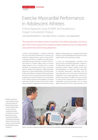
- 1. ©2016 TOSHIBA MEDICAL SYSTEMS56 | VISIONS27 1) Children’s Health and Exercise Research Centre, University of Exeter, UK 2) Bristol Congenital Heart Centre, The Bristol Heart Institute, University Hospitals, Bristol NHS Foundation Trust, Bristol 3) National Institute for Health Research (NIHR) Cardiovascular Biomedical Research Unit, Bristol Heart Institute, University of Bristol 4) Toshiba Medical Systems Europe, B.V. SPORTS MEDICINE ULTRASOUND 2D WMT Currently, echocardiography is however performed only at rest and exercise performance, if assessed, uses traditional cardio-pulmonary exercise testing (CPET). A limitation of CPET is its inability to provide data on myocardial functional responses to exercise, the domi- nant process to increase cardiac output to enhance oxygen delivery1 and it is therefore only an indirect description of myocardial reserve. Data is particularly sparse in children’s and adolescent’s athlete population groups. But as sports professionalism and training levels in elite youth athletes is reaching that of their adult peers, the search for non-invasive, discriminative and predictive imaging tools to assess the health of the young athlete, for this rapidly increasing youth population, has gained importance. Equally, the question about how to assess exercise limitations is applicable and as important in many paediatric disease groups e.g., congenital heart disease (CHD) and novel methodologies and protocols to assess cardiac function during exercise are needed. In search for echocardiographic assessment tools during exercise myocardial deformation imaging or 2D Wall Motion Tracking (WMT) has emerged as a potentially suitable imaging modality during stress echocardiography as it allows for direct assessment of myocardial performance. Importantly, some 2D strain parameters such as strain rate are relatively load independent2 and correlate positively to invasive con- tractility measurements3. 2D WMT is also more sensitive to mild functional impairment than traditional echo- cardiographic functional parameters and can assess regional myocardial function. Image acquisition of 2D Guido E. Pieles DPhil, MD, MRCPCH 2,3), Craig A. Williams, PhD, MSc 1), Carla Witkam – Van den Maegdenbergh 4) Exercise Myocardial Performance in Adolescent Athletes The assessment of cardiac function is essential in the athletic population not only as part of the screening process for underlying cardiac disease, but also to longitudinally assess performance and training adaptations. A Novel Approach Using 2D WMT and Simultaneous Oxygen Consumption Analysis Figure 1: Set-up for simultaneous performance of exercise echocardiography and cardio-pulmonary exercise testing
- 2. VISIONS27 | 57ULEU160048 strain data is time efficient and easy to obtain. Unlike Tissue Doppler Imaging, 2D WMT also shows angle independency, paramount when acquiring echocar- diographic images during exercise. Echocardiographic hardware and software however have to be robust and able to allow tracking at high heart rates and challeng- ing conditions during exercise and further development work is required. Combined, 2DWMT and CPET can provide a comprehen- sive and direct description of cardio-pulmonary exercise response and might also help differentiate between normal and pathological myocardial function during exercise. Here we present pilot data of a synergistic and time efficient comprehensive assessment of cardio- pulmonary response to incremental exercise, using 2D WMT to assess myocardial function in conjunction with simultaneous assessment of metabolic gas analysis by CPET, in adolescent elite athletes. METHODS 14 male adolescent professional football academy players (mean age 15.4 ± 0.8 y) underwent echo- cardiography at rest, during exercise and recovery while completing an incremental CPET on a recumbent cycle ergometer (Fig. 1). Echocardio- graphy at rest was performed following England Football Association screening guidelines. LV myocardial performance was serially assessed during exercise and recovery measuring LV peak systolic longitudinal (LV Sl) and LV peak systolic circum- ferential (LV Sc) 2D strain. The study was conducted in collaboration with our research partners Toshiba Medical Systems and Manchester United Football Club Youth Academy, UK. EXERCISE ECHOCARDIOGRAPHY AND 2D WMT Exercise echocardiography was performed using the Artida for image acquisition and UltraExtend ACP soft- ware for 2D WMT analysis. Analysis is also possible using the AplioCV system and the on-board analysis software. A LV focused 4-chamber view (Fig. 2) and a paraster- nal short axis view (Fig. 3) were captured for 2D WMT analysis at rest, at several exercise stages and at recovery. Three cardiac cycles were acquired at rates of 30–90 frames per second in raw DICOM format and analysis was performed on one manually selected cardiac cycle. The endo- cardial borders were manually contoured at end- diastole with the range of interest adjusted to include the whole myocardium. Peak strain was defined as the maximal deformation of a segment in systole and represented as a percentage of the original size. Standard nomenclature was used to describe the Figure 2: Representative 2D image sequence during exercise for LV Sl. A: rest at HR = 87 bpm; B: 50 W at HR = 90 bpm; C: 100 W at HR = 117 bpm Figure 3: Representative 2D image sequence during exercise for LV Sc. A: rest at HR = 84 bpm; B: 50 W at HR = 96 bpm; C: 100 W at HR = 99 bpm
- 3. ©2016 TOSHIBA MEDICAL SYSTEMS58 | VISIONS27 LV segments and LV peak systolic longitudinal strain recorded for the three lateral and three septal segments. Circumferential peak systolic strain was measured at the base of the LV. Mean or global values for circumferential and longitudinal strain were calculated for each level only if good tracking was obtained in a minimum of four segments. An incremental CPET on a recumbent cycle ergometer to volitional exhaustion was performed by all participants during imageacquisition. ASSESSMENT OF MYOCARDIAL PERFORMANCE BY 2D WMT DURING CARDIO-PULMONARY EXERCISE TESTING We tested if LV myocardial performance can be quantita- tively assessed by 2DWMT during exercise and if 2DWMT can describe myocardial performance during exercise. Image acquisition for 2D WMT was robust up to a power output of 150 W. The performance of 2D WMT during exercisestagesrevealedseveralimportantrelationshipsof cardiac exercise adaptations. 2D WMT analyses showed a linear increase in myocardial performance with increasing power output and exercise stage (Fig. 4). LV peak systolic global longitudinal (Sl) and LV peak systolic global cir- cumferential (Sc) strain showed a linear relationship with significant differences across increasing exercise stages up to 150 W compared to rest (p 0.01). This indicates that, besides stroke volume and HR increase, intrinsic increase of myocardial performance is an important mechanism of cardiac output increase during exercise. FORCE-FREQUENCY RELATIONSHIP DURING EXERCISE LV Sl peak also significantly correlated to HR max (r = 0.59, p = 0.03) directly confirming the classic concept of a posi- tive force-frequency relationship (FFR) – a key relation- ship for the normal adaptation of cardiac function during exercise (Fig. 5). RELATIONSHIP BETWEEN CARDIAC FUNCTION DURING EXERCISE AND RECOVERY Cardiac recovery response is an important marker of fitness and we therefore also assessed 2D strain at 2 min and 6 min of recovery and found a significant correlation between LV Sl peak and LV Fig. 4: Left: LV Peak systolic global longitudinal (Sl) (left) and LV Peak systolic global circumferential (Sc) against power output, with linear regression lines. Right: LV Peak systolic global longitudinal (Sl) and LV Peak systolic global circumferential (Sc) against stage, with linear regression lines. Ten stages as follows: Rest, 0 W = unloaded pedalling, 25 W, 50 W, 75 W, 100 W, 125 W, 150 W, 2 min rec Fig. 5: LV Peak systolic global longitudinal (Sl) (left) and LV Peak systolic global circumferential (Sc) strain (right) in relation to HR with linear regression lines showing a positive force-frequency relationship over different exercise stages
- 4. VISIONS27 | 59 Sl rec (r = 0.57, p = 0.04) and LV Sc peak and LV Sc rec (r = 0.56, p = 0.04). These findings point towards a direct relationship between myocardial performance during exercise and recovery and this will need to be explored further, as recovery 2D strain parameters could serve as a useful tool in assessing cardiac function and reserve. MYOCARDIAL PERFORMANCE METABOLIC RELATIONSHIP Our combined methodology also allows for assessment of the relationship between myocardial performance and exercise oxygen consumption. In our small cohort we found only a weak correlation between LV Sl peak and LV Sc peak to VO2max, (r = -0.20 – 0.40, p 0.05) and larger populations will need to be studied to assess the relationship between myocardial exercise performance and metabolic exercise parameters in more detail. DISCUSSION 2D WMT echocardiography during exercise is feasible to describe myocardial performance and in com- bination with simultaneous CPET can enhance our under- standing and interpretation on the complex cardiac and metabolic exercise adaptations during exercise and recov- ery.To our knowledge this is the first time that the relation- ship between myocardial performance as measured by 2D WMT and the metabolic exercise parameters have been assessed simultaneously in adolescent elite athletes. We have determined that LV myocardial performance increases significantly and incrementally through different exercise stages without reaching a plateau. We have also described an accentuated force-frequency relationship during exercise. The exercise force- frequency relationship has not been demonstrated using 2D strain during exercise. Direct measurement of the force-frequency relationship during exercise stress could particularly be of importance to discover early ventricular dysfunction in patients with near normal resting function. Overall, our data indicate that myocardial perfor- mance assessment by 2D WMT is a sensitive and responsive tool for the quantification of cardiac adaptation during exercise and in recovery. The advantage of our combined protocol compared to other methods, e. g., inotropic stimulation or pacing to increase myocardial performance, lies in its non-invasiveness and more importantly has a higher external validity, in that it mimics physical activity and its effect on cardiac performance. LIMITATIONS It should be noted that 2D strain assesses only unidi- rectional myocardial deformation forces and cannot therefore capture the complex multi-dimensional and directional cardiac myofibre deformation4. We have attempted to address the multi-dimensional LV myocar- dial deformation by analyzing the two most widely used deformation planes, longitudinal and circumferential strain analysis. The recent development of 3D WMT5 will allow us to address this limitation in the future. Image optimization during exercise to obtain adequate 2DWMT data should include reduction of artefacts, noise and image window focus with the view to obtain sufficient frame rates. CONCLUSION Direct assessment of ventricular function parameters by using 2D WMT during exercise can be utilized to directly describe myocardial exercise performance and can overcome the limited predictive value of exercise capacity on myocardial function. In the clinical setting, this protocol could serve as a tool to better quantify myocardial reserve, which is an important concept in patient risk stratification of ventricular dysfunction. Our current study as introduced in this paper will use 2D WMT to compare myocardial performance in three paediatric groups, non-trained but healthy children, elite youth athletes and children with CHD to deter- mine the mechanisms of exercise limitations and cardiac dysfunction in children with CHD. ACKNOWLEDGEMENTS This study is part of a research partnership between Toshiba Medical Systems UK, Manchester United Football Club and the Universities of Bristol and Exeter. The research partnership between Toshiba and the University of Bristol is a contractual research partner- ship that determines the independence of the research from either parties’ interests. The research is supported by the University of Exeter and the University of Bristol NIHR Biomedical Research Unit for Cardiovascular Disease. GEP holds a National Institute for Health Research (NIHR) Academic Clinical Lectureship. We would like to thank the athletes and staff at Manchester United Football Club Youth Academy. ULEU160048 Credits Mark Hitchman, Tim Palarm Mair Howe References 1. Bassett, D. R., Jr. and E. T. Howley (2000).“Limiting factors for maximum oxygen uptake and determinants of endurance performance.”Med Sci Sports Exerc 32(1): 70–84. 2. Weidemann, F., F. Jamal, M. Kowalski, T. Kukulski, J. D’Hooge, B. Bijnens, L. Hatle, I. De Scheerder and G. R. Sutherland (2002). “Can strain rate and strain quantify changes in regional systolic function during dobutamine infusion, B-blockade, and atrial pacing-implications for quantitative stress echo- cardiography.”J Am Soc Echocardiogr 15(5): 416–424. 3. Kovacs, A., A. Olah, A. Lux, C. Matyas, B. T. Nemeth, D. Kellermayer, M. Ruppert, M. Torok, L. Szabo, A. Meltzer, A. Assabiny, E. Birtalan, B. Merkely and T. Radovits (2015). “Strain and strain rate by speckle tracking echocardiography correlate with pressure-volume loop derived contractility indices in a rat model of athlete’s heart.”Am J Physiol Heart Circ Physiol: ajpheart 00828 02014. 4. Ferferieva, V., A. Van den Bergh, P. Claus, R. Jasaityte, P. Veulemans, M. Pellens, A. La Gerche, F. Rademakers, P. Herijgers and J. D’Hooge (2012). “The relative value of strain and strain rate for defining intrinsic myocardial function.”Am J Physiol Heart Circ Physiol 302(1): H188–195. 5. Seo, Y., T. Ishizu and K. Aonuma (2014).“Current status of 3-dimensional speckle tracking echo.