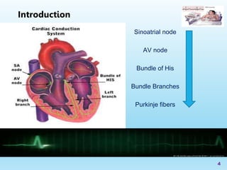
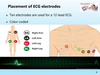
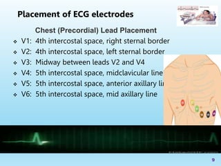
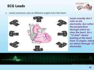
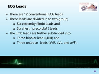
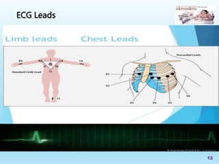
Basics of Electrocardiography (ECG)
Birhanu Chekol (BSc. MSc.)
Senior Anesthetist and Paramedic specialist
3/13/2023
1
 Objectives
 Introduction
 Cardiac conduction System
 Placement of electrodes
 ECG lead
 Summary
 References
2
Outline
Upon completion of this lecture, you will be able to:-
 Define ECG
 Describe the cardiac conduction system
 Identify the waves of the ECG
 Explain the different types of ECG leads
 Describe the importance of prehospital ECG
3
Objectives
Sinoatrial node
AV node
Bundle of His
Bundle Branches
Purkinje fibers
4
Introduction
 The electrocardiogram (EKG) is a representation of the
electrical events of the cardiac cycle.
 ECG is the gold standard for the noninvasive diagnosis of
cardiac diseases and may occasionally be the only marker
for the presence of heart disease
 Each event has a distinctive waveform
5
Introduction
 A typical ECG tracing is a repeating cycle of three electrical
entities:
a. P wave
b. QRS complex
c. T wave
6
Introduction
With ECG we can identify…
 Arrhythmias
 Myocardial ischemia and infarction
 Pericarditis
 Chamber hypertrophy
 Electrolyte disturbances (i.e. hyperkalemia, hypokalemia)
7
Introduction
 Ten electrodes are used for a 12-lead ECG.
 Color-coded
8
Placement of ECG electrodes
Chest (Precordial) Lead Placement
 V1: 4th intercostal space, right sternal border
 V2: 4th intercostal space, left sternal border
 V3: Midway between leads V2 and V4
 V4: 5th intercostal space, midclavicular line
 V5: 5th intercostal space, anterior axillary line
 V6: 5th intercostal space, mid axillary line
9
Placement of ECG electrodes
 Leads (cameras) view at different angles from the heart.
10
ECG Leads
Leads actually don’t
refer to the
electrodes, but rather
the perspectives
through which we
view the heart. So a
"12-lead" means
looking at the heart
from 12 angles, which
we obtain through 10
electrodes.
 There are 12 conventional ECG leads
 These leads are divided in to two group;
 Six extremity (limb) leads and
 Six chest ( precordial ) leads.
 The limb leads are further subdivided into:
 Three bipolar lead (I,II,III) and
 Three unipolar leads (aVR, aVL, and aVF).
11
ECG Leads
12
ECG Leads
 Early recognition and fast, appropriate treatment can
prevent the extension of an MI
 Early recognition = early intervention
 An important diagnostic tool will also be the patient’s
general appearance
13
Why Are We doing Pre-hospital
ECG’s?
 An electrocardiogram is a painless, noninvasive way to
help diagnose many common heart problems.
 The prehospital 12-lead ECG is associated with better
survival in patients with acute coronary syndrome (ACS)
 Early recognition = early intervention
14
Summary
 Ecgmadesimple.com
 Book: ECGs for the Emergency Physician – Amal Mattu
 lifeinthefastlane.com/ecg-library
15
References
16
3/13/2023
Thank
you!!!

Basics of Electrocardiography.pptx

  • 1.
    Basics of Electrocardiography(ECG) Birhanu Chekol (BSc. MSc.) Senior Anesthetist and Paramedic specialist 3/13/2023 1
  • 2.
     Objectives  Introduction Cardiac conduction System  Placement of electrodes  ECG lead  Summary  References 2 Outline
  • 3.
    Upon completion ofthis lecture, you will be able to:-  Define ECG  Describe the cardiac conduction system  Identify the waves of the ECG  Explain the different types of ECG leads  Describe the importance of prehospital ECG 3 Objectives
  • 4.
    Sinoatrial node AV node Bundleof His Bundle Branches Purkinje fibers 4 Introduction
  • 5.
     The electrocardiogram(EKG) is a representation of the electrical events of the cardiac cycle.  ECG is the gold standard for the noninvasive diagnosis of cardiac diseases and may occasionally be the only marker for the presence of heart disease  Each event has a distinctive waveform 5 Introduction
  • 6.
     A typicalECG tracing is a repeating cycle of three electrical entities: a. P wave b. QRS complex c. T wave 6 Introduction
  • 7.
    With ECG wecan identify…  Arrhythmias  Myocardial ischemia and infarction  Pericarditis  Chamber hypertrophy  Electrolyte disturbances (i.e. hyperkalemia, hypokalemia) 7 Introduction
  • 8.
     Ten electrodesare used for a 12-lead ECG.  Color-coded 8 Placement of ECG electrodes
  • 9.
    Chest (Precordial) LeadPlacement  V1: 4th intercostal space, right sternal border  V2: 4th intercostal space, left sternal border  V3: Midway between leads V2 and V4  V4: 5th intercostal space, midclavicular line  V5: 5th intercostal space, anterior axillary line  V6: 5th intercostal space, mid axillary line 9 Placement of ECG electrodes
  • 10.
     Leads (cameras)view at different angles from the heart. 10 ECG Leads Leads actually don’t refer to the electrodes, but rather the perspectives through which we view the heart. So a "12-lead" means looking at the heart from 12 angles, which we obtain through 10 electrodes.
  • 11.
     There are12 conventional ECG leads  These leads are divided in to two group;  Six extremity (limb) leads and  Six chest ( precordial ) leads.  The limb leads are further subdivided into:  Three bipolar lead (I,II,III) and  Three unipolar leads (aVR, aVL, and aVF). 11 ECG Leads
  • 12.
  • 13.
     Early recognitionand fast, appropriate treatment can prevent the extension of an MI  Early recognition = early intervention  An important diagnostic tool will also be the patient’s general appearance 13 Why Are We doing Pre-hospital ECG’s?
  • 14.
     An electrocardiogramis a painless, noninvasive way to help diagnose many common heart problems.  The prehospital 12-lead ECG is associated with better survival in patients with acute coronary syndrome (ACS)  Early recognition = early intervention 14 Summary
  • 15.
     Ecgmadesimple.com  Book:ECGs for the Emergency Physician – Amal Mattu  lifeinthefastlane.com/ecg-library 15 References
  • 16.

Editor's Notes

  • #7 In rare case like hypokalemia, U wave can be seen
  • #14 EKG’s sent to the ED before patient arrival allows for the right personnel to be available to properly care for the patient in the most time efficient manner