Prevalence and associated risk factor of bovine calves coccidiosis in nekemt city, oromia, western ethiop.pdf
•
0 likes•2 views

Prevalence and associated risk factor of bovine calves coccidiosis in nekemt city, oromia, western ethiopia Authors:Walkite Furgasa , Sosina Dawit , Shibiru Wako and Adisu Dube Int J Biol Med Res. 2023; 14(4): 7660-7664 | Abstract | PDF File

Recommended
Recommended
More Related Content
Similar to Prevalence and associated risk factor of bovine calves coccidiosis in nekemt city, oromia, western ethiop.pdf
Similar to Prevalence and associated risk factor of bovine calves coccidiosis in nekemt city, oromia, western ethiop.pdf (20)
More from BioMedSciDirect Publications
More from BioMedSciDirect Publications (11)
Recently uploaded
Recently uploaded (20)
Prevalence and associated risk factor of bovine calves coccidiosis in nekemt city, oromia, western ethiop.pdf
- 1. BioMedSciDirect Publications Int J Biol Med Res.2023 ;14(4):7660-7664 Contents lists available at BioMedSciDirect Publications Journal homepage: www.biomedscidirect.com International Journal of Biological & Medical Research International Journal of BIOLOGICAL AND MEDICAL RESEARCH www.biomedscidirect.com Int J Biol Med Res Volume 14, Issue 4, Oct 2023 Copyright 2023 BioMedSciDirect Publications IJBMR - ISSN: 0976:6685. All rights reserved. c ARTICLE INFO ABSTRACT Keywords: Bovine calf Coccidiosis Nekemte Oocyst Prevalence Risk factor 1. Introduction Parasitic diseases are a major constraint in animal health and productionthroughoutthetropicandsub-tropicalcountriesofthe world (Bruhn et al., 2011). Parasitic disease caused by helminthes, protozoa and arthropods can cause more economic losses than disease caused by bacteria and viruses but their impact is not clear to animal owners (Juyal and Single, 2011). Bovine coccidiosis occursworldwideandusuallyaffectscattleunderoneyearold,but it occasionally seen in yearlings and adults. Bovine coccidiosis has been observed in almost all areas where cattle are raised and is usually most common and important in calves younger than one year (Daugschies and Najdrowsk, 2005). Coccidiosis spreads from one animal to another by contact with infected feces and is one of the most alarming problems for calf rearing industry. The development of clinical coccidiosis in cattle mainly depends on factors like species of Eimeria, age of infected animal, number of oocysts ingested, presence of concurrent infections and type of production system and management practices (Daugschies and Najdrowsk,2005). Eimeria infections are one of the most common and important diseases of cattle worldwide. It is a protozoan disease that has a special place among the many parasite species for different reasons caused by various species of Eimeria, widespread distribution regardless of the climatic conditions, results major economic losses in animal husbandry worldwide (Almeida et al., 2011;Nisar-Khanetal.,2013). About 11 species of Eimeria have been identified and documented to cause disease in cattle (Coetzer and Justin, 2004) and the most common pathogenic species in cattle are E. bovis and E. zuernii (Maas, 2007). The other species have been shown experimentallytobemildlyormoderatelypathogenic,buttheyare not considered important pathogens. All calves kept under traditional management systems are exposed and become infected early in life. As coccidiosis spreads from one animal to another by contact with infected feces, it is one of the serious problemsforcalfrearingindustry(Lucasetal.,2006). Adult animals are usually asymptomatic carriers that often serve as a source of infection for juvenile animals, which are more susceptible to infection. In association with other enteropathogens,coccidiahavebeenindicatedasanimportant Coccidiosis is a parasitic disease caused by a small, single celled parasite, called a protozoa, that lives inside the cells of an infected animal's intestinal tract and is one of the most common and important disease of calves in the world. A Cross-sectional study was conducted in and around nekemt town from January 2022 up to September 2022 to determine the prevalence and associated risk factors of infection in calves. Faecal samples were collected from a total of 384 calves less than 24 month of age and examined for the oocysts of coccidian. Detailed information of the age, sex, breed, management system and hygienic status were considered as risk factors. Centrifugal faecal floatation technique using sugar solution was used to detect coccidian oocyst. From the total calves included, 176 calves were demonstrated for the presence of coccidian infection. The most of diarrheic calves show a positive result for Eimeria infection .Coccidian oocyst was detected in calves from 1 to 2 year of age but greater prevalence was observed in age categories less than 6 month of age. Statistically significant association (p<0.05) between prevalence of coccidiosis and age, breeds and husbandry system was observed in the study. There was a statistically significant association (P<0.05) between prevalence of coccidiosis and the hygienic status of the calf. Accordingly, calves with poor hygienic condition showed significantly higher prevalence than calves which have relatively better hygienic condition However, there was no statistically significant association (p>0.05) with sex. In conclusion, the study revealed that calf coccidiosis was prevalent in and around nekemte town and consequently affects the productivity of the sector. Hence, appropriate diseasepreventionandcontrolmeasuresarerequiredtobeundertakentoreduceitseffect. Original article PREVALENCE AND ASSOCIATED RISK FACTOR OF BOVINE CALVES COCCIDIOSIS IN NEKEMT CITY, OROMIA, WESTERN ETHIOPIA a a b b Walkite Furgasa , Sosina Dawit , Shibiru Wako and Adisu Dube * Corresponding Author : Wollega University, School of Veterinary Medicine Email: walkiteharamaya@gmail.com Copyright 2023 BioMedSciDirect Publications IJBMR - All rights reserved. c Walkite Furgasa a Wollega University, school of veterinary Medicine b East wollega Agricultural office, Guto Gida Veterinary Clinc
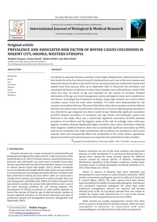
- 2. causeofdiarrheaincalvesonthefirstfewmonthsoftheirage(Abebe et al., 2008). They develop only in the intestinal epithelial cells, leading to mucosa damage and the appearance of clinical signs, malnutrition, weakness, anaemia, diarrhoea and haemorrhagic faeces(Yuetal.,2011). The development of clinical coccidiosis in cattle mainly depends on factors like species of Eimeria, age of infected animal, number of oocysts ingested, presence of concurrent infections and type of production system and management practice (Alula et al., 2013). Diagnosis of coccidiosis depends on the discovery of oocysts on faecal examination using direct smear, flotation or McMaster's techniques. Studies have demonstrated that the prevalence of Eimeria species in cattle varies between different regions and age of animal(Alemayehuetal.,2013). The prevalence, species composition, and importance of bovine coccidiosis have been documented in various countries of the world. In Ethiopia, different finding reported that young age and poor hygienic status of the farms were strongly associated with infection of coccidiosis in dairy farms., However, in another study by Alula agreed that age was a significant factor but breed, body condition, sex, and management system were not significantly associated with thedisease(Dawidetal.,2012). According to Dawid et al 2012), although coccidiosis is an important cause of calf morbidity and mortality in Ethiopia and very little attention has been given to this disease. Nekemte is one of the cities of Oromia where urban and periurban dairy farms dwell to provide milk for population of the city. Reports on coccidiosis are scarce to assess the magnitude of this disease and no original informationaboutthediseaseinthepresentstudy.Inviewofthelack of authentic information available regarding the prevalence of Coccidiosis affecting calves in the study area. Therefore the objectives of the present study were: To estimate the prevalence of coccidiosis affecting calves in Nekemte city and to estimate the associatedriskfactorsofcoccidiosisindairycalves MATERIALSANDMETHODS StudyArea The study was conducted in Nekemte town, East Wollega zone, Oromia regional state, Western Ethiopia from January 2022 up to September2022.Nekemteliesatlatitudeof90o5'Nandlongitudeof 36° 33' E with an elevation of 1,960 to 2,170 meters above sea level. Distance of the district is 331 km from Addis Ababa. The climatic condition alternates with long summer rainfall (June to September), short rain season (March to April) and winter dry season (December to February). The maximum and minimum annual rainfall and daily temperature range are between 2,200to 1,500mm and 15°Cto 27°C respectively. This area is characterized by mixed farming system, which is engaged in agricultural and livestock production. The calves population of the town is 4812 cattle, 851 sheep, 359 goats, 81 horses, 21 mule, 851 donkeys and 4850 chickens. The majority of the people of the town belong to the O r o m o e t h n i c community and Afan Oromo (the Oromo language) is the widely spoken language in the area(EWAO,2013). StudyPopulation The study was conducted on calves younger than 24 months by dividing in to three groups: Birth up to 6 months, 6-12 months and 12-24 months which were determined by asking the owner of the animal orally (Mihreteab et al., 2012). This range of age was selected because the disease is more common in young animal. Epidemiological information with respect to their age, sex, breed, management system, and date of sample collection, hygienic states and kebele or name of the farm was collected. Simple random sampling was used to select the study animals from farms and from small holder. Hygienic status of calf pens and the calves themselves were assessed based on housing system (ventilation, stocking and sanitation) and body parts of the calves and was conveniently categorizedaspoor,mediumandgood(Mihreteabetal.,2012). StudyDesign A Cross-sectional study was conducted in and around nekemte town from November 2021 to March 2022 to determine the prevalence and associated risk factors of Coccidiosis in calves. Active data was generated from randomly selected calves with regard to age, breed, sex, Husbandry system, and hygienic states were consideredasriskfactorstotestforoccurrenceofcoccidiosis. SampleSizeDetermination Simple random sampling method was used to select the calves from target population. Since there was no similar work done in the area previously, expected prevalence was taken as 50% and the confidenceintervalchosenas95%andprecision5%.Bysubstituting these values in the formula, the sample size founded to be 384. Thus, the sample size is calculated according to (Thrusfield, 2007) as follows: Where, n=required sample size, d=absolute precision (usually 0.05)Pexp=expectedprevalence DataCollection A total of 384 faecal samples were collected during the entire period of the study. About 10-20 g fresh faecal sample was collected per rectum from each calf using sterile disposable plastic gloves. Each sample was placed in a clean plastic container labeled and transported to Veterinary parasitology laboratory, School of Veterinary Medicine, Wollega University on the same day of collection, and preserved at refrigeration temperature until processing within 24 hours of arrival. At the time of sampling, the name of the farm, date of sampling, age, sex, breed, fecal consistency and hygienic status of farm was recorded for each calf on a recording format. A 3g portion of each of the 384 fecal samples collected was weighed out using a balance and put in a mortar. After grinding with pistil 40ml of sugar solution was added, mixed thoroughly and poured into a 100-ml glass beaker through a strainer. Then it was rinsed and the solution was poured into 15-ml centrifuge tubes but not filled. After centrifugation at 1200 rpm for 5 min, more sugar solutionwasaddeduntilaconvexmeniscuswasformedontopofthe tube.Aglasscoverslipwasplacedontopofeachtubeandwasleftfor 30min.Then,eachglasscoverslipwasbrisklyliftedupandplacedon a clean glass slide, not allowing formation of air bubbles. The entire area under each cover slip was examined under a binocular microscopeat40×magnification(Hendrix,1998). 7661 Walkite Furgasa et al. /Int J Biol Med Res.14(4):7660-7664 Figure : Map of the study Area Source: Arc Map GIS 10.4 2 2 n = (1.96) pexp (1-pexp)/ d
- 3. DataManagementandAnalysis Data collected from study sites were coded and entered in to a Microsoft excel spread sheet program for analysis. Statistical analysis was done on Statistical Package for Social sciences (SPSS) software version 16. Descriptive statistics like percentage was used to express prevalence while chi-square (χ2) test was used to compare the association of coccidiosis with different risk factors. In all the cases, 95% confidence level and 0.05 absolute precision errorswereconsidered.Ap-value≤0.05wasconsideredstatistically significant. RESULT Out of 384 faecal samples examined, 176 were positive for Eimeria oocysts with the overall prevalence of 36.4%. Even if coccidian oocyst was detected on all age groups the highest prevalence was recorded in those calves found in the range from one to sixth month of age. There was a statistically significant difference (P<0.05) in the prevalence of coccidiosis among the various age group (Table 1). The prevalence of coccidiosis was a bit higher in cross breed calves than in local breed calves. However, the breed of the calves was significantly associated (P < 0.05) with prevalence of coccidiosis. There was no statistically significant association (P>0.05)betweensexandcoccidianinfection. There was a statistically significant association (P<0.05) between prevalence of coccidiosis and the fecal consistency of the calf. Accordingly, calves with diarrheic condition showed higher prevalence than calves which have soft and normal fecal consistency (Table2). Table : Prevalence of coccidiosis in calves in relation to consistency of feces Table : Prevalence of coccidiosis in calves in relation to host factors There was a statistically significant association (P<0.05) between prevalence of coccidiosis and the hygienic status of the calf. Accordingly, calves with poor hygienic condition showed significantly higher prevalence than calves which have relatively betterhygieniccondition(Table3).Coccidianinfectionsaccordingto management system have significant difference with Intensive, semi-Intensive, and extensive husbandry systems with higher prevalence on intensive system than other systems and the lowest prevalence was observed on calves belongs to semi-intensive system. DISCUSSION The overall prevalence of coccidiosis in and around Nekemte basedoncoprologicalexaminationwas36.4%andthiscurrentstudy was in line with the prevalence study of calf coccidiosis in kombolcha which is 32% (Alemayew et.al., 2013). However, the prevalence was lower than the previous findings reported in Addis Ababa and bishoftu by (Abebe et al., 2008) (68.1%), in Pakistan by (Muhammad et al., 2010) (47.09%), in the coastal plain area of Georgia (USA) by (Ernst et al., 1987) (82.28%) and in sub-humid tropical climate by (Rodriguez-Vivas et al., 1996) (87.8%). This variation is most likely attributed to the differences in agro-ecology, management types and husbandry practices of the study animals in differentareas(Radostitsetal.,2006). The study has revealed that the prevalence of Eimeria has significant association with breed of calves. There was statistically significant association (P<0.05) between breed and coccidia infection. Higher prevalence of the disease condition was observed in exotic breed when compared to local and cross breed. This might be in relation to genetic factors as exotic breed are more susceptible to a disease. This finding agrees with the report of (Abebe et al., 2008) and (Alemayew et al., 2013). There was also no statistically significant association (P>0.05) between sex and coccidia infection. The prevalence in female calves was similar to that of males in this study. This finding agrees with the report of (Abebe et al., 2008) and (Alemayewetal.,2013). Age of the calves was significantly associated (P<0.05) with the risk of infection by coccidiosis and the highest prevalence was recorded in those calves with youngest age groups (1 to 6months). This is in contrast to (Abebe et al., 2008) who reported that risk of infection by Eimeria species appeared to increase with the age of the examined calves. However, this observation in the current study was in line with (Dennis et al., 2012), (Perfield, 2010) and (Mihreteab et al.,2012).whonotedthatyounganimallessthan6monthswere Table : Prevalence of coccidiosis in calves in relation to husbandrysystemandhygienicstatus 7662 Walkite Furgasa et al. /Int J Biol Med Res.14(4):7660-7664
- 4. more susceptible than adults. Stress factors like weaning and change ofdietcanincreaselevelofinfectionandincidenceofthediseasedue to stress-induced immune suppression (Kaufman, 1996; Radostitis et.al., 2007). In addition to this, coccidiosis is a self- limiting disease in adult and spontaneous recovery without specific treatment is common when the multiplication stage of the coccidian has passed (Radostitis et.a., 2007). Based on this, previous exposure might have a contribution to the development of certain level of immunity of older calves as compared to younger that did not experience previous exposure (Faber et al.,. 2010). The influence of husbandry system from this study also shows that a significant association between prevalence of coccidian infection and different husbandry system which is agree with (Kennedy and Kralka 1987), but strongly disagreeswiththeworkofAlemayewinKombolchaonprevalenceof bovine coccidiosis (Alemayew et al., 2013). Coccidiosis is mostly a disease of young animals kept under intensive management systems when there is stress, overcrowding, housing under conditions of poor hygiene, food changes, nutritional deficiencies, and adverse weather conditions which are favorable for the survival of oocysts and therefore higher infection rates when compared to extensive farmingsystems(Vorsterand Mapham,2012). The strong association of the infection with coccidiosis in relation to the hygienic status of calve has been demonstrated in this study. This observation agrees with (Mihreteab et.al, 2012). Calves with poor hygiene showed significantly higher prevalence than calves which have relatively better hygiene. This could imply that poorsanitationincalvehousingareasaswellaspoormanagementof housing favours infection with coccidiosis. Obviously, poor ventilation, heavy stocking, cows present with calves, and soiled bedding were regarded as risk factors for coccidiosis (Daugschies and Najdrowski 2005; Radostitis et al., 2007; Vorster and Mapham, 2012). Positive correlation between fecal consistency and the occurrence of diarrhea was detected in the present study. During investigation, most of diarrheic calves show a positive result for Eimeriainfection.Theseagreewith(Pandit,2009).Theresultsofthe present study confirmed the importance of coccidiosis among the causesofcalfdiarrheainthestudyarea. CONCLUSIONANDRECOMMENDATION This study has revealed that the prevalence of calves coccidiosis in and around nekemte town was 36.4%, which can be taken as high rate of infection. The prevalence of coccidiosis has no significant association with sex and body condition of animals examined during the study period. However, the disease has a significant association (P<0.05) with breed, age, Husbandry system and hygienic status. Breed, age, husbandry system and hygienic status of calves were the major risk factors for the prevalence of coccidiosis in this area. In conclusion, the study revealed that calf coccidiosis was prevalent in and around nekemte town and consequently affects the productivity of the sector. In line with the above conclusion the following recommendation are forwarded: Calves should get colostrum in the first 24 hrs of their life to ensure their immune status,Sanitary prophylaxis should be maintained, Stressful conditions which triggers the disease occurrences should be avoided, Coccidiostats shouldbeusedinrationearlyforpreventionandAwarenesscreation for Livestock producers for the improvement of hygienic status of calve EthicsApprovalandConsenttoParticipate The current study was conducted after permitted ethical approval and a statement given by the Research Review Committee ofWallagaUniversityonNovember9,2021(MinutesNo.12/2021). Acknowledgments The authors acknowledged Wollega University, school of veterinarymedicinefortheircontributionforthisresearch. AuthorContributions Authors made a significant contribution during this Msc thesis workfromitsbeginningtoend. 7663 References 1. Abebe, R., Wossene, A. and Kumsa, B. (2008): Epidemiology of Eimeria infections in calves in Addis-Ababa and Debre- Zeit dairy farms, Ethiopia. InterJApplResVetMed.(6),Pp24-30. 2. Alemayehw, A, Mohammed, .N, and Timketa, B. (2013): Prevalence of bovine coccidian in Kombolcha district of south Wollow Ethiopia. J Vet Med and AnimalHealthPp41-45. 3. Almeida, A., Magalhaes, V., Muniz, E. and Munhoz, A. (2011): Frequency of species of the genus Eimeria in naturally infected cattle in Southern Bahia, NortheastBrazil.BrazilianJVetParasitol.20,Pp78-81. 4. Alula, A., Nuru, M. and Belina, T. (2013): Prevalence of bovine Coccidia in Kombolcha district of South Wollo, Ethiopia, J. Vet. Med. Anim. Hlth., 5(2), Pp 41-45. 5. Bangoura, B. and Bardsley, K. (2020): Ruminant coccidiosis. Vet Clin North AmFoodAnim.Pract.36,Pp187–203. 6. Bangoura, B., Mundt, C., Schmäschke, R., Westphal, B., Daugschies, A. (2012): Prevalence of Eimeria bovis and Eimeria zuernii in German cattle herds and factorsinfluencingoocystexcretion.ParasitolRes110(2),Pp875-881. 7. Belli, S., Smith, N., Ferguson, D. (2006): The coccidian oocyst: a tough nut to crack!TrendsParasitol22(9),Pp416-23. 8. Bruhn, F., Lopes, A., Demeu, F., Perazza, A., Pedrosa., M. and Guimaraes., M. (2011): Frequency of species of Eimeria in females of the holstein friesian breed at the post-weaning stage during autumn and winter. Rev. Bras. Parasitol.20(4),Pp303-307. 9. Burton, J., Madsen, A., Chang. L., Weber, P., Buckham, K., Dorp, R. and Hickey, M. (2005): Gene expression signatures in neutrophils exposed to glucocorticoids: a new paradigm to help explain “neutrophil dysfunction” in parturient dairy cows. Veterinary Immunology and Immunopathology. 105, Pp197-219. 10. Chapman, H. (1974a): The effects of natural and artificially acquired infectionsofcoccidiainlambs.ResVetSci16(1),Pp1-6. 11. Chapman, H. (1974b): The immunity of lambs to coccidia acquired in the fieldandbyartificialinfection.ResVetSci16(1),Pp7-11. 12. Coetzer, J. and Justin, R. (2004): Infectious Diseases of Livestock. Second Edition3,OxfordUniversitypress.Pp319–331. 13. Coico,R.,Sunshine,G.(2009):Immunology:ashortcourse.Wiley-Blackwell, Hoboken,NewJersey.Pp8-100. 14. Curt, A. (2005): Senior Extension Associate, Department of Agricultural and Biological Engineering, PRO-DA Biology and Management Conference, Program, Cornell University. Dairy Calves and Heifers. Biology and ManagementConference,JanuaryPp25-27. 15. Daugschies, A. and Najdrowski, M. (2005): Eimeriosis in cattle: current understanding.J.Vet.Med.BInfect.Dis.Vet.PublicHealth,52,Pp417-427. 16. Davis,L.,Rand,G.,Bowman,D.andSchizonts,F.(1962):microgametocytesof Eimeria auburnensis Christensen and Porter, 1939, in calves. J. Protozool. 9, Pp424-427. 17. Dawid, F., Amede, Y., Bekele, M. (2012). Calf coccidiosis in selected dairy farmsofDireDawa,EasternEthiopia.GlobalVeterinaria.9,Pp460-464. 18. Debela, E. (2002): Epidemiology of gastro-intestinal helminthiasis of Rift Valley goats under traditional husbandry system in Adami Tulu district, Ethiopia.Eth.J.Sci.,25,Pp35-44. Walkite Furgasa et al. /Int J Biol Med Res.14(4):7660-7664
- 5. 7664 19. Dennis, M., Georgekg. and Gerald, M. (2012): An investigation of factors associated with the prevalence of bovine coccidiosis and its spatial epidimology in Busia Bungoma and siaya countrys,Unversity of Nairobi Kenyapp1-2. 20. Dubey, P., Wouda, W. and Muskens, J. (2008).Fatal intestinal coccidiosis in a three week old buffalo calf (bubalusbubalus).American Society of Parasitologists.94(6),Pp1289–1294. 21. Ernst, J., Stewart, B. and Witlock, D. (1987): Quantitative determination of coccidian oocysts in beef calves from the coastal plain area of Georgia (USA). Vet.Parasitol,23,Pp1-10. 22. EWAO,(2013):EthiopiaWollegaArctecture.OfficeArcMapGIS10.4. 23. Faber, J., Kollmann, D., Heise, A., Bauer, C., Failing, K., Burger, H., Zahner, H. (2002): Eimeria infection in cows and their calves: oocyst extraction and levels of specific serum and colustrum antibodies. Veterinary Parasitology. 104,Pp1-17. 24. Fayer,R.(1980):Epidimologyofprotoaninfection:ThecoccidianVeterinary ParasitologyParasitology.6,75-103. 25. Ferguson, D., Hutchison, W., Dunachie, J. and Siim, J., Chr. (2005): Ultrastructural study of early stages of asexual multiplication and microgametogonyofToxoplasmagondiiinthesmallintestineofthecat.Acta PathMicrobiolScand,SectB82,Pp167-181. 26. Fox, J. (1997): Results of recent field trials using decoquinate coccidiostat. Agri-Pract.Pp4-10. 27. Fox, J. (2017): Bovine coccidiosis: A review, including field safety studies withdecoquinateforprevention.ModernVetPracPp599-603. 28. Gillhuber, J., Rügamer, D., Pfister, K. & Scheuerle, M. (2014). Giardiosis and other enteropathogenic infections: a study on diarrheic calves in Southern Germany.BMCResearchNotes,7:112-120. 29. Hendrix, C. (1998): Diagnostic veterinary parasitology. 2nd ed. Alabama: Auburn University. Pp15-27. Hendrix, C. (1998): Diagnostic veterinary parasitology.2nded.Alabama:AuburnUniversity.Pp15-27. 30. Juyal, P. and Singal, L. (2011): Herbal immunomodulatory and therapeutic approaches to control parasitic infection in livestock.Department of veterinary parasitology college of veterinary science Punjab agricultural universityLudhiana-1441004-india.Pp.1-8. 31. Kaufman, J. (1996): Parasitic infection of domestic animals, a diagnostic manual.Germany:birkhauservelag.Pp.24-27. 32. Kennedy, .J. (2001):Coccidiosisin cattle. Government of Alberta agricultural andruraldevelopment.16,Pp1-2. 33. Kennedy, J. (2011): Coccidiosis in cattle.Government of Alberta agricultural andruraldevelopment.2,Pp1-4. 34. Kennedy, J. and Kralka, A.(1987): A survey of Eimeria species in cattle in centralAlberta. 35. Lassen, B. and Ostergaard, S. (2012): Estimation of the economical effects of Eimeria infections in Estonian dairy herds using a stochastic model. Prev Vet Med.106,Pp258–265.Availablefrom. 36. Lucas, A., Sweeker, W., Seaglia, G., Lindsay, D and Zajac, M. (2006): Variation in Eimeria composition on weaning beef heifers. J Parasitol. (92), Pp 1115- 1117. 37. Maas, J. (2007): Coccidiosis in Cattle. University of California Department of VeterinaryViews.CaliforniaCattlemen'sMagazine.Pp1-2. 38. Merck, C., (2005): Coccidiosis in Ruminants ; 9th edition,Merc and co.,Pp 163-166,16 39. Mihreteab, B., Ferid, D. and Yeshitila, A. (2012): Calf coccidiosis in selected dairyfarmsofDireDewa,EasternEthiopiaGlobalVeterinaria9(4):460-464. 40. Mosmann, T., Sad, S. (1996): The expanding universe of T-cell subsets: Th1, Th2andmore.ImmunologyToday.(12),Pp128-146. 41. Nagwa,I.,Toaleb,M.,FaragalLa,M.,El-Moghazy.andSoad, Hassan,E.(2011): Diagnosis of Eimeriosis in Cattle by ELISA Using Partially Purified Antigen. WorldApp.Sci.J.12,Pp33-38. 42. Nisar-Khan, M., Rehman, T., Sajid, M., Abbas, R., Zaman, M., Sikandar, A. and Riaz, M. (2013): Determinants influencing prevalence of coccidiosis in Pakistanibuffaloes.PakistanVetJ.(33),Pp287-290. 43. Pandit, B. (2009): Prevalence of Coccidiosis in Cattle in Kashmir valley. ISSN 4(1),Pp973-6980. 44. Pence, M. (2011): Coccidiosis in cattle. University of Georgia, collage of veterinarymedicine.1-3. 45. Quigley, J. (2001): A Review of coccidiosis in calves. Calf notes. Com. (17), Pp 1-6. 46. Radiostitis, M., Constable, P. and Hinchliff, W. (2007): Disease associated withprotozoa,veterinarymedicineatextbookofthediseaseofhorse,sheep, pig,andgoat.10thed.London:HarcourtpublishersLtd.Pp1498-1506. 47. Radostits, O, Constable, P. (2006): Veterinary Medicine. A Text Book of the Disease of Cattle, Horse, Sheep Pigs and Goats. 10th Ed., Sanders, Edinburgh, Pp.969-984. 48. Rodríguez-Vivas, I., Acosta, J., Alpizar, and L. (1996):Epidemiologicalfactors associated to bovine coccidiosis in calves (Bosindicus) in a subhumidtropical. 49. Scott P. (2011). Coccidiosis in cattle. East of England Development Agency Pp1-2. 50. Step, L., Streeter, N., Kirkpatrick, G. (2002): Bovine coccidiosis-A review. The BovinePractitioner.(36),Pp126-135. 51. Taylor, M., Coop, L. and Wall, L. (2007): Veterinary parasitology. 3rd ed. Singapore:Blackwellpublishedltd.Pp39-73. 52. Thrusfield, M. (2007): Veterinary epidemiology. 3rd ed. London: Blackwell Science.Pp.222-234. 53. Vorster, J. and Mapham, P. (2012): Review on coccidiosis,Vetdiagnostix Veterinary Pathology services available at WWW onlinevets.co.za Pp 1- 11Veterinary Laboratory Agency, 2009.Coccidiosis in cattle.Available at Pp 1-2. 54. Yu SK, M., Huang, N., Jia, YQ., Lin, Q. (2011): Prevalence of coccidial infection in cattle in Shaanxi province, Northwestern China. J Anim Vet Adv. (10), Pp 2716-2719. All rights reserved. Copyright 2023 BioMedSciDirect Publications IJBMR - ISSN: 0976:6685. c Walkite Furgasa et al. /Int J Biol Med Res.14(4):7660-7664