1. Awnish Gupta (awnish.k.gupta@gmail.com) Research/Publication
Graphene Research
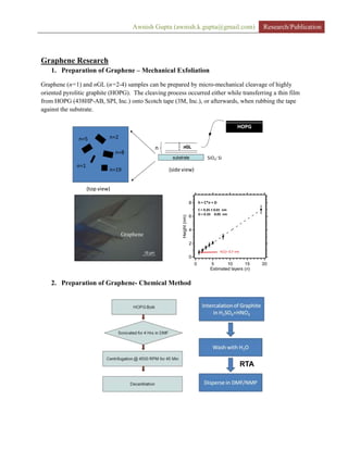
1. Preparation of Graphene – Mechanical Exfoliation
Graphene (n=1) and nGL (n=2-4) samples can be prepared by micro-mechanical cleavage of highly
oriented pyrolitic graphite (HOPG). The cleaving process occurred either while transferring a thin film
from HOPG (438HP-AB, SPI, Inc.) onto Scotch tape (3M, Inc.), or afterwards, when rubbing the tape
against the substrate.
8
6
4
2
0
Height(nm)
20151050
Estimated layers (n)
h = C*n + D
C = 0.35 ± 0.01 nm
D = 0.33 0.05 nm
h(1)= 0.7 nm
substrate
n nGL
substrate
n nGL
SiO2: Si
n=2
n=1
n=8
n=19
n=5
HOPG
(top view)
(side view)
Graphene
2. Preparation of Graphene- Chemical Method
RTA
2. Awnish Gupta (awnish.k.gupta@gmail.com) Research/Publication
substrate n=3 nGL
3. Dispersive Raman Scattering from n-Graphene Layer Films
Resonant Raman scattering studies of n-graphene layer films (nGLs; n=1-4). We follow the
scattering process to sixth order and observe many new Raman peaks which participate in double
resonance (DR) Raman scattering. Many of the Raman bands exhibit linear dispersion, i.e.,
d/dE=constant, where is the Raman peak frequency and E is the excitation photon frequency.
This behavior will be shown to stem from DR, the dispersion of the electronic and phonon states
involved, and also on the order of the scattering. For example, we find values in the range of -
20< d/dE <170 cm-1
. The band dispersions also exhibit an interesting dependence on the
number of layers n and can be used to map out phonon dispersion of nGLs for comparison with
theoretical calculations
3. Awnish Gupta (awnish.k.gupta@gmail.com) Research/Publication
Phonon dispersion (dots) (n=1) based on 2nd
order Raman spectra. Theoretical curves (solid lines) are taken
from Saito et. al., PRL (2002)
A simple strategy to determine n in nGLs was also determined as follows:
1. Choose few thin flakes on a particular substrate by looking under an optical
microscope.
2. Find a flake which has a single 2D (~ 2700 cm-1) peak, to ensure that the layer
being observed is a 1GL flake.
3. Measure the G-band intensity on an absolute scale (counts/sec-mW).
4. Measure the G-band intensity of an unknown nGL.
5. Assign n by IG = n IG(n=1).
6. Assignment of n could be verified by frequency shift of 2D3 and 2D2.
4. Temperature Dependent Raman Scattering from n-Graphene Layer Films
Results from a study of Raman G-band ~ 1585 cm-1 as a function temperature T and on (n=1,2,3)
were obtained. Data for the frequency and linewidth will be discussed in terms of contributions from
the electron-phonon interaction, negative lattice expansion, and multi-phonon processes. The T-
behavior depends strongly on whether the nGL is supported on Si substrate or freely suspended. The
presumption is that the suspended films exhibit intrinsic behaviour, while the supported films do not.
Results for the change in the G-band frequency over the range 80<T<800 K for n=1,2,8 supported on
SiO2/Si and graphite (HOPG) are shown. A noticeable quadratic T-dependence is observed over this
wide range of temperature.
4. Awnish Gupta (awnish.k.gupta@gmail.com) Research/Publication
Temperature dependent G-band frequency shift for nGLs supported on Si/SiO2
5. Raman Scattering from Incommensurately Stacked Bi-Layer Graphene
Despite the weak interlayer interaction between graphene layers, our Raman scattering studies on
incommensurately stacked (IS) bi-layer grapheme (n=2) reveal an altogether different Raman
spectrum commensurately stacked (CS) n=2 films as opposed to IS n=2 films. We find that IS
layers activate a new band (“I-band”) for sp2
bonded carbon near ~1350 cm-1
which has two
components I1 and I2 (Figure 3): I1 is dispersive at ~50 cm-1
/eV, while I2 exhibits very weak
dispersion(Figure 4). These Raman peaks are not due to ordinary D-band scattering in sp2
carbons
that is normally associated with defects. From the shape of the 2nd
order 2D band at ~2700 cm-1
,
we see evidence for a large decrease in the electronic subband splitting due to IS layers. A theory
is proposed to explain these new results in IS n=2 graphene.
Raman bands near ~1350 cm-1
for n=1 monolayer (ML), and n=2 C and I stacked films. The ML-
Edge spectrum exhibits normal D-band scattering.
5. Awnish Gupta (awnish.k.gupta@gmail.com) Research/Publication
Dispersion of I1 and I2 Raman bands
6. Localized Raman Scattering from Graphene Edges
TEM and Raman scattering show that a typical grapheme edge prepared by micromechanical
cleavage from HOPG is, on average very straight, but at short range, can be seen meandering by
about ~ 2 nm and thereby presenting a mixture of zigzag and armchair local symmetries.
Nevertheless, we find that these “real” edges exhibit polarized scattering as if the absorption and
re-radiation of the photons were made by a line antenna aligned along the average direction of the
edge. Scanning the excitation beam from off the sample and then across the edge allows the
observation of the onset of the G-band with distance, as well as a D-band localized within w~ 50
nm of the edge. A value for w can be estimated by analyzing the convolution of the Gaussian
laser beam profile with the localized scattering which is assumed, for simplicity, to be constant
and localized within w of the edge.
6. Awnish Gupta (awnish.k.gupta@gmail.com) Research/Publication
TEM of a graphene edge showing edge roughness ~ 1-3 nm.
7. Raman Scattering from Narrow Graphene Ribbons
Raman data collected on vary narrow (ribbon width ~ 2-3 nm) one- or two-layer graphene
nanoribbons (GNRs). New G-bands, more similar to that observed in 1.2-1.6 nm diameter single-
walled nanotubes than grapheme are observed. The GNRs were synthesized by H. Dai and co-
workers at Stanford. We discussed the activation of these new Raman modes in narrow GNRs in
terms of transverse phonon confinement. Interestingly, several of these GNRs were observed to
NOT exhibit a D-band.
Raman spectrum of 3-nm wide GNR showing extra peaks in the wings of the dominant G-band
7. Awnish Gupta (awnish.k.gupta@gmail.com) Research/Publication
Carbon Nanotube Research
1. Growth of large scale SWNTs via CVD:
To synthesis large scale SWNTs, preparation of catalytic nanoparticles is equally important. We
prepared nanoparticles for SWNTs growth via a slightly different method than described in [Alan
M. Cassell et al, “Large Scale CVD Synthesis of Single walled Carbon Nanotubes”, J. Phys.
Chem. B 1999, 103, 6484]. In figure 1, we showed step by step synthesis of nanoparticles for
SWNTs growth. After the last step we obtained a very fine power of grayish-yellow color.
(Vigorous Stirring)
(Wait for 1 hour)
(Stirring for 15 min)
1 gm Alumina Nanoparticles + 50 ml ethanol
.51 ml Concentrated HF ( 52 % HF)
.02 gm Ammonium molibdunate ((NH4)6 Mo7O24.xH2O)
&
.134 gm Ferric Sulphate (Fe2 (So4) 3.xH2O)
Heat 90-100 C for 1 hour under N2 flow and
then leave it to become dry
Ground in a Mortar
Calcinate at 400-500 C for 1 hour
Figure 1: Synthesis of nanoparticles for SWNTs growth.
Growth of SWNTs is done by CVD process using CH4 as a carrier gas and step by step
process is described in figure 2. The process is slightly modified from previously published
mehod by Avetik R. Hurutyunyan [“CVD Synthesis of SWNT under soft conditions” , Nano
Letters 2002, vol 2, no 5, 525]. We had also successfully grown SWNTs in large scales using
alcohol (CH3OH and C2H5OH) as well using the same nanoparticles. In both the process (CH4
8. Awnish Gupta (awnish.k.gupta@gmail.com) Research/Publication
and Alcohols), we obtained a fluffy black powder after the growth. TGA analysis showed that
after burning the nanotubes in air, 30% (by weight) material left consisting of mainly metal
nanoparticles.
Catalyst sample 40-80 mg
CATALYST REDUCTION STEP:(10 sccm H2 + 90 sccm He) for 10-15 hours at 500 oC
Change He to 350 sccm Ar and then increase temp by 10 o
C/min up to 850 o
C.
Flow (350 sccm Ar + 40 sccm CH4 + 10 sccm H2) for 1 hour
Close the CH4 and H2 valves and Cool the furnace to the room temp with Ar gas
flowing
Figure 2: Growth process of large scale SWNTs.
These nanotubes were characterized by Raman, TGA and TEM. In figure 3, we show
Raman spectrum of SWNTs prepared by CVD process and using CH4 as a carrier gas. Raman
spectrum shows clearly the presence of RBM, D, G and 2D band. Raman technique is a non
destructive technique to differentiate between different structures of sp2 carbons. The Shape of
G-band (showing G+
and G-
) and presence of RBM is important for SWNTs. RBM band can be
used to estimate the diameter distribution of nanotubes. Calculated diameter of nanotubes (based
on RBM) is shown as an inset. Small ID/IG established that nanotubes do not have large defect
densities.
700
600
500
400
300
200
100
0
Intensity
25002000150010005000
wave number (cm-1)
T band [1590.2]
Peak 2 & 3
RBM
No Notch ?
G'
Raman Shift (cm-1 )
Intensity(a.u.)
T-Band
2D(G’)-Band
RBM-Band
D-Band
160.5 - 1.53 nm
231.2 - 1.03 nm
252.6 - 0.94 nm
261.6 - 0.90 nm
274.8 - 0.86 nm
286.1- 0.82 nm
9. Awnish Gupta (awnish.k.gupta@gmail.com) Research/Publication
Figure 3: Raman spectrum of SWNTs.
2. Purification and debundling of arc-SWNTs:
SWNTs grown with arc-discharge method have impurities consisting of amorphous
carbon, metal nanoparticles, carbon shells etc. To remove all the impurities, our group has
developed an effective method. In-short, we first do dry oxidation at ~400 C (temperature and
time determine by TGA analysis, shown in figure 4 (c)) for 15 min ( ~ 120 cc/min dry air) which
burns the amorphous carbon and carbon shells covering the metal nanoparticles, we then do acid
reflux (HCl and HNO3) to remove the metal contents in the sample.
Raman Spectra From
Carbolex Nanotubes
Dry Oxidation
Raw Soot
PurificationDebundling
(a) (b)
(c)
10. Awnish Gupta (awnish.k.gupta@gmail.com) Research/Publication
Figure 4: (a) Raman spectra of arc produced nanotubes. (b) A schematic scheme for purification
and debundling of nanotubes. (c) TGA of as received material (upper) showing two peaks
originating from the amorphous carbon (low temperature peak) and SWNTs (high temperature
peak) and TGA of material after dry oxidation showing the significant decrease in the intensity
of peak originating from amorphous carbon.
3. Growth of isolated SWNTs:
Growth of isolated SWNTs is important to understand the (n,m) dependence on the
physical properties of SWNTs. Preparation of catalytic particles (figure 5) for isolated
SWNTs growth is performed by the method first described by Halfner et al. The method
involves preparing a ferric nitrate solution in IPA and dipping the substrate for 1 min and
later washing with hexane. We grown isolated nanotubes at 900 C and used CH4 as a
carrier gas. Nanotubes are grown on Si, SiO2 and quartz substrate by this method. Figure
5 (right) shows an AFM height image showing the isolated nanotubes grown by this
method.
• Catalyst Particles:
Fe(III)NO3 in anhydrous IPA 2mg/ml) [ 1 min]
+ Wash with hexane [1 min]
• Growth: 40 sccm CH4 @ 900 oC
1 hr1 hr
550
Time
H2+Ar+CH4
900
20 o
C/m
20 oC/m
H2+Ar Ar • AFM Images (Height Bar- 3nm)
• Nanotubes can be grown on Si,
SiO2/Si and Quartz surface also
Figure 5: Preparation of catalytic nanoparticles and growth of isolated SWNTs.
Density of nanotubes can be controlled by concentration of ferric nitrate, diameter of
nanotubes can be controlled by temperature and length of nanotubes can be control by
mixing ethane (C2H6) with CH4 during the growth process. In figure 6 (a), we show the
density control of isolated nanotubes per micron2
area onto the substrate. Left image is
20x20 microns2
while the right image is 4x4 microns2
. In figure 6(b), we show the
diameter control of nanotubes. Left column show nanotubes grown at 1100 C while right
column show nanotubes grown at 900 C. At 900 C, we find the diameter distribution to
be ~ 0.5-2.0 nm while most probable diameter at 0.8 and 1.2 nm. Diameter distribution of
11. Awnish Gupta (awnish.k.gupta@gmail.com) Research/Publication
nanotubes is measured with AFM height. At 1100 C, we find nanotubes diameter
distribution of ~ 1.0 – 7.0 nm with most probable diameters of ~ 3.0 nm. It was
interesting to see nanotubes as large as 7.0 nm (shown in HRTEM, isolated tube at the
bottom image) which is larger diameter ever reported.
(a) 0.5 mg/ml 2.0 mg/ml
0
5
10
15
20
25
0 0.2 0.4 0.4 0.6 0.8 1 1.2 1.4 1.61.8 2 2.22.4
Counts
Diameter (nm)
Nanotube Diameter Distribution
16
Growth: 1100 C Growth: 900 C
(b)
(a) 0.5 mg/ml 2.0 mg/ml
0
5
10
15
20
25
0 0.2 0.4 0.4 0.6 0.8 1 1.2 1.4 1.61.8 2 2.22.4
Counts
Diameter (nm)
Nanotube Diameter Distribution
16
Growth: 1100 C Growth: 900 C
(b)
(a) 0.5mg/ml 2.0mg/ml
0
5
10
15
20
25
0 0.20.40.40.60.8 1 1.21.41.61.8 2 2.22.4
Counts
Diameter(nm)
Nanotube Diameter Distribution
16
Growth: 1100 C Growth: 900 C
(b)
7 nm
2 nm
Figure 6. (a) Density control (b) Diameter control
4. SWNTs-FET: Preaparation and Electrical properties:
12. Awnish Gupta (awnish.k.gupta@gmail.com) Research/Publication
We studied the effect of nanotube-nanotube junction on the electrical properties of nanotubes
prepared by CVD. In figure 7, we show schematic of two kinds of devices studies here: (a)
shows a schematic of device where nanotubes are connecting source and drain via
percolating network and (b) shows a schematic of device with one or few nanotubes (no
junctions) connecting the source and drain. Device shown in fig 7(b) is realized by
controlling the density of nanotubes and controlling the spacing between the electrodes. We
prepared both of these devices by shadow mask techniques to avoid any chemical exposure
(i.e., photoresist etc.)
10 µm 50 nm - 1 µm
Si (back gate)
VG
Au
S D
Percolating network
Si (back gate)
VG
S D
1 or few tubes
(a) (b)
Figure 7. (a) Schematic of FET device prepared by SWNTs percolating network (b)
Schematic of FET devices showing one or few SWNTs connecting the S-D electrode.
SWNTs are grown by CVD and devices are prepared by shadow mask techniques to avoid
any chemical exposure.
4 (a). Device Characteristics (percolating network)
To prepare this kind of devices, we grew high density, short ( ~ 4 microns in length)
SWNTs on Si/SiO2 substrate. Device is prepared by putting a standard TEM grid (shown in
8(a)) on the substrate and depositing contact pads (source-drain) through hole. Fig 8(b)
shows the density of grown nanotubes. We observed few interesting behaviors in these
devices: (1) we observed hysteresis in I-V measurement ( VG=0) (fig 8c). This hysteresis
behavior can be attributed to the charge trapping [Marty et al Nano Lett 3, 1115 (2003)]. At very
high current densities ( which will depend on the number of nanotubes connecting S-D) we did not
observe very pronounce hysteresis. (2) We observed a presence of conduction peaks at room
13. Awnish Gupta (awnish.k.gupta@gmail.com) Research/Publication
temperature (RT). These conduction peaks (marked fig 8h, also present in 8g) has previously seen
only at low temperature in kinked isolated nanotube [Dekker et al, Science, Vol. 293, 76 (2001)] and
explained in terms of construction of quantum dot between the kinks. Our results also indicate that the
nanotubes cross junction is acting as a quntum dot by sharp bending when one nanotube will lie upon
another nanotube [Nojeh et al Nano Letters , Vol 3, No. 9, 1187-1190 (2003)]. Experiments on single
cross are in progress to verify this.
2 µm5 µm
0
50
100
150
0 5 10
Current(micro-amp)
Voltage (V)
I-V Characteristics
10 V to -10 V Scan
-10 V to 10 V Scan
-2.0E-09
-1.5E-09
-1.0E-09
-5.0E-10
0.0E+00
5.0E-10
1.0E-09
1.5E-09
-0.2 -0.1 0 0.1 0.2
Current(Amp)
Voltage(V)
UG @ different temp
9 K 20 K
35 K 50 K
75 K 100 K
125 K 150 K
200 K
0
50
100
150
200
250
0 0.05 0.1 0.15
Conducatnce(X10^-9)
1/T(K)
Conducatce Vs Temp
Conductance@0V
Conductance (MIN)
18
20
22
24
26
28
30
32
34
-12 -8 -4 0 4 8 12
Conductance(x10-6)
Gate Voltage
10-6dI/dV(Fit)
20100-10-20
Voltage (V)
5.0
4.5
4.0
3.5
3.0
2.5
2.0
10
-6
I/V
dI/dV Fit
I/V
(a) (b)
(c) (d) (e)
(f) (g) (h)
VG
VG=0
RT
RT
VG=0
RT
RT
Figure 8 (a) Shadow mask technique (b) AFM of percolating network (c) Hysteresis behavior of
device (d) Temperature dependent I-V characteristics (e) Temperature dependent conduction (f) Gate
voltage dependence, VD-S ~ 1V (g) Observation of conduction peaks at room temperature for device 1
which are originating from cross junction nanotubes (h) conduction peaks for device 2.
4(b). Device Characteristics (one of few nanotubes)
Devices with only one or few nanotubes connecting the source-drain electrods were prepared by
the same way as in section 4(a) but on low density and longer nanotubes. We also controlled the gap
between the electrodes by depositing two sources during contact-pads deposition. A schematic is
shown in fig 9(a). fig 9 (b) shows an optical image of the contact pads. We could control the spacing
14. Awnish Gupta (awnish.k.gupta@gmail.com) Research/Publication
between source and drain from 10’s of nanometers to few microns. Unlike high density percolating
network devices, we did not see ohmic contact in I-V characteristics (see Fig 9c and 9d). During gate
voltage measurements we observed a clear turn on and turn off state similar to reported for isolated
nanotubes (fig 9e). Occasionally, we also observed the presence of multimode transport. By this
device preparation method, we could prepare 1 or few SWNTs FET devices without exposure to the
chemicals.
Source 1 Source 2
Shadow mask
Gap
Initial spacing between pads
Si/SiO2/SWNT
Modified spacing
42 µm
5 µm
5 µm
1.5 µm
-6.0E-08
-4.0E-08
-2.0E-08
0.0E+00
2.0E-08
4.0E-08
6.0E-08
8.0E-08
-4 -2 0 2 4
Ids(Amp)
Vds (V)
c2008c1908
Vds-Ids…
-4.E-09
-2.E-09
0.E+00
2.E-09
4.E-09
6.E-09
-10 -5 0 5 10
Current(Amp)
Voltage(V)
c1909c1809-II
Vds-Ids
0.0E+00
5.0E-08
1.0E-07
1.5E-07
-100 -50 0 50 100
DrainCurrent(Amp)
Gate Voltage (V)
c2006c1906
Vds:1V
0.E+00
4.E-09
8.E-09
-80 -40 0 40 80
DrainCurrent(Amp)
Gate-Voltage (V)
Vds: .2 V
multimode transport ?
(a) (b)
(c) (d)
(e) (f)
Figure 9. (a) Schematics of method employ to decrease the spacing between electrodes (b) Optical
and AFM image of device, (c), (d) I-V characteristics of two devices (e), (f) FET behavior of two
devices. (f) Also shows the signature of multimode transport.
5. Boron-doped SWNTs:
In figure 10, we show comparison of Termo Electric Power (TEP) measurements in undoped and B-
doped ( 3% wt) SWNTs mats. Data is collected using home-made apparatus. In figure 10a and 10b, we
show the change in TEP upon vacuum degassing or undoped nanotubes and B-doped nanotubes. While
15. Awnish Gupta (awnish.k.gupta@gmail.com) Research/Publication
undoped nanotubes show –ve TEP value after degassing for few hours ( after oxygen removal), B-doped
nanotubes remain a +ve value even after degassing for few days. In fig 10c, we show the TEP change
with temperature after degassing for one day. TEP value goes approaches zero at zero temperature but
always remains negative. In the case of B-doped SWNTs, TEP decrease with decreasing temperature but
goes from +ve value to –ve value at ~ 30 K. It is very strange behavior as at zero temperature TEP should
go back to zero value.
-40
-20
0
20
40
TEP(V/K)
120010008006004002000
Time (min)
-60
-50
-40
-30
-20
-10
0
TEP(V/K)
5004003002001000
Temp (K)
Cooling
Heating
26
24
22
20
18
16
TEP
3500300025002000150010005000
Time (min)
TEMP 200 C
-8
-6
-4
-2
0
2
4
TEP(microV/K)
6050403020100
Temp (C)
20
15
10
5
0
-5
TEP(microV/K)
30025020015010050
Temp (C)
Heating
Cooling
Undoped SWNTs 3% Boron doped SWNTs
Degassing @ 200C Degassing @ 200C
TEP vs. T
TEP vs. T
(a) (b)
(c) (d)
16. Awnish Gupta (awnish.k.gupta@gmail.com) Research/Publication
Nanowire Research
1. Growth of NWs
Different growth models have already in place to understand the growth mechanism of a
particular nanostructure. Knowledge and understanding of phase diagram of different materials and their
compounds should be sufficient to synthesize a nanostructure (specially nanowires) but still it takes a few
attempts to optimize the growth conditions or replicate someone else’s work into a laboratory. As a
curious graduate student I worked on a short project involving the growth and physical properties
measurements of ZnO nanowires. ZnO is a wide bandgap semiconductor and of immense importance for
solid state laser. Later I worked on the nanomanipulation of several different kind of wire to tune the
desired physical properties. In figure 1, I show a SEM image of ZnO nanowires. ZnO nanowires are
grown with CVD and growth process is self catalytic VLS process. ZnO can form several other
nanostructures (i.e., nanobelts, nanocombs, nanoshees, nano-tetrahydral-pyramids etc.) can control
growths of only one dominant structures can be achieved by controlling the growth parameters.
5 μm5 μm5 μm
17. Awnish Gupta (awnish.k.gupta@gmail.com) Research/Publication
2. Electrical Properties of Nanostructures:
In figure 2(a), (b) and (c), we show the difference in electrical properties of different structures of the
same material (ZnO). I-V data is collected using Keithley-4200.
NW-2-2-I1-I2-2
-1.5E-6
-1.0E-6
-5.0E-7
0.0E+0
5.0E-7
-20. -15. -10. -05. 00. 05. 10. 15. 20.
V (Volts)
I(Amps)
Voltage
NW-2-2-I6-J6-5
-1.0E-07
0.0E+00
1.0E-07
2.0E-07
3.0E-07
4.0E-07
5.0E-07
6.0E-07
7.0E-07
-20. -15. -10. -5. 0. 5. 10. 15. 20.
V (Volts)
I(Amps)
Voltage
NW-2-2-F7-G7-3
-1.5E-8
-1.0E-8
-5.0E-9
0.0E+0
5.0E-9
1.0E-8
-20. -15. -10. -5. 0. 5. 10. 15. 20.
V (Volts)
I
Voltage
(a)
(b)
(c)
18. Awnish Gupta (awnish.k.gupta@gmail.com) Research/Publication
3. Nano-Manipulation of Nanowires:
In figure 3(a), I show a nanomanipulation of a GaP nanowire to study the physical properties as function
of wire length. Surprisingly, we observed the lasing from short nanowires during Raman measurements.
In figure 3(b) and (c) we show that two wire can be join as a affect of charging. Notice the difference
between the dotted ellipse and arrow.
2 μm2 μm
(a) (b)
(c)
19. Awnish Gupta (awnish.k.gupta@gmail.com) Research/Publication
Undergraduate Research
The Indian Institute of Technology Bombay (IIT-B) July. 2001 – April. 2002
Powai, INDIA (at The Department of Physics (www.iitb.ac.in ))
Growth and Characterization of metal filled carbon nanotubes: Extensive literature search was done
to understand the critical parameters for nanotubes growth. Metal filled MWNTs were successfully grown
using plasma CVD and their TEM and EDS data were analyzed. A growth process is developed to fill
these Transition metals (Ni and Co) to almost 50% tubes.
The Physical Research Laboratory (PRL) May. 2001 – July. 2001
Ahemedabad, INDIA ( www.prl.ernet.in )
Ultrashort pulsed beam propagation: This project involves the theoretical understanding of production
of ultra short pulses ( few cycles, one cycle and ½ cycle) in collaboration with an experimental group at
PRL. Propagation and optical properties of these femto (sub-femto) second pulses are studied in vacuum,
dispersive linear and non-linear media.
The Indian Institute of Technology Bombay (IIT-B) July. 2000 – Dec. 2000
Powai, INDIA (at The Department of Physics (www.iitb.ac.in ))
Tunneling Magnetoresistance (TMR) and its applications: Theoretical project involving concept of
spin dependent tunneling, GMR and TMR. Tested accepted theories for new materials and determining
the expected value of TMR.
Centre for advanced technology (CAT) Dec. 2000- Jan. 2001
Indore, India (www.cat.ernet.in)
Fifth school on the physics of beams: The School was on experimental high energy physics. Topic
covered were accelerator physics, transverse and longitudinal beam dynamics, cyclotron, synchrotron,
storage rings, free electron lasers along with experimental demonstrations of superfish for r.f. cavity,
beam position monitor, assembly of FEL magnets and dipole magnetic field measurements.
Indian Institute of Sciences (IISc) Sep. 2001
Banglore, India ( www.iisc.ernet.in)
National workshop on nanomaterials: Synthesis, Modification and Characterization by ion beam:
This workshop was on techniques involving fabrication of nanocrystalline particles and other
nanostructures and modification of thin films using ion beam.
20. Awnish Gupta (awnish.k.gupta@gmail.com) Research/Publication
The Indian Institute of Technology Bombay (IIT-B) Nov. 2000
(http://www.me.iitb.ernet.in/yantriki)
Yantriki 2000 – National robotics competition organized by IIT Bombay, INDIA. We designed a robot
to play soccer on water against an opponent team. We lost to the winner on pre-quarterfinal round.
The Indian Institute of Technology Bombay (IIT-B) Nov. 2001
(http://www.civil.iitb.ernet.in/~lastraw)
Last-straw 2001 - “bridging the gap” competition organized by IIT Bomaby, INDIA. We required to
make a bridge using straws for highest load testing.
21. Awnish Gupta (awnish.k.gupta@gmail.com) Research/Publication
Publications
1. J Huang, B Wang, I Lahiri, A. K. Gupta, PC Eklund and W Choi, “Tuning Electrical Conductance of
Serpentine Single‐Walled Carbon Nanotubes” Advanced Functional Materials 20 (24), 4388-
4393(2010)
2. A. K. Gupta, Y. Tang, V. H. Crespi and P. C. Eklund, “Nondispersive Raman D band activated by
well-ordered interlayer interactions in rotationally stacked bilayer graphene” Physical Review B 82
(24), 241406 (2010)
3. A. K. Gupta, C. Nisoli, P. E. Lammert, V. H. Crespi and P. C. Eklund, “ Curvature-Induced D-Band
Raman Scattering in Graphene” Journal of Physics: Condensed Matter 22 (33), 334205 (2010)
4. SH Cheng, K Zou, F Okino, HR Gutierrez, A. K. Gupta, N Shen, PC Eklund, JO Sofo and J. Zhu,
“Reversible fluorination of graphene: evidence of a two-dimensional wide bandgap semiconductor”
Physical Review B 81 (20), 205435 (2010)
5. B Wang, A. K. Gupta, J Huang, H Vedala, Q Hao, VH Crespi, W Choi and P. C. Eklund, “Effect of
bending on single-walled carbon nanotubes: A Raman scattering study” Physical Review B 81 (11),
115422 (2010)
6. J. Wu, A. K. Gupta, H. R. Gutierrez and P. C. Eklund, “Cavity-enhanced stimulated Raman
scattering from short GaP nanowires” Nano letters 9 (9), 3252-3257 (2009)
7. H. E. Romero, P. U. Joshi, A. K. Gupta, H. R. Gutierrez, M. W. Cole, S. Tadigadapa and P. C.
Eklund, “Adsorption of Ammonia on Graphene” Nanotechnology 20 (24), 245501(2009)
8. A. K. Gupta, ”Raman scattering from n-graphene layers (nGLs; n= 1, 2, 3...)” Thesis Pages-223,
The Pennsylvania State University (2009)
9. A. K. Gupta, T. J. Russin, H. R. Gutierrez and P. C. Eklund,” Probing Edge Defects in Graphene via
Raman Scattering” ACS Nano, 3 (1), 45-52 (2008)
10. A. K. Gupta, Y. Tang, T. J. Russin, V. H. Crespi and P. C. Eklund,” Raman Scattering from
incommensurately Stacked Graphene Bi-Layer” (submitted to Nano Letters)
11. U. J. Kim, G. R. Gutierrez, A. K. Gupta and P. C. Eklund, “ Raman Scattering Study of the Thermal
Conversion of Bundled Carbon Nanotubes into Graphitic Ribbons” Carbon, 46 (5), 729-740 (2008)
12. P. Joshi, A. Gupta, S. Tadigadapa and P. C. Eklund,” Electrical Properties of back-gated n-graphene
layers films”, Proceedings of SPIE-The International Society for Optical Engineering (2007)
13. A. Gupta, G. Chen, P. Joshi, S. Tadigadapa, P. C. Eklund, “ High Frequency raman scattering from
n-graphene layers”, Nano Letters, Vol. 6, No.12, 2667-2673 (2006)
22. Awnish Gupta (awnish.k.gupta@gmail.com) Research/Publication
14. P. Joshi, A. Goyal, A. Gupta, S. Tadigadapa, P. Eklund, "Improvement of the Elastic Modulus of
Micromachined Structures using Carbon Nanotubes", Proceedings of SPIE - The International
Society of Optical Engineering, Jan. 2005, San Francisco, California.
15. A. Goyal, S. Tadigadapa, A. Gupta, P.C. Eklund, “Use of Single Wall Carbon Nanotubes (SWNTs)
to Increase the Quality Factor of an AT-cut micromachined Quartz Resonator”, Applied Physics
Letters, 87, 204102, 2005.
16. A. Goyal, S. Tadigadapa, A. Gupta, P.C. Eklund, “Improvement in Q-factor of AT-Cut Quartz
Crystal Resonators using Single Wall Carbon Nanotubes (SWNTs)”, Proceedings of 2005 IEEE
International Ultrasonics Symposium, Sep. 18-21, 2005, Rotterdam, Netherlands.
17. A. Goyal, S. Tadigadapa, A. Gupta, P.C. Eklund, “Micromachined Quartz Resonator Functionalized
with Single Wall Carbon Nanotubes (SWNTs) for Sensing Applications”, Proceedings of IEEE
Sensors 2005, the 4th
IEEE Conference on Sensors, Oct. 31- Nov. 3, 2005, Irvine, California.
18. P. Joshi, N. Duarte, A. Goyal, A. Gupta, S. Tadigadapa, P.C. Eklund, “Improvement of the elastic
modulus of micromachined structures using carbon nanotubes”, MRS Proceedings, Volume 875,
O1.5, MRS Spring Meeting, March 28 – April 1, 2005, San Francisco, USA.
23. Awnish Gupta (awnish.k.gupta@gmail.com) Research/Publication
Conference Presentations#
1. A. K. Gupta et.al, “ Confined Phonons in sp2 NanoCarbons” in ICMS-ICMR Winter School on
New Carbon Materials, Dec. 8- 13, JNCASR, Banglore, India (2008) – Invited Talk
2. Peter C. Eklund, A. K. Gupta, H. R. Gutierrez, T. Russin “Resonant Raman Scattering from
Graphene and Narrow Graphene Ribbons” IUMRS-ICEM , July 28 – Aug 1, Sydney, Australia
(2008) - Invited Talk
3. A. K. Gupta, T. J. Russin and P. C. Eklund,” Temperature-Dependent Raman Scattering from n
(n=1,2,3…) Graphene Layers” MRS Spring Meeting, March 24 – 28, San Francisco, CA (2008)
4. A. K. Gupta, Y. Tang, T. J. Russin, V. H. Crespi and P. C. Eklund,” Raman Scattering from
Incommensurately Stacked Bi-Layer Graphene” MRS Spring Meeting, March 24 – 28, San
Francisco, CA (2008)
5. A. K. Gupta, Y. Tang, T. Russin, V. H. Crespi and P. C. Eklund “Incommensuratey Stacked
Graphene Bi-Layer: A Raman Study” APS Meeting. March 10- 14, New Orleans, LA (2008)
6. A. K. Gupta, and P. C. Eklund “Dispersive Raman Scattering from n=1-4 Graphene Layer
System” APS Meeting. March 10- 14, New Orleans, LA (2008)
7. A. K. Gupta, H. R. Gutierrez and P. C. Eklund “Probing Edge Defects in n=1,2.. Graphene
Layer System via Raman Scattering” APS Meeting. March 10- 14, New Orleans, LA (2008)
8. A. K. Gupta, T. Russin and P. C. Eklund “ Anharmonic Effects in Raman Scattering from Few-
Layer Graphene Systems ” APS Meeting. March 10- 14, New Orleans, LA (2008)
9. Q. Lu, J. Wu, A. K. Gupta and P. C. Eklund “Enhanced Raman Scattering Near the Tip of
Semiconducting Nanowires” APS Meeting. March 10- 14, New Orleans, LA (2008)
10. J. Wu, A. K. Gupta and P. C. Eklund “Non-Linear Raman Scattering from Semiconducting GaP
Nanowires” APS Meeting. March 10- 14, New Orleans, LA (2008)
11. A. K. Gupta, T. J. Russin, P. Joshi, H. R. Gutierrez, G. Chen and P. C. Eklund “Phonons in n-
Graphene Layers”, PASI (Novel Materials for Micro- and Nano Electronics) Renaca, Chile
(2007) – Invited talk
12. A. K. Gupta, G. Chen and P. C. Eklund “1st, 2nd and 3rd order Raman scattering from n-
Graphene Layers supported on Si/SiO2 substrates”, APS Meeting, March 5-9, Denver, CO
(2007)
13. A. K. Gupta and P. C. Eklund “Raman Scattering Probe of Graphene-Substrate Interactions”,
APS Meeting, March 5-9, Denver, CO (2007)
24. Awnish Gupta (awnish.k.gupta@gmail.com) Research/Publication
14. A. K. Gupta, T. J. Russin, H. R. Gutierrez and P. C. Eklund “Generation of Carbon Scrolls from
Graphene”, APS Meeting, March 5-9, Denver, CO (2007)
15. P. Joshi, A. K. Gupta, S. Tadigadapa and P. C. Eklund “Electrical and Optical Properties of
Supported n-Graphene Layers Films”, SPIE-sensors, San Franscisco, CA (2007)
16. A. K. Gupta, G. Chen, P. Joshi, S. Tadigadapa and P. C. Eklund “A non destructive technique
(RAMAN) to count the layers of graphene in nGL films” in PASI, June 18 – June 30, Costa Rica
(2006)
17. A. K. Gupta and P. C. Eklund “Raman Scattering from few layered graphene films”, in APS
meeting, March 13-17 2006, Baltimore, MD (2006)
18. A. K. Gupta, K. W. Adu, H. R. Gutierrez, Q. Xiong, J. Wu, X. M. Liu and P. C. Eklund “Novel
low dimensional systems for new technologies” Materials Day, Penn State University, University
Park, Pennsylvania (2006)
19. D. Narehood, K. Adu, Y. Chen, A. K. Gupta, X. M. Liu and P. C. Eklund“Carbon nanotubes-
polymer composite for EMI shielding” Materials Day, Penn State University, University Park,
Pennsylvania (2006)
20. A. K. Gupta, Q. Xiong, U. J. Kim, K. W. Adu, H. R. Gutierrez, J. Wu, X. M. Liu and P. C.
Eklund “Chemical Sensors for NanoFilaments” Materials Day, Penn State University, University
Park, Pennsylvania (2005)
21. U.J. Kim, C.A. Furtado, H.R. Gutierrez, X.M. Liu, A. Gupta and P.C. Eklund “The effects of
tube-wall functional groups on the dissolution of individual SWNTs” APS meeting, March 22-
26, Montreal, Quebec, Canada (2004)
#
List of authors is (in general) in order of contribution to a particular work. First author in the list
presented the work.