neck exam.pptx cervical lymph node examination
•Download as PPTX, PDF•
0 likes•15 views

The lymph node examination is performed with circular motion, identifying pain, and swollen ganglia or induration. For the anterior cervical lymph node exam, palpate the lymph nodes in the neck using circular motion over the underlying tissues in each area.

Recommended
Recommended
More Related Content
Similar to neck exam.pptx cervical lymph node examination
Similar to neck exam.pptx cervical lymph node examination (20)
More from FAZAIA RUTH PFAU MEDICAL COLLEGE ,KARACHI,PAKISTAN
More from FAZAIA RUTH PFAU MEDICAL COLLEGE ,KARACHI,PAKISTAN (20)
Recently uploaded
Recently uploaded (20)
neck exam.pptx cervical lymph node examination
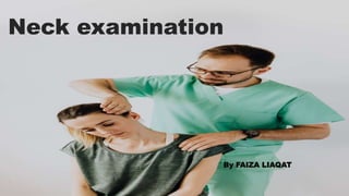
- 1. Neck examination By FAIZA LIAQAT
- 2. Anatomy of neck The neck consists of seven cervical vertebrae, supporting structures like muscles, blood vessels, and nerves. Key components include the trachea, esophagus, and thyroid gland.
- 3. I. Submental and submandibular nodes. II. Upper third sternocleidomastoid (SCM) muscle. III. Middle third SCM (between hyoid and cricoid. IV. Lower third SCM (between cricoid and clavicle). V. Posterior to SCM (posterior triangle). VI. Midline from hyoid to manubrium. Cervical lymph node levels
- 5. Neck masses Inflammatory Congenital Traumatic Neoplastic
- 6. Swellings on the bases of location
- 8. Examination of neck Intro (WIIPPPPEE) Wash your hands Introduce yourself Identity of patient – confirm Permission (consent and explain examination) Pain? Position sitting in chair with room behind the chair for the examiner to stand Privacy Expose neck and clavicles (patient may need to tie hair back/ remove necklace)Equipment – have a glass of water to hand
- 9. Inspection From front and sides Lumps/ asymmetry Scars (thyroidectomy/ parathyroidectomy scars using a pen torch) Skin changes, facial plethora (SVC obstruction) Distended neck veins (SVC obstruction) If a neck lump is seen: Ask patient to1) swallow (a thyroid lump or thyroglossal cyst) 2)tongue protrusion (thyroglossal cyst) Palpation Palpate: anterior Trachea (For tracheal deviation) Carotid pulse (one side at a time) Palpate: posterior Explain to the patient that you will be moving behind them to palpate their neck. Take this opportunity to inspect the back of the neck. Thyroid gland Palpate one lateral lobe at a time then isthmus (nodules and thrills)Ask the patient the swallow Anterior and posterior triangles Parotid glands Lymph nodes Percussion Over sternum for retrosternal goiter) Auscultation Carotid bruits Thyroid bruits Any other neck lumps (if pulsatile with bruit suspect carotid artery aneurysm
- 10. Thyroglossal Cyst Most common congenital neck mass 50% present before age 20 Midline (75%) or near midline (25%) Elevates on swallowing/protrusion of tongue Surgery
- 11. Branchial cyst Cystic mass Behind the anterior margin of the SCM muscle, below mandible Remnant of 2nd Branchial clefts Appear at any age (mostly 15-25) Painless swelling Hard, smooth, not very mobile Full of yellowish golden material, cholesterol crystals Can not be reduced or compressed May have small sinus tract into tonsillar fossae No associated LAP
- 12. Branchial fistula (or sinus) First branchial fistula Second branchial fistula Third branchial fistula Fourth branchial fistula If its end is closed it is called a sinus. Ranula Cystic swelling floor of mouth(ranula little frog) Mucus extravasations from sublingual salivary gland May extend through FOM muscles into neck "plunging ranula"
- 13. Rare tumor of the chemoreceptor tissue of the carotid body (chemodectoma) 40-60 years of age Painless slowly growing pulsating lump Upper part of the ant triangle Solid, hard, pulsating spherical or irregular mass Can move from side to side but not up and down Carotid body tumor
- 14. Cystic hygroma (lymphangioma) Collection of lymphatic sacs which contain clear colorless lymph Congenital Present at birth or within the first years of life Commonly found at the base of the neck, occupying large space Lobulated and flattened cysts Smooth and very close to skin and contain clear fluid transillumination
- 15. Lymphoma Site: any cervical lymph node, common in posterior triangle No tenderness Solid and rubbery Smooth, discrete and well defined (not matted) Mobile
- 16. Investigations Blood test Ultrasound Xray CT scan FNAC Thyroid function test