1. Assessing an Engineered Periosteum in Reconstructing a Critical-Sized Femur Defect in Mice
Raimundo Romero1
, Laura S. Chubb2
, John K. Travers3
, Emilie Asbury3
, Attie Pennybaker3
, Ruth Rose2
, Nicole
P. Ehrhart1,2
, and Matt J. Kipper1,3
School of Biomedical Engineering1
, Department of Clinical Sciences2
, Department of Chemical and Biological
Engineering3
, Colorado State University
Introduction: Load-bearing critical-sized bone defects require immediate support through cortical bone
allografts. To prevent allograft rejection and disease transmission, bone allografts undergo extensive processing,
stripping away proteins and cells key to bone healing. Complications such as non-union and fracture lead to
allograft failure rates of up to 60% after 10 years1
. Many strategies to improve allograft healing have been
attempted. To date, there are no allograft treatments or coatings that have elevated bone allograft healing to the
clinical gold standard—bone autografts. We hypothesize that a polysaccharide-based engineered periosteum can
improve cortical bone allograft healing.
Materials and Methods: Chitosan was acquired from Novamatrix (Sandvika, Norway). Heparin sodium from
porcine intestinal mucosa was purchased from Celsus Laboratories (Cincinnati, OH). Chitosan was methylated to
make N,N,N-trimethyl chitosan (TMC) following a method previously reported2
. Mouse mid-diaphyseal femur
allografts (4mm) were harvested from 8 week old BALB/c mice. The allografts were cleansed and frozen prior to
surface modification with a chitosan nanofiber (NF) engineered periosteum as previously described3
. rhFGF-2 and
rhTGF-β1 were purchased from R&D Systems (Minneapolis, MN). Growth factors were reconstituted and
adsorbed for 1 hour under gentle agitation the day before implantation. Passage 3 mouse luciferase-expressing
adipose-derived stem cells (Luc-ASCs) were seeded onto coated allografts at 500,000 cells in 30 μl─1
, allowed to
attach, moved to new wells, and cultured overnight. Allografts were implanted into a 4-mm left mid-diaphyseal
femoral defect in 6-8 week old C57BL/6 mice, and were stabilized with an intramedullary pin. Experimental
groups consisted of uncoated allografts, allografts with NF+FGF-2+TGF-β1, allografts+ASCs, and allografts with
NF+FGF-2+TGF-β1+ASCs. Mice were monitored for 6 weeks. Luc-ASCs were longitudinally tracked in mice
periodically. At post-operative week 6, mice were euthanized, femurs were excised, formalin fixed, and prepared
for microcomputed tomography (μCT) analysis to measure new bone formation. New bone formation between
experimental groups was compared with an ANOVA and Tukey’s test (p < 0.05). Femurs were then prepared for
histological analysis by decalcification and paraffin embedding followed by subsequent sectioning and H&E
staining. A blinded histological analysis was performed by a board-certified veterinary pathologist to assess graft
incorporation. All animal experiments were approved by Colorado
State University’s Institutional Animal Care & Use Committee.
Results and Discussion: Luc-ASCs persist at the defect site at least 7
days. Luc-ASCs peak bioluminescence signal is observed at day 4
post-implantation, suggesting the Luc-ASCs respond to bioactive
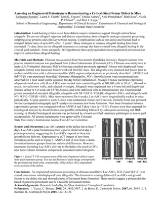
growth factor delivery. Representative μCT images of new bone
formation can be seen in Figure 1. ANOVA test of new bone volume
formation between groups found no statistical differences. However,
treatments including Luc-ASCs delivery to the defect site result in 24%
new bone volume formation compared to uncoated control allografts.
Figure 1. (A) μCT 3-D reconstructions of the mid-diaphyseal defect region
from each treatment group. The top and bottom of each image correspond to
the proximal and distal ends, respectively, of the defect. (B) Longitudinal
cross sections of the defect.
Conclusions: An engineered periosteum consisting of chitosan nanofibers, Luc-ASCs, FGF-2 and TGF-β1 was
coated onto mouse mid-diaphyseal bone allografts. This biomimetic coating delivered Luc-ASCs and growth
factors to the defect site and showed a trend of increased bone formation. These results suggest a polysaccharide-
based engineered periosteum can be a feasible periosteum mimic.
Acknowledgements: Research funded by the Musculoskeletal Transplant Foundation.
References: 1. Yazici, C. Biomat. 2008, 29. 3882-3887. 2. de Britto, D. Carbohydr Polym. 2007, 69. 305-310. 3.
Romero, R.; Carbohydr Polym 2015, 122.144-51.