Prolonged Simvastatin Treatment Provided a Decrease in Apoptotic, Inflammator...
Final poster
1. Stimulating Osteoblastic Activity Following Exposure to Ionizing Radiation by Increasing Blood Calcium and Vitamin D
Steve Marcello, Ryan Dziuba, Shelby Hall
Seton Hall University, Department of Biological Sciences, South Orange, NJ
Steve.Marcello@student.shu.edu
Introduction:
• An astronaut is expected to have low-dose 1H, helium,
and HZE particles like iron pass through them.
• IR levels above 1 Gy can case osteopenia by increasing
osteoclastic activity by modifying progenitor cells.
• During extended missions beyond the protection of
Earths' magnetosphere, astronauts are exposed to
radiation ranging between 1-2 Gy over a several days,
or for larger solar particle events (SPEs) 1-2 Gy in 8-
24 hours.
• At these doses there is significant cancellous bone loss,
damage to progenitor cells, and increased osteoclasts
present on cancellous bone.
• Current technology does not shield astronauts from IR
so therapies upon return are vital to ensure health bone
formation following exposure >1 Gy.
• It is the aim of this study to return bone density to
levels consistent with levels prior to exposure through
supplementation of 500mg of calcium and 400IU of
vitamin D for 10 weeks. This will induce osteoblastic
activity influenced by PTH with the secretion of
alkaline phosphatase in response to increased blood
calcium levels increasing.
Materials and Methods:
• 25 mice were separated into 5 groups of 5 mice each
and observed for 10 weeks
• Each group was assigned a different treatment; 1-no
radiation, 2-received IR only, 3-received IR plus
vitamin D 400IU 2x/day, 4-recievd IR plus 500mg
2x/day calcium, 5-received IR plus calcium and
vitamin D at 500mg and 400IU 2x/day, respectively.
• Type of IR used to treat cells, 56Fe, 2 Gy, 600
MeV/ion for 24 hours.
• Following exposure alkaline phosphatase test are
conducted on all groups using p-nitrophenyl
phosphate (10mM) as substrate, MgCl2 (2 mM), 2-
amino-2-methylpropanol/HCl buffer (0.5 M), and an
appropriate amount of extract.
• One unit of alkaline phosphatase was quantified by
the Lowry method and was conducted weekly for 10
weeks, as well as directly after 24 hour exposure to
IR
• In addition, structural integrity was checked weekly
using MicroCT.
Table 1- Treated groups show higher rates of osteoblast
precursor cells
Table 2- ALP activity shows recovery trend of treated
groups after 10 weeks of IR
Table 3- MicroCT analysis of radiation induced
osteopenia in mouse trabecular bone. Labeled by
group number.
Results:
• MicroCT (Table 3) shows the level of osteopenia in each group
after 10 weeks, showing Group 5 as the most similar to the control,
Group 1.
• Group 2 exhibited the worst effects, showing severe osteopenia
(Table 3) and decreased ALP levels comparative to Group 1.
• Group 3 showed slight improvement over group 2. However,
because Calcium levels were not supplemented, the Vitamin D did
not improve Alkaline Phosphatase Levels significantly.
• Group 4 showed more promising results than Group 3, due to the
increased calcium in the subjects. As seen in Table 4, Alkaline
Phosphatase rose with increased calcium levels. However, Calcium
uses vitamin D to enter the bloodstream so with out supplementing
with vitamin D, mean calcium serum levels did not increase
significantly.
• Group 5 showed the greatest degree of improvement with prolonged
increase to blood calcium PTH recruits ALP to increase osteoblastic
progenitors.
Conclusion:
• It was the aim of this study to show that supplementation of 800mg
calcium and 400IU of vitamin D significantly increases blood
calcium levels, stimulating the parathyroid to release PTH. This is
believed to recruit ALP to increase bone deposition in trabecular
bone.
Significance:
• We believe that supplementation of 500mg Calcium and 400IU of
Vitamin D daily is an effective treatment to counteract the
decreased ALP activity and subsequent reduced osteoprogenitor cell
differentiation following exposer to the levels of ionizing radiation
astronauts may experience during extended space travel.
References:
Leslie Silk, D. G. (2015). ionizing particle radiation as a modulator of endogenous bone marrow cell reprogramming:
implications for hematological cancers. International journal of sport nutrition and exercise metabolism .
Sujatha Muralidharan1, S. P. (2015). ionizing particle radiation as a modulator of endogenous bone marrow cell
reprogramming: implications for hematological cancers. Frontiers in Oncology .
Xiangming Zhang, P. W. (2015 ). Radiation activated CHK1/MEPE pathway may contribute to microgravity-induced bone
density loss. Elsevier.
Yasaman Shirazi-Fard, J. S.-S. (2015 ). Mechanical loading causes site-specific anabolic effects on bone following exposre
to ionizing radiation. Elsevier .
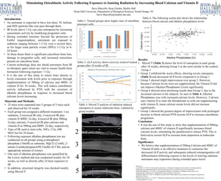
Alkaline Phosphatase (IU)
Table 4- The following scatter plot shows the relationship
between blood calcium and alkaline phosphatase levels
Calcium(mg/L)
1
2
3
4
5