4. Dr. Krishna, PGIMS, Rohtak
SHOCK
*A generalized state of decreased tissue perfusion
*If prolonged it may lead to irreversible damage of the life
supporting organs
Causes:
I. Cardiogenic
II. Neurogenic
III. Hypovolaemic
9. Dr. Krishna, PGIMS, Rohtak
1st rule = Limb crushed severely (>6hrs)
= Amputation
-Above the compression or crushed injury
-Before compression is released
Dialysis
10. Dr. Krishna, PGIMS, Rohtak
Venous thrombosis & Pulmonary
Embolism
Commonest complication of Trauma & Surgery
Most frequently
Calf veins
Less frequent in proximal thigh & pelvic veins
Pulmonary Embolism
From Proximal thigh & pelvis
Incidence = 5% & Fatal = 0.5%
11. Dr. Krishna, PGIMS, Rohtak
VTE
The primary cause in surgical
HYPERCOAGULABILITY of the Blood
due to activation of Factor X by Thromboplastin from
damaged tissues
Thrombosis occurs → secondary factors involved
Stasis
Pressure
Prolonged immobility
Endothelial damage
Increase in No. & stickiness of platelets
17. Dr. Krishna, PGIMS, Rohtak
Pulmonary Embolism
Difficult to diagnose =only minority have symptoms (chest pain,
dyspnea, hemoptysis)
• So high risk patients should be
examined for pulmonary consolidation
• X-ray
• Lung scintigraphy
• Pulmonary angiography
• Normal D-dimer has almost 100%
negative predictive value (virtually
excludes PE)
• CT
20. Dr. Krishna, PGIMS, Rohtak
Rx of Pulmonary Embolism
Cardiorespiratory resuscitation
Vasopressor for shock
Oxygen
Large dose heparin (15 000 units)
Streptokinase (dissolve clot)
Antibiotics (prevent lung infection)
21. Dr. Krishna, PGIMS, Rohtak
TETANUS
Tetanus organism live only in dead tissue → exotoxin → blood & lymph
to CNS →anterior horn cell
Will develop
Tonic clonic contraction
Jaw & face (trismus & risus sardonicus)
Neck & trunk
Diaphragm & Intercostal muscle → spasm → ASPHYXIA
https://www.youtube.com/watch?v=2baVlK5Uvyc
22. Dr. Krishna, PGIMS, Rohtak
Prophylaxis
Active immunization (tetanus toxoid)
Booster doses (immunized patients)
Non Immunized patients
Wound toilet & antibiotics
If wound contaminated →antitoxin
23. Dr. Krishna, PGIMS, Rohtak
Treatment for Tetanus
IV antitoxin
Heavy Sedation
Muscle Relaxant drug
Tracheal Intubation
Controlled respiration
24. Dr. Krishna, PGIMS, Rohtak
GAS GANGRENE
By clostridial infection (esp C. perfringens)
Anaerobic with low oxygen tension
Produce toxins → destroy cell wall → tissue necrosis → Spreading
25. Dr. Krishna, PGIMS, Rohtak
Clinical Features
Within 24 hours
Intense pain
Swelling
Brownish discharge
Pulse rate increased
Characteristic smell sweetish & mousy odor
Little or no pyrexia
Gas formation not marked
Toxaemic → coma → DEATH
26. Dr. Krishna, PGIMS, Rohtak
Prevention
Deep penetrating wound should be EXPLORED
ALL dead tissue → completely EXCISED
Doubt about tissue viability → leave it OPEN
No antitoxin
27. Dr. Krishna, PGIMS, Rohtak
Treatment for gas gangrene
The key = EARLY DIAGNOSIS
General measures (fluid, IV antibiotics)
Hyperbaric oxygen (limiting spread)
Decompression of wound
Removal of all dead tissue
Amputation (advanced case)
28. Dr. Krishna, PGIMS, Rohtak
FAT EMBOLISM
Only minority patients with
circulating fat globules will develop
POST TRAUMATIC RESPIRATORY
DYSFUNCTION
Source of fat emboli = bone
marrow
Usually in MULTIPLE CLOSED
FRACTURE
But other condition also reported
(burns, renal infarction,
cardiopulmonary operation)
29. Dr. Krishna, PGIMS, Rohtak
Closed/open
Fracture
Fat in bone
marrow
escapes
Formation of
fat globules in
vessels
Fat embolus
Stick in
pulmonary
bed
Trigger
clotting
cascade
30. Dr. Krishna, PGIMS, Rohtak
Features
After 1-2 days of trauma
Usually young adults with LL fracture
Early warning signs
Rise in temperature & pulse rate
More pronounced case(classical triad)
Breathlessness
Mild mental confusion
Petechia (chest & conjuntival fold)
31. Dr. Krishna, PGIMS, Rohtak
Most severe case
Marked respiratory distress →coma →ARDS
no definitive test, but hypoxia <60mmHg after major trauma is
suspicious
32. Dr. Krishna, PGIMS, Rohtak
Gurd’s Criteria
Major Features
(at least 1)
Minor Features
(at least 4)
Lab Features
(at least 1)
-Respiratory Insufficiency
-Cerebral involvement
-Petechiae Rash
-Pyrexia
-Tachycardia
-Retinal changes
-Jaundice
-Renal changes
-Fat macroglobulinemia
-Anaemia
-Thrombocytopenia
-High ESR
34. Dr. Krishna, PGIMS, Rohtak
Treatment
Mild case
Monitoring of blood PO2
Signs of hypoxia (<8kPa @ 60mmHg)
Oxygen
If severe
ICU with sedation & assisted ventilation
Swan ganz Catheterization (monitor cardiac Fx)
Fluid balance
Supportive
Heparin-thromboembolism
Steroids-pulmonary oedema
Aprotinin - prevent aggregation of chylomicrons
35. Dr. Krishna, PGIMS, Rohtak
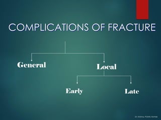
Early (& immediate) Complications
Local Visceral Injury
Vascular Injury
Nerve Injury
Compartment Syndrome
Hemarthrosis
Infection
Gas gangrene
36. Dr. Krishna, PGIMS, Rohtak
Local visceral Injury
Fracture around the trunk are often Complication by injury to the
adjacent viscera:
Etc: Pelvic fracture
Etc : Rib fracture → penetration to the lungs → Pneumothorax
Bladder & urethral rupture
This requires Emergency Rx → chest tube insertion
38. Dr. Krishna, PGIMS, Rohtak
Vascular injury
commonly – knee, femoral shaft, elbow
& humerus
Artery may be cut, torn, compressed or
contused
Intima may be detached, thrombus
block, artery spasm
Effect → ↓↓ bld flow coz Ischemia leads
to tissue death & peripheral gangrene
39. Dr. Krishna, PGIMS, Rohtak
Vascular Injury - Clinical features
Pt with ischemia may have 5 P’s:
- paraesthesia/numbness
- pain
- pallor
- pulselessness
- paralysis
Investigate if suspect vascular injury : CT Angiogram
40. Dr. Krishna, PGIMS, Rohtak
Treatment
Emergency treatment
All bandages/splints removed
X-Ray The fracture again
Circulation reassessed for next half hour
If no improvement, do vessels exploration
Suture torn vessels, vein grafting, if thrombosed do
endarterectomy
Aim: to restore bld flow
41. Dr. Krishna, PGIMS, Rohtak
Nerve Injury
Variable degree of motor & sensory loss along the distribution of
the nerve
May be neurapraxia, axonotmesis or neurotmesis
43. Dr. Krishna, PGIMS, Rohtak
Nerve Trauma Effect
Axillary Dislocation of shoulder Deltoid paralysis
Radial # of humerus Wrist drop
Median Supracondylar # of humerus Pointing index
Ulnar # medial epicondyl humerus Claw hand
Sciatic Post dislocation of hip Foot drop
Common
peroneal
Knee dislocation # neck of
fibula
Foot drop
44. Dr. Krishna, PGIMS, Rohtak
In closed injuries – nerve is seldom severed &
spontaneous recovery should be awaited
In open fractures – complete lesion (neurotmesis): the
nerve is explored during wound debridement &
repaired
Acute nerve compression – occur with fracture or
dislocation around the wrist. C/o numbness in median
& ulnar dist. If no improvement >48 hours → after
fracture reduction, do nerve exploration &
decompression