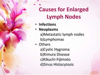
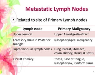
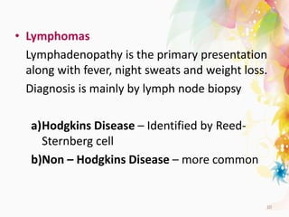
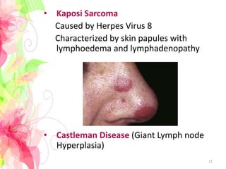
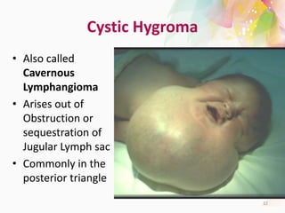
Differential
Diagnosis of
Enlarged
Cervical Lymph
Nodes
Apoorva Kottary 28
1
Causes for Enlarged
Lymph Nodes
• Infections
• Neoplasms
a)Metastatic lymph nodes
b)Lymphomas
• Others
a)Cystic Hygroma
b)Kimura Disease
c)Kikuchi-Fijimoto
d)Sinus Histocytosis
2
3
Lymph Node Drainage
Submandibular Tongue, Submandibular Gland, Lips, Mouth,
Conjunctiva
Submental Lower Lip, Floor of Mouth, Tip of Tongue, Skin of
Cheek
Jugular Tongue, Tonsil, Pinna, Parotid
Posterior Cervical Scalp & Neck, Skin of Arms & Pectoralis, Thorax,
Cervical and Axillary Lymph Nodes
Sub Occipital Scalp and Head
Post Auricular Exernal Auditus Meatus, Pinna, Scalp
Pre Auricular Eyelids And Conjuctiva, Temporal Region, Pinna
Right Supraclavicular Mediastinum, Lungs, Esophagus
Left Supraclavicular Thorax, Abdomen Wall, Breast 4
Infections
• Bacterial – Coccal infections, Influenza,
Tularemia, Typhoid
• Viral - HIV, Syphilis, Herpes, Hepatitis, Cat
scratch Disease
• Parasitic – Toxoplasmosis, Trypanosomiasis,
Histoplasmosis, Leishmaniasis
• Mycobacterial - Tuberculosis
5
Tubercular Lymphadenitis
• May affect any
Lymph node in the
neck and axial
region but chiefly
the Supraclavicular
lymph node
• Show Caseation and
matting
6
Infectious Mononucleosis
• Caused by Ebstein-
Barr Virus
• Enlarged posterior
cervical and axillary
lymph nodes.
• Associated with
tonsillitis,
pharyngitis and
spleenomegaly
7
Neoplastic Lesions
• Metastatic Lymph Nodes
• Lymphomas
• Kaposi Sarcoma
• Castleman disease
8
Metastatic Lymph Nodes
• Related to site of Primary Lymph nodes
Lymph node Primary Malignancy
Upper cervical Upper AerodigestiveTract
Accessory chain in Posterior
Triangle
Nasopharyngeal malignancy
Supraclavicular Lymph nodes Lung, Breast, Stomach,
colon, Kidney, Ovary, & Testis
Occult Primary Tonsil, Base of Tongue,
Nasopharynx, Pyriform sinus
9
• Lymphomas
Lymphadenopathy is the primary presentation
along with fever, night sweats and weight loss.
Diagnosis is mainly by lymph node biopsy
a)Hodgkins Disease – Identified by Reed-
Sternberg cell
b)Non – Hodgkins Disease – more common
10
• Kaposi Sarcoma
Caused by Herpes Virus 8
Characterized by skin papules with
lymphoedema and lymphadenopathy
• Castleman Disease (Giant Lymph node
Hyperplasia)
11
Cystic Hygroma
• Also called
Cavernous
Lymphangioma
• Arises out of
Obstruction or
sequestration of
Jugular Lymph sac
• Commonly in the
posterior triangle
12
• Treatment –
Surgical Excision with
preservation of neural
and vascular tissue
Recurrence rate is
about 5% when whole
tumour is removed and
about 50% when part
of it is left behind.
13
• Kimura Disease
Chronic inflammatory Disorder with painless
unilateral inflammation of cervical lymph
nodes
• Kikuchi – Fujimoto Disease
Necrotizing lymphadenitis chiefly affecting
the cervical Lymph nodes
• Sinus Histiocytosis (Rosai – Dorfman
Disease)
Rare Histiocytic disorder with over
production of WBCs
14
Factors that will aid
diagnosis
• Size
• Pain / Tenderness
• Consistency
• Matting
• Location
15
THANK YOU
16

Cervical Lymphadenopathy

  • 1.
  • 2.
    Causes for Enlarged LymphNodes • Infections • Neoplasms a)Metastatic lymph nodes b)Lymphomas • Others a)Cystic Hygroma b)Kimura Disease c)Kikuchi-Fijimoto d)Sinus Histocytosis 2
  • 3.
  • 4.
    Lymph Node Drainage SubmandibularTongue, Submandibular Gland, Lips, Mouth, Conjunctiva Submental Lower Lip, Floor of Mouth, Tip of Tongue, Skin of Cheek Jugular Tongue, Tonsil, Pinna, Parotid Posterior Cervical Scalp & Neck, Skin of Arms & Pectoralis, Thorax, Cervical and Axillary Lymph Nodes Sub Occipital Scalp and Head Post Auricular Exernal Auditus Meatus, Pinna, Scalp Pre Auricular Eyelids And Conjuctiva, Temporal Region, Pinna Right Supraclavicular Mediastinum, Lungs, Esophagus Left Supraclavicular Thorax, Abdomen Wall, Breast 4
  • 5.
    Infections • Bacterial –Coccal infections, Influenza, Tularemia, Typhoid • Viral - HIV, Syphilis, Herpes, Hepatitis, Cat scratch Disease • Parasitic – Toxoplasmosis, Trypanosomiasis, Histoplasmosis, Leishmaniasis • Mycobacterial - Tuberculosis 5
  • 6.
    Tubercular Lymphadenitis • Mayaffect any Lymph node in the neck and axial region but chiefly the Supraclavicular lymph node • Show Caseation and matting 6
  • 7.
    Infectious Mononucleosis • Causedby Ebstein- Barr Virus • Enlarged posterior cervical and axillary lymph nodes. • Associated with tonsillitis, pharyngitis and spleenomegaly 7
  • 8.
    Neoplastic Lesions • MetastaticLymph Nodes • Lymphomas • Kaposi Sarcoma • Castleman disease 8
  • 9.
    Metastatic Lymph Nodes •Related to site of Primary Lymph nodes Lymph node Primary Malignancy Upper cervical Upper AerodigestiveTract Accessory chain in Posterior Triangle Nasopharyngeal malignancy Supraclavicular Lymph nodes Lung, Breast, Stomach, colon, Kidney, Ovary, & Testis Occult Primary Tonsil, Base of Tongue, Nasopharynx, Pyriform sinus 9
  • 10.
    • Lymphomas Lymphadenopathy isthe primary presentation along with fever, night sweats and weight loss. Diagnosis is mainly by lymph node biopsy a)Hodgkins Disease – Identified by Reed- Sternberg cell b)Non – Hodgkins Disease – more common 10
  • 11.
    • Kaposi Sarcoma Causedby Herpes Virus 8 Characterized by skin papules with lymphoedema and lymphadenopathy • Castleman Disease (Giant Lymph node Hyperplasia) 11
  • 12.
    Cystic Hygroma • Alsocalled Cavernous Lymphangioma • Arises out of Obstruction or sequestration of Jugular Lymph sac • Commonly in the posterior triangle 12
  • 13.
    • Treatment – SurgicalExcision with preservation of neural and vascular tissue Recurrence rate is about 5% when whole tumour is removed and about 50% when part of it is left behind. 13
  • 14.
    • Kimura Disease Chronicinflammatory Disorder with painless unilateral inflammation of cervical lymph nodes • Kikuchi – Fujimoto Disease Necrotizing lymphadenitis chiefly affecting the cervical Lymph nodes • Sinus Histiocytosis (Rosai – Dorfman Disease) Rare Histiocytic disorder with over production of WBCs 14
  • 15.
    Factors that willaid diagnosis • Size • Pain / Tenderness • Consistency • Matting • Location 15
  • 16.