SfN 2015 - Anil Sharma - Genetic tools to study sensory motor circuits FINAL
1. Genetic tools to study sensory motor circuits
Anil Sharma1
, Haohao Wu1
, Carmelo Bellardita2
, Yang Xuan1
, Konstantinos Meletis1
, Ole Kiehn2
, Francois Lallemend1
1
Department of Neuroscience, Karolinska Institutet, Stockholm, Sweden ; 2
Mammalian locomotor laboratory, Department of Neuroscience, Karolinska Institutet, Stockholm, Sweden
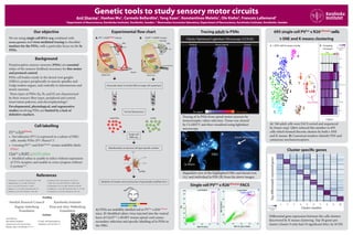
Cluster specific genes
695 single cell PVCre
x R26tdTomato
cells
t-SNE and K means clustering
References
1. de Nooij, J.C., et al. 2013. Neuron 77, 1055–1068
2. Hippenmeyer, S., et al. 2005. PLoS Biol 3
3. Lee, J., et al. 2012. PLoS One 7, e45551
4. Stepien, A.E., et al. 2010. Neuron 68, 456-472
5. Takatoh, J., et al. 2013. Neuron 77, 346–360
6. Usoskin, D. 2015. Nat. Neurosci. 18, 145-153
7. Wall, N.R., et al. 2010. PNAS 107, 21848-21853
8. Wickersham, I.R., et al. 2007. Neuron 53, 639-647
9. Windhorst, U., et al. 2007. Brain Res. Bull. 73, 155-202
10. Zampieri, N., et al. 2014. Neuron 81, 766–778
Experimental flow chart
Background
Proprioceptive sensory neurons (PSNs) are essential
relays of the sensory feedback necessary for fine motor
and postural control.
PSNs cell bodies reside in the dorsal root ganglia
(DRGs), project peripherally to muscle spindles and
Golgi tendon organs, and centrally to interneurons and
motor neurons.
Three types of PSNs (Ia, Ib, and II) are characterised
by their sensory fibre types, peripheral and central
innervation patterns, and electrophysiology8
.
Developmental, physiological, and regenerative
studies involving PSNs are limited by a lack of
definitive markers.
Our objective
We are using single cell RNA-seq combined with
mouse genetics and virus mediated tracing to elucidate
markers for the PSNs, with a particular focus on the Ia
PSNs.
Cell labelling
PVcre
x R26tdTomato
:
• Parvalbumin (PV) is expressed in a subset of DRG
cells, mostly PSNs (PV+
/Runx3+
)1
.
• Crossing PVCre
and R26tdTomato
strains indelibly labels
PSNs2
.
ChatCre
x RGθT + EGFP-rabies:
• Modified rabies is unable to infect without expression
of TVA receptor, and unable to cross synapses without
G protein3,6,7
.
Tracing adult Ia PSNs
A) PSNs are indelibly labelled red in PVCre
x R26tdTomato
mice. B) Modified rabies virus injected into the ventral
horn of ChATCre
x RGΦT mouse spinal cord causes
secondary infection and specific labelling of Ia PSNs in
the DRG.
Tracing of Ia PSNs from spinal motor neurons by
monosynaptic rabies infection. Tissue was cleared
by CLARITY and then visualised using lightsheet
microscopy.
Magnified view of the highlighted DRG and dorsal root
(A), and individual Ia PSN (B) from the above images.
Single cell PVCre
x R26tdTomato
FACS
A) 768 adult cells were FACS sorted and sequenced
by Smart-seq2. Q&A reduced this number to 695
cells which formed discrete clusters by both t-SNE
and K means. B) Canonical markers identify PSN and
cutaneous mechanoreceptors.
Differential gene expression between the cells clusters
discovered by K means clustering. Top 50 genes per
cluster (cluster 9 only had 10 significant hits), by SCDE.
Funding
Swedish Research Council
Ragnar Söderberg
Foundation
Karolinska Institutet
Knut and Alice Wallenberg
Foundation
Contact
Anil Sharma
Karolinska Institutet
Department of Neuroscience
Retzius väg 8, Stockholm 171 77
E-mail: anil.sharma@ki.se
Telephone: 08-524 863 74
DRG
Clarity Optimised Lightsheet Microscopy (COLM)
No signal High signal
Ventral Dorso-lateral Dorsal
Cluster number
Topdifferentiallyexpressedgenes
1 2 3 4 5 6 7 8 9 10
-3 -2 -1 0 1 2 3
Gene expression
Z-score
2 4 6 8
Cluster
1 2 3 4 5 6
Etv1
Whirlin
Runx3
Parvalbumin
TrkC
TrkB
TrkA
7 8 9 10
Log2
counts per
million
A t-SNE with K means overlay B Grouping
of clusters
PSNs Cut.?
2
10
9
8
7
64
3
5
1
Non PSNs
DRG
A
Ia fibers
Axon bifurcation
B
Muscle
MS
GTO
DRG
Motor
neurons
Interneurons
Ia PSN
Ib PSN
II PSN
II PSN
Spinal cord
PVCre
x R26tdTomato
mouseA B
Interneurons
Secondary
rabies
infection
EnvA-ΔG-EGFP-Rabies
DRG
Ib PSN
Ia PSN
ChAT+ Motor
neurons
Spinal cord
ChATCre
x RGΦT mouse
+ EGFP-rabies tracing
PCA1
PCA2
PCA3
Ia PSNs
5’UTR GENE IRES-venus-pA TTX IRES-cherry-pA
5’UTR GENE IRES-Cre 3’ UTR
Ia/Ib/II PSNs Ia PSNs
Dissociate adult (3 month) DRG to single cell suspension
Sort by fluorescence
Single cell
RNA-seq
Bioinformatics to discover cell type specific markers
Validation of markers and construction of genetically modified mice