Neurotoxicity assay on 2D and 3D culture using High Content Screening (HCS) t...
Mouse retina count poster draft Final
1. The mammalian retina contains neuronal cells as well as a
number of non-neuronal glial cells. The different types of glial
cells include Müller glia, retinal astrocytes, and microglia.
Müller glial cells and astrocytes nourish neurons, and
microglia act as sentinels that respond to injury or disease
within the nervous system. The long-term goal of our
laboratory has been to study interactions between microglia,
Müller glia, and astrocytes in healthy and diseased tissue. The
focus of the present study was to develop a technique that
would allow the laboratory to study changes in cell number in
retinal flat mounts and cultures. Immunohistochemistry (IHC)
was performed to fluorescently label mature murine retinal
tissue. Retinal flat-mounts were stained with SOX2, a nuclear
marker for macroglial cells (Müller cells and retinal astrocytes)
and cholinergic amacrine cells, NeuN, a nuclear marker for
ganglion cell neurons, and counter-stained with Hoechst
solution to label all nuclei. Pure cultures of mouse microglial
cells treated with liposomal clodronate (a drug which
specifically targets and ablates microglia) and vehicle were
counter stained with Hoechst solution. Cell counts were
performed on the images of the fluorescently labeled samples
using Image-J software. Convolutions were used to filter
images of immune-labeled cultures and retinal flat-mounts to
make the images clear enough to capture cell number. The cell
count assistance protocol yielded acceptable cell count results
of the stained cells and determined a detectable difference in
the number of clodronate treated cells versus vehicle treated
control cells. The images produced of the retinal flat-mounts
were analyzed to determine the percentage of SOX2 positive
Müller glia in the mature murine retinal tissue. A modified
Image J program could be used to determine cellular number in
cultures and retinal flat mounts.
Standardizing Methods and Procedures for Mouse Retinal Flat-mounts and Glial
Cell Counts
Richard Anderson III1, Subramanian Dharmarajan1, Teri L Belecky-Adams1
1Department of Biology, Indiana University-Purdue University Indianapolis, INDIANA
Indiana University-Purdue University Indianapolis
Abstract ResultsMethods
Cell Counts
The image processing software Image-J was used to count the
cells in the IHC stained images taken by confocal microscope.
In order to prepare the images for cell counting, the
background of the image had to be taken out and contrast
enhanced. Excess particles, pixels, and coloration in the image
called “noise” was taken out, and the color maxima were
found and correlated to cell number.
Introduction
The retina is a multilayered neuronal tissue consisting of
different types of neuronal cells as well as non neuronal cells,
termed glial cells. The primary glial cells of the retina, Müller
glia, together with the retinal astrocytes make up the
macroglial population in the retina. The retina is also populated
by microglia which constitute the resident macrophages of the
neural tissue. Müller glia are the predominant glial cells of the
retina and make up about “16% of the mouse retina” (1).
Müller glia play an important role during development of the
retina and also in supporting the normal function of the retina.
Microglia are “the resident immune cells of the CNS which
normally respond to neuronal damage and remove the
damaged cells by phagocytosis” to maintain homeostasis (2).
The purpose of this study is to develop a technique to
determine cell numbers in fluorescently labeled cells in vitro
and in vivo in retinal flat mounts.
References
1. Chang-Jin Jeon, et al. (1998). "The Major Cell Populations
of the Mouse Retina" The Journal of Neuroscience ,
18(21):8936–8946
2. Dheen ST, et al. (2007). "Microglial activation and its
implications in the brain diseases." Curr Med Chem
14(11):1189-97
3. http://www.jiscdigitalmedia.ac.uk/infokit/colour-
management/understanding-colour
4. http://www.di.uq.edu.au/sparqihc: University of
Queensland, Australia
Results
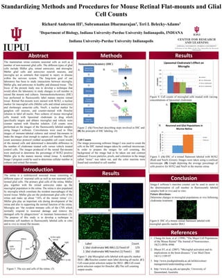
Figure 3: (A) Microglial cells labeled with specific marker
IBA1. (B) Hoechst counter stain label showing all nuclei. (C)
Cell count point selection output for IBA1. (D) Cell count
point selection output for Hoechst. (E) The cell counting
output results.Figure 1: The eye and cells of the retina. (3)
Figure 2: (A) Flowchart describing steps involved in IHC and
(B) the principle of IHC labeling. (4)
A. B.
C.
Conclusion
Figure 5: (A) IHC of a retinal flatmount labeled with SOX2
(Red) and NeuN (Green). Images were taken using a confocal
microscope. (B) Graph depicting the average percentage of
cells positive for SOX2 and NeuN in the murine retina
Figure 6: IHC of a mouse retinal flatmount labeled with
microglial specific marker IBA1
Image-J software’s maxima counter can be used to assist in
the determination of cell number in fluorescently labeled
samples both in vivo and in vitro.
Future directions:
Determine changes in microglial populations in vivo following
clodronate treatment
Retinal Flat
Mount Prep
Tissue
Fixation
Blocking
Primary
Antibodies
Secondary
Antibodies
Hoechst
counter stain
D.
Figure 4: Cell counts of microglial cells treated with varying
concentrations of liposomal clodronate
Immunohistochemistry (IHC)
A. B.
E.
A.
B.
Label Count
1 10ul clodronate MG IBA1 (1) fixed.tif 173
1 10ul clodronate MG hoechst (1) fixed.tif 192