
Recommended
More Related Content
What's hot
What's hot (20)
Similar to Fungal Species Isolation in Dermatophytosis
Similar to Fungal Species Isolation in Dermatophytosis (20)
Recently uploaded
Recently uploaded (20)
Fungal Species Isolation in Dermatophytosis
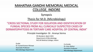
- 1. MAHATMA GANDHI MEMORIAL MEDICAL COLLEGE, INDORE Synopsis Thesis for M.D. (Microbiology) “CROSS SECTIONAL STUDY FOR ISOLATION AND IDENTIFICATION OF FUNGAL SPECIES FROM ALL CLINICALLY SUSPECTED CASES OF DERMATOPHYTOSIS IN TERTIARY CARE HOSPITAL OF CENTRAL INDIA” Principle Investigator- Dr . Ananya Verma Guide: Dr. Suneel Kumar Ahirwar Associate Professor Microbiology Department Co-Guide: Dr. Anju Mahor Assistant Professor Microbiology Department PG Resident, Batch 2021 Department of Microbiology M.G.M Medical College, Indore
- 2. INDEX 1. TITLE 2. INTRODUCTION AND RATIONALE 3. AIM AND OBEJECTIVE 4. METHODOLOGY STUDY CENTER STUDY DESIGN STUDY DURATION 5. SAMPLE SIZE 6. INCLUSION AND EXCLUSION CRITERIA 7. STATISTICAL METHOD / STUDY ANALYSIS 8. SOURCE OF FUNDING 9. MASTER CHARTS AND DUMMY TABLES 10. REFERENCES
- 3. TITLE “CROSS SECTIONAL STUDY FOR ISOLATION AND IDENTIFICATION OF FUNGAL SPECIES FROM ALL CLINICALLY SUSPECTED CASES OF DERMATOPHYTOSIS IN TERTIARY CARE HOSPITAL OF CENTRAL INDIA”
- 4. Introduction • Dermatophytes are the most common cause of superficial cutaneous fungal infections of human body mainly involving the keratinised tissues like skin ,hair and nails . • It includes three genera- ● Epidermophyton ● Microsporum ● Trichophyton They are collectively known as Dermatophytes. ● The tinea infections are prevalent globally, but they are common in tropics and in geographical areas with higher humidity , overpopulation, and poor hygienic living conditions. ● An increasing frequency of Dermatophytosis has been observed during last two decades especially in immunocompromised patients such as AIDS, Diabetes mellitus, cancer and organ transplantation patients.
- 5. • There has been a significant increase in the incidence of chronic, relapsing and recurrent cases of superficial Dermatophytosis in India that are often unresponsive to conventional drugs and doses of recommended antifungal treatment. According to WHO, the prevalence rate of superficial mycotic infections worldwide has been found to be 20-25%. The recent prevalence of Dermatophytosis in India ranges from 36.6-78.4%.
- 6. RATIONALE OF THE STUDY ● This study is being conducted for the first time at MGM Medical College, Indore . ● This study will reveal most common fungal species causing Dermatophytosis in central India . ● It will also help clinicians to start antifungal treatment as early as possible.
- 7. AIM AND OBJECTIVE ● To Identify the causative fungal species from all clinically suspected cases of Dermatophytosis in a tertiary care hospital of Central India .
- 8. METHODOLOGY Study centre: Mahatma Gandhi Memorial Medical College and M.Y. Hospital, Indore Duration of Study: 1 year after approval of study by institutional scientific and Ethical Committee. Study design-Cross sectional study
- 9. Sample: All clinically suspected cases of Dermatophytosis from out patient department of Dermatology. Inclusion criteria: All age groups of either sex with clinical features of Dermatophytosis. Exclusion Criteria: Nil Sample size: Minimum 400 samples
- 10. Materials and Methods ● Sample taken from the infected areas like skin , nail and hair of patients of all age group from out patient department of Dermatology.
- 11. Procedure Planned Sample received from out patient department of dermatology [like skin scrapping, nail clipping, hair] Direct Microscopic Examination (10-20%, or 40% KOH Mount) Fungal culture (SDA Media and DTM) Incubate at 25 ̊̊C and 37 ̊̊C for 4 weeks and will be observed at regular intervals. Growth Lactophenol Cotton Blue Mount Slide Culture Identification of Species
- 12. Statistical Analysis Plan • Data will be entered in excel sheet and will be analysed using open sources software. • Continuous data will be expressed in terms of mean and standard deviation. • Categorical data will be expressed in proportion and percentage. • Appropriate test of significance will be applied wherever necessary and p-value <0.05 will considered as statistically significant. Source of Funding: None
- 13. Master Chart Serial No. Lab No. OPD No. Age Sex Sample Clinical diagnosis Fungal isolates Species 1. 2. 3. 4. 5.
- 14. DUMMY TABLES Table 1: Demographic Profile of Dermatophytosis SERIAL NO. AGE MALE(%) FEMALE(%) TOTAL(n) 1. 1-10 YEARS 2. 11-20 YEARS 3. 21-30 YEARS 4. 31-40 YEARS 5. 41-50 YEARS 6. 51-60 YEARS 7. >60 YEARS
- 15. TABLE 2: Fungal Species Isolates SERIAL NO. FUNGAL SPECIES SKIN HAIR NAILS TOTAL NO. OF ISOLATES 1. 2. 3. 4.
- 16. References: • 1.Chandra j. Textbook of medical mycology. New Delhi. Jaypee Publishers;2018;4:179-182 • 2.Havlickova B, Czaika VA , Epidemiology trends in skin mycosis worldwide. Mycosis 2008;4:2-15 • 3.kannan P,Janaki, Selvi GS Prevalence of dermatophytes and other fungal agents isolated from clinical samples. Indian j. Med microbiology. 2006;24:212-5. • 4.Uthansingh K, Sahu , Debata NK. Isolation and identification of fungus associated with skin and nail scalps. Apollo Med. 2019;16:16-21 • 5.Rajagopalan et al. BMC Dermatology;2018:18:6.
- 17. THANK YOU…. `