
Recommended
More Related Content
What's hot
What's hot (20)
Similar to Oesophagus by dr. vineeta
Similar to Oesophagus by dr. vineeta (20)
Recently uploaded
Recently uploaded (20)
Oesophagus by dr. vineeta
- 1. DR. VINEETA N KUMAR 1 OESOPHAGUS 10/15/2020
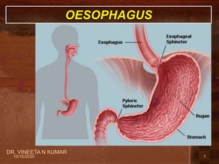
- 2. ESOPHAGUS • It is a tubular structure about 25 cm long. • It begins as the continuation of the pharynx at the level of the 6th cervical vertebra. • It pierces the diaphragm at the level of the 10th thoracic vertebra to join the stomach. • It is divided into 3 parts: • 1- Cervical. • 2- Thoracic. • 3- Abdominal. Abdominal thoracic Cervical 210/15/2020 DR. VINEETA N KUMAR
- 3. DR. VINEETA N KUMAR 3 CERVICAL PART • Posteriorly: • Vertebral column. • Laterally: • Lobes of the thyroid gland. • Anteriorly: • Trachea and the recurrent laryngeal nerves. RELATIONS 10/15/2020
- 4. DR. VINEETA N KUMAR THORACIC PART • In the thorax, it passes downward and to the left through superior then to posterior mediastinum • At the level of the sternal angle, the aortic arch pushes the esophagus again to the midline. 10/15/2020
- 5. DR. VINEETA N KUMAR ANTERIOR RELATIONS • Trachea • Left recurrent laryngeal nerve, rt pulmonary ar,diaphragm • Left principal bronchi,Left atrium Thoracic part 10/15/2020
- 6. DR. VINEETA N KUMAR 6 POSTERIOR RELATIONS • Bodies of the thoracic vertebrae • Thoracic duct • Azygos vein • Right posterior intercostal arteries • Descending thoracic aorta (at the lower end) Thoracic part 10/15/2020
- 7. LATERAL RELATION • On the Right side: • Right mediastinal pleura • Terminal part of the azygos vein. • On the Left side: • Left mediastinal pleura • Left subclavian artery • Aortic arch • Thoracic duct DR. VINEETA N KUMAR 710/15/2020
- 8. ESOPHAGUS AND LEFT ATRIUM • There is a close relationship between the left atrium of the heart and esophagus. • What is the clinical application? • A barium swallow in the esophagus will help the physician to assess the size of the left atrium (dilation) as in case of long standing mitral stenosis or heart failure.
- 9. 9 RELATIONS IN THE ABDOMEN • In the Abdomen, the esophagus descends for 1.3 cm and joins the stomach. • Anteriorly, left lobe of the liver. • Posteriorly, left crus of the diaphragm. • Fibers from the right crus of the diaphragm form a sling around the esophagus. • At the opening of the diaphragm, the esophagus is accompanied by: – The two vagi – Branches of the left gastric vessels – Lymphatic vessels. 10/15/2020 DR. VINEETA N KUMAR
- 10. ESOPHAGEAL CONSTRICTIONS • The esophagus has 4 anatomic constrictions. • The first is at the junction with the pharynx(pharyngeoesophageal junction). • The second is at the crossing with the aortic arch and the left main bronchus. • THIRD when it crosses left bronchus. • The fourth is at the junction with the stomach. • They have a considerable clinical importance.
- 11. 1. They may cause difficulties in passing an esophagoscope. 2. In case of swallowing of caustic liquids (mostly in children), this is where the burning is the worst and strictures develop. 3. The esophageal strictures are a common sites of the development of esophageal carcinoma. 4. In this picture what is the importance of the scale?
- 12. DR. VINEETA N KUMAR 12 ARTERIAL SUPPLY • Upper third by the inferior thyroid artery. • The middle third by the thoracic aorta. • The lower third by the left gastric artery. 10/15/2020
- 13. VENOUS DRAINAGE • The upper third drains in into the inferior thyroid veins. • The middle third into the azygos veins. • The lower third into the left gastric vein, which is a tributary of the portal vein. • Brachiocephalic vein 10/15/2020 DR. VINEETA N KUMAR 13
- 14. DR. VINEETA N KUMAR 14 LYMPH DRAINAGE • The upper third is drained into the deep cervical nodes. • The middle third is drained into the superior and inferior mediastinal nodes. • The lower third is drained in the celiac lymph nodes in the abdomen. 10/15/2020
- 15. DR. VINEETA N KUMAR 15 NERVE SUPPLY • It is supplied by sympathetic fibers from the sympathetic trunks. • The parasympathetic supply comes form the vagus nerves. • Inferior to the roots of the lungs, the vagus nerves join the sympathetic nerves to form the esophageal plexus. • The left vagus lies anterior to the esophagus. • The right vagus lies posterior to it. 10/15/2020
- 16. 16 The abdominal cavity is divided into 9 compartments: by: 2 vertical and 2 horizontal planes Vertical planes: 2 Midclavicular lines. Horizontal planes: Subcostal and Intertubercular lines. 8th costal L 1 10/15/2020 DR. VINEETA N KUMAR
- 17. APPLIED ANATOMY • Oesophageal constrictions • Oesophageal varices • Achalasia cardia • Tracheo oesophageal fistula • Oesophagitis • Gerd DR. VINEETA N KUMAR 1710/15/2020