1. Breast Cancer Stem Cells: A Look into the Cellular and Molecular Structure
Amber Rigdon
Abstract
Breast cancer stem cells have been identified as tumor-initiating cells. They have high expression
of the cell surface marker CD44 and low or no expression of the cell surface marker CD24.
Levels of palmitoleic acid are lower, but are at a higher intensity in breast cancer stem cells
compared to non-stem cancer cells. Breast cancer stem cells have been found to undergo
epithelial-mesenchymal transition processes that are controlled by the co-expression of Oct-4
and Nanog. Identification of these pathways lead to the idea that Curcumin could lead to a break
through drug therapy, targeting breast cancer stem cells.
Introduction
Breast cancer is the single most common malignancy in women and is the most common cause
of death from a malignant disease in women1. Most cases of cancer originate from a single cell
that begins to proliferate and form new tumors. For years scientists studying tumor formation
have questioned, what makes one cell form tumors versus another cell2. One theory has been that
any single tumor cell has the ability to be a tumor-initiating cell (T-IC) and form new tumors.
Another theory stated that only a few cells in a tumor had this capability to become TI-Cs which
proliferate and form new tumors3.
These TI-Cs had been found to be similar to normal, non cancerous stem cells. Normal
human stem have the ability to go down many different pathways as they mature2. For example,
human mammary gland stem cells have the ability to either proliferate to form new
Page ! of !1 11
2. Rigdon
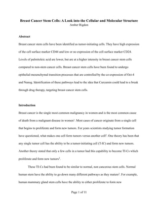
cells of differentiate into progenitor cells. They can form either ductal progenitor cells that
become mature mammary ducts, or they can become lobuloalveolar progenitor cells that become
mature mammary alveoli (Figure 1)2. When cells are mutated and become TI-Cs this process is
altered and tumors grow. These TI-Cs proliferate much more that normal stem cells, giving rise
to a tumor. Besides the growth of a tumor, due to the increased self-renewal, the cell
differentiation is also altered. These alterations in the alveoli and ducts along with the rapid cell
growth of the TI-Cs is thought to be the cause of breast cancer (Figure 2)2.
Figure 1. The normal pathway of a mammary gland stem cell (MGSC). The MGSC either undergoes self-
renewal or differentiation. If the cell differentiates, it becomes either a ductal or lobuloalveolar progenitor
and then becomes a mature duct or mature alveoli respecitvely. Figure taken from reference 2.
Page ! of !2 11
3. Rigdon
Figure 2. The pathway of a mutated breast cancer-initiating cell (BrCa-IC). The BrCa-ICs undergo self
renewal far more often than normal MGSCs. The differentiation is also altered, which is thought to cause
breast cancer. Taken from reference 2.
Beyond simply identifying these cells, it has been important to discover the structure and
characteristics to better understand them. Cell surface markers and biomarkers can distinguish
one cell type from another, so by identifying the cell markers in breast cancer stem cells it is
possible to use these markers as targets for therapy. By discovering cell surface markers and
biomarkers, cancer stem cells can be isolated and further studied. Different regulatory pathways
that are often disrupted in breast cancer cells can be studied to determine what pathways and how
many need to be disrupted to cause the formation of breast cancer stem cells. This review will
examine whether the identification of biomarkers and disrupted pathways in breast cancer stem
cells can lead to the development of targeted treatments. Since breast cancer stem cells, like
normal stem cells, proliferate and differentiate into many cell types, the possibility of targeting
breast cancer stem cells to prevent metastasis in breast cancer will also be discussed.
Page ! of !3 11
4. Rigdon
Breast Cancer Initiating Cells
Initially it had been observed that a small number of cells, called breast cancer stem cells,
are capable of becoming T-ICs2. Transplantation of human breast cancer cells into
immunocompromised mice showed that only the cells with CD44+ and CD24-/low formed tumors
whereas the cells with CD44- and CD24+ did not (Table 1)4. Based on this study, it has been
determined that the adhesion molecules CD44 and CD24 could be used to differentiate the
population of tumorigenic breast cancer stem cells from the non stem cancer cells that did not
form tumors, depending on whether they are positive or negative for these markers.
Table 1. Tumorigenicity Markers
Mice injected with CD44+ and CD24-/low all produced tumors, but no significant tumors have been found
in the mice injected with CD44- and CD24+. Many of the lineage markers found in normal cells have not
been present in these tumorigenic cells4.
It has been known that the small amount of these tumorigenic cells, also known as breast
cancer stem cells, undergo similar processes of normal stem cells, such as proliferation and
giving rise to diverse cell types. These findings are vital to the development of therapeutic
techniques. By finding out more about the structure and makeup of these cancer stem cells,
including their cell surface makers and more, it could lead to the possibility of targeting them
specifically in chemotherapy4.
CD44+ and CD24-/low Tumors
CD44- and CD24+ No Tumors
Page ! of !4 11
5. Rigdon
Palmitoleic Acid
The make up of cancer stem cells had been of great curiosity to many scientists since it could
lead to a breakthrough treatment for breast cancer. Cell membranes are made up mostly of lipids,
which are important to many biological processes. The cell membrane of breast cancer stem cells
has been studied in the hope that they could find something that distinguished it from non stem
cancer cells5.
Abnormal lipid synthesis has been linked to cancer, and other diseases. When studying
the cell membrane of breast cancer stem cells it has been found that most of the fatty acids did
not show a significant difference between cancer stem cells and non stem cancer cells. However,
the cancer stem cells have been found to have a significant decrease in palmitoleic acid, but the
palmitoleic acid that is present is at a higher intensity. It has been found that there has been a
59.8% ratio between cancer stem cells and non stem cancer cells for palmitoleic acid5.
It has been determined that although both cell types can synthesize palmitic acid, only
non stem cancer cells extensively produce palmitoleic acid, consistent with the findings that non
cancer stem cells have higher levels of palmitoleic acid than cancer stem cells. Similar findings
have been discovered in leukemia stem cells and may lead to the development of drug therapies
that target cancer stem cells specifically, by taking advantage of this difference in palmitoleic
acid formation5.
Oncogenic Transformation of Epithelial Cells
In order to target breast cancer stem cells in treatments, it is important to know as much as
possible about those cells. Most breast cancers originate from epithelial cells, but it is important
Page ! of !5 11
6. Rigdon
to identify and understand what specific mutations lead to the formation of breast cancer. It is
known that many mutations can lead to breast cancer, but a single mutation hasn’t been identified
to be common in all cases. Many mutations coexist in breast cancer cells, but it is also unknown
how many mutations are required for human mammary epithelial cells to become cancerous6.
Regulatory pathways that are found to often be disrupted have been chosen and
synthetically disrupted in human mammary epithelial cells that have no known pathology.
Disruption of the regulatory pathway of p53, a tumor suppressor gene and disruption of the Ras-
signaling pathway have been simulated. They also simulated the activation of telomerase activity
that allows breast cancer cells to maintain their telomeres. When just one pathway was disrupted,
no tumors formed. It had also been found that the Ras needed high over expression, for tumors to
form. The disruption/activation of all three of these simultaneously lead to significant tumor
formation6.
Table 2. Formation of subcutaneous tumors in nude mice
hTERT simulates telomerase activity activation (allows breast cancer to keep telomeres). V is the control
of the pBabe vector (Ras disruption derivative). LT simulates disruption of p53 pathway. Ras-puro, Ras-
hygro & Ras-zeo are different disruptions in the Ras pathway by use of selectable markers (all from the
vector pBabe). Table modified from reference 6.
Genotype No. tumors/injection Ras overexpression
hTERT, V 0/3 -
hTERT, Ras-puro 0/6 12.0
LT, V, V 0/3 -
LT, hTERT, V 0/6 -
LT, Ras-puro 0/3 12.0
LT, hTERT, Ras-hygro 0/24 3.5
LT, hTERT, Ras-zeo 1/15 7.2
LT, hTERT, Ras-puro 14/27 12.0
Page ! of !6 11
7. Rigdon
This experiment suggested that the co-disruption of these regulatory pathways and the
activation of telomerase, all of which are commonly altered in naturally arising tumors can lead
to human mammary epithelial cells becoming breast cancer cells. This suggests the possible
formation of breast cancer stem cells6.
Epithelial-Mesenchymal Transition
Since certain regulatory pathways have been discovered to, when disrupted, cause
tumorigenicity, they have been used to determine a possible origin of breast cancer stem cells6. It
is known that cancer stem cells have high CD44 levels and low levels of CD24 and vice-versa is
true of non stem cancer cells4. The knowledge of the pathway disruptions and the CD expression
has been used to try and find the origin of breast cancer stem cells by attempting to generate
CD44+CD24-/low cancer stem cells from CD44lowCD24+ non stem cancer cells7.
This study, repeated the previous study, disrupting the pathways, but also further
analyzed the phenotype of these cells using the cell surface markers CD44 and CD24. They,
however evaluated these not only in terms of breast cancer cells, but breast caner stem cells,
looking at their stem-like characteristics. The cells were allowed to grow so the phenotypes could
be determined. The cells in the single cell cloning assay have been found to contain homozygous
CD44lowCD24+ cells, heterozygous CD44+CD24-/low cells and CD44lowCD24+ cells and
homozygous CD44+CD24-/low. This shows that CD44+CD24-/low cancer stem cells can originate
from CD44lowCD24+ non stem cancer cells since they can generate both homozygous and
heterozygous clones7.
Page ! of !7 11
8. Rigdon
The CD44+CD24-/low cells have been found to have low or undetectable levels of the
epithelial markers E-cadherin and β-catenin and high levels of mesenchymal markers vimentin
and fibronectin. The opposite has been true of the CD44lowCD24+ cells. This suggests that the
cells underwent a epithelial-mesenchymal transition process(EMT)7. These findings suggest it
might be possible that normal stem cells are capable of giving rise to cancer stem cells if they
undergo a mutation disrupting one or more of the regulatory pathways mentioned6,7.
Oct-4 and Nanog Promoting EMT
Significant evidence suggests that breast cancer stem cells undergo EMT during their formation7.
Oct-4 (necessary for self-renewal in embryonic stem cells) and Nanog (a transcription factor)
induce expression of each other and are thought to be involved in EMT in breast cancer stem
cells. Both of these have been found to be expressed in, and are biomarkers in, cancer stem cells.
They have been linked to pancreatic, lung and colorectal cancers with poor prognosis, but
whether they in fact play a role in EMT in breast cancer stem cells has been unknown8.
The non-tumor cells from the breast cancer patients had no detectable Oct-4 and Nanog
expression. However, most of the breast cancer stem cells showed Oct-4 and/or Nanog
expression. Although, expression has not been dependent on age or stage of the tumor, it showed
significant correlation with the tumor size, histological grade, lymph node status and molecular
subtype. The co-expression of Oct-4 and Nanog promoted mesenchymal marker expression, but
the study stated that further experiments needed to be carried out in order to be certain that the
Oct-4 and Nanog expression directly influenced the EMT processes8. These findings show that
Page ! of !8 11
9. Rigdon
the co-expression of Oct-4 and Nanog could be used as a biomarker to predict the outcome of
breast cancer patients and help determine a more accurate prognosis8.
Curcumin Inhibiting Breast Cancer Stem Cells
Curcumin is a plant phenol that has many anti-tumor effects and is capable of targeting cancer
stem cells. In previous studies Curcumin has been found to be an anti-cancer agent in many
different ways. The analysis of how Curcumin targets breast cancer stem cells could lead to the
development of a break though breast cancer treatment9.
It had previously been determined that breast cancer stem cells undergo an EMT process
and E-cadherin and β-catenin are down-regulated7. Since the down-regulation of these epithelial
markers is indicative of cancer stem cells, it has been thought that the up-regulation would
prevent the EMT process from occurring and inhibit breast cancer stem cell migration9.
It has been discovered that Curcumin does in fact inhibit the down-regulation of E-
cadherin and β-catenin which inhibits the EMT pathway, preventing the migration of cancer
stem cells. This discovery is very promising in future treatments for cancer. By inhibiting the
migration of cancer stem cells with the combination of chemotherapy or radiotherapy it could
lead to a more effective treatment for invasive breast cancers9.
Conclusion
The knowledge of breast cancer stem cells has vastly expanded in the past decade. It is not only
just known that only certain cells have the ability to become TI-Cs2, but that they have high
CD44 levels and low levels of CD24, which can be used to differentiate these breast cancer stem
Page ! of !9 11
10. Rigdon
cells from non stem cancer cells. The discovery of these cell surface markers led to many
breakthroughs within the field of breast cancer stem cells4.
It is known that breast cancer stem cells have lower levels of palmitoleic acid, but it is
seen at a higher intensity5. It has also been found that disruptions in the p53 and Ras pathways
along with the activation of telomerase leads leads to the human mammary epithelial cells
undergoing the EMT process6. During this process E-cadherin and β-catenin are down-regulated
and Oct-4 and Nanog were expressed7,8. It is thought that Oct-4 and Nanog could be used in the
future to help better determine the outcome of cancer patients and develop a better prognosis8.
The plant phenol Curcumin has been found to have multiple anti-cancer agents. Curcumin is able
to up-regulate E-cadherin and β-catenin, which could prevent human mammary epithelial cells
from undergoing the EMT process9.
Many breakthroughs have been found in the field of breast cancer stem cells, and many
more are to come. These breakthroughs allow the development of targeted treatments. By
targeting breast cancer stem cells it would lead to a much more effective treatment. Since breast
cancer stem cells are the only cells that have tumor initiating capabilities, targeting them could
prevent metastasis. This field is very promising in the development of treatments and even a
possible cure for breast cancer.
Page ! of !10 11
11. Rigdon
References
1. Gaffan J, Dacre J, Jones A (2006) Educating undergraduate medical students about oncology: a
literature review. Journal of Clinical Oncology 24: 1932-1939.
2. Dick J (2003) Breast cancer stem cells revealed. Proceedings of the National Academy of Sciences
100: 3547-3549.
3. Reya T, Morrison S, Clarke M, Weissman I (2001) Stem cells, cancer, and cancer stem cells. Nature
414: 105-111.
4. Al-Hajj M, Wicha M, Benito-Hernandez A, Morrison S, Clarke M (2003) Prospective identification
of tumorigenic breast cancer cells. Proceedings of the National Academy of Sciences 100:
3983-3988.
5. Wake M, Ide Y, Ishizaki I, Nagata Y, Masaki N, et al. (2014) Single-cell time-of-flight secondary ion
mass spectrometry reveals that human breast cancer stem cells have significantly lower content of
palmitoleic acid compared to their counterpart non-stem cancer cells. Biochime Advance Publication
Oct 14.
6. Elenbaas B, Spirio L, Koerner F, Fleming M, Zimonjic D, et al. (2001) Human breast cancer cells
generated by oncogenic transformation of primary mammary epithelial cells. Genes & Development
15: 50-65.
7. Morel AP, Lièvre M, Thomas C, Hinkal G, Ansieau S, et al. (2008) Generation of Breast Cancer Stem
Cells through Epithelial-Mesenchymal Transition. PLoSONE 3: 1-7.
8. Wang D, Lu P, Zhang H, Luo M, Zhang X, et al. (2014) Oct-4 and Nanog promote the epithelial-
mesenchymal transition of breast cancer stem cells and are associated with poor prognosis in breast
cancer patients. Oncotarget Advance Publication Oct 14.
9. Mukherjee S, Mazumdar M, Chakraborty S, Manna A, Saha S, et al. (2014) Curcumin inhibits breast
cancer stem cell migration by amplifying the E-cadherin/β-catenin negative feedback loop. Stem Cell
Research & Therapy Advance Publication Oct 14.
Page ! of !11 11