1. Bioactivity-guided isolation and structure elucidation of an anti-amyloid substance from
Psychotria stenostachya
Arcadius V. Krivoshein1,* and Alison Shottek2
1Department of Basic & Social Sciences and 2BS Program in Pharmaceutical Sciences, Albany College of Pharmacy & Health Sciences, Albany, N.Y.
How do amyloid (A ) peptides aggregate? How can we affect the equilibrium between monomeric and oligo/polymeric forms
(Fig. 1) of A peptides? Answering these questions would hold a promise to novel therapeutics for Alzheimer’s disease and other
diseases involving intrinsically disordered proteins.
We were interested to see if extracts of Peruvian medicinal plants (such as those of Psychotria genus) may contain anti-amyloid
small molecules. We report here screening of plant extracts for the ability to prevent amyloid formation, and isolation and
identification of one such molecule, hydroquinone, from P. stenostachya extract. We also report separation of P. remota extract by
HPLC and preliminary MS/MS experiments aimed to identify one of its active constituents.
As a model system, we used ovalbumin (Fig. 2) that forms amyloid-like aggregates upon heating (Azakami et al., 2005). Ovalbumin
aggregates were quantified using Thioflavin T (Fig. 3) – a fluorescent dye that specifically binds to amyloid aggregates.
Presented at the Pacifichem 2015 meeting, December 15-20, 2015, Honolulu, HI (ORGN 2011)
Fig. 1. Aggregation pathways of A peptides
(from Pryor et al., 2012)
Small molecules in extracts of certain Psychotria
species prevent ovalbumin amyloid formation
The anti-amyloid activity of P. stenostachya
extract is localized in a single HPLC peak
Fig. 5. Sample equivalent to 1.25 mg of P. stenostachya extract was loaded on a
Waters XTerra Prep MS C8 column (7.8 100 mm, 5 m) eluted with a gradient of
AcCN in aqueous 0.05% TFA at flow rate of 2.3 ml/min. 1.4-ml fractions were
collected, dried in vacuo, redissolved in PBS, and assayed for their ability to prevent
ovalbumin aggregation.
Fig. 4. Effect of plant extracts (at concentrations equivalent to 2.5 mg/ml of ethanolic
extracts) on heat-induced formation of amyloid-like aggregates of ovalbumin.
Introduction
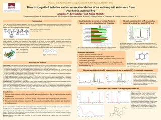
Spectral data for P. remota Fr. 4 suggest procyanidin A2
Conclusions:
• Various plant extracts exhibit non-specific anti-amyloid activity due to high-molecular-weight
components
• Extracts of P. stenostachya, P. remota, and P. alba contain anti-amyloid small molecules
• The anti-amyloid substance present in P. stenostachya extract has been isolated and identified
as hydroquinone
References: Azakami, H., et al. (2005) J. Agric. Food Chem. 53, 1254-7; Le Vine, H., III (1993) Prot. Sci. 2, 404-10; Pryor, N. E., et al. (2012) Int. J.
Mol. Sci. 13, 3038-72.
To whom correspondence should be addressed: Arcadius V. Krivoshein, PhD, Albany College of Pharmacy & Health Sciences, Bioscience Research
Building 104A, 106 New Scotland Avenue, Albany, NY 12208, arcadius.krivoshein@acphs.edu
Fig. 2. Three-dimensional
structure of ovalbumin (from
RCSB Protein Data Bank)
Plant extracts: Plants collected in Peruvian Amazon were dried and extracted with 70% (v/v) EtOH at room temperature three times. The extracts were
filtered and dried in a rotary evaporator at 55 C. The solid ethanolic extracts were stored at -20 C in the extract bank at the Centro de Investigación de
Productos Naturales de la Amazonía, Universidad Nacional Agraria de la Selva (CIPNA-UNAS, Tingo Maria, Peru).
For screening experiments (Fig. 4), the solid ethanolic extracts were dissolved at 50 mg/ml in MeOH and stored at -20 C. Prior to use in the
aggregation assays, the MeOH extracts were diluted 10-fold with PBS and clarified by centrifugation. In some experiments, the extracts were subjected
to ultrafiltration through a 3-kDa cut-off membrane (Millipore Amicon Ultra devices).
For HPLC separations, the solid ethanolic extracts were dissolved at 50 mg/ml in PBS, clarified by centrifugation, and subjected to ultrafiltration
through a 3-kDa cut-off membrane (Millipore Amicon Ultra devices).
Ovalbumin aggregation assay: Ovalbumin (Grade III, Sigma A-5378) at 2 mg/ml concentration in PBS - alone or in the presence of a plant extract
(2.5 mg/ml final concentration) - was heated for ten minutes at 85 C. ThT assay (Le Vine, 1993) was performed on Perkin Elmer 650-15 fluorescence
spectrophotometer using the excitation wavelength of 440 nm and the emission wavelength of 485 nm. Final concentration of ThT in the assay was 20
M, and final concentration of ovalbumin – 0.04 mg/ml.
Preparative HPLC was conducted on a Waters Breeze 2 HPLC system consisting of binary pump, temperature-controlled autosampler, and PDA
detector. Chromatographic fractions were collected using Gilson 201 fraction collector and lyophilized.
Single-crystal X-ray diffraction analysis was performed for us in the laboratory of Dr. Tatiana V. Timofeeva (New Mexico Highlands University, Las
Vegas, NM). The measurements were conducted at 100 K on a Bruker-AXS SMART APEX II CCD diffractometer using graphite-monochromatized
MoKα radiation (λ = 0.7107 Å). The structure was solved by direct methods and refined by means of full–matrix least–squares with anisotropic
approximation for all non-hydrogen atoms using SHELXL–97 program.
Materials and methods
Fig. 3. Thioflavin T (ThT) dye
Acknowledgements: We thank Dr. Manuel Sandoval (CIPNA-UNAS, Tingo Maria, Peru) for the gift of plant extracts, Mr. Carlos Ordonez and Dr.
Tatiana V. Timofeeva (New Mexico Highlands University, Las Vegas, NM) for the X-ray diffraction analysis, and Dr. Alexander Shekhtman (University
at Albany, SUNY, Albany, NY) for NMR analysis.
Fig. 6. Single-crystal X-ray diffraction analysis of the active fraction
from Fig. 5 crystallized from MeOH. An ORTEP diagram is shown.
(Data by C. Ordonez and T. V. Timofeeva)
Structure elucidation:
• UV-Vis spectrum: maximum at 288 nm
• ESI MS: poor ionization in either positive or negative mode
• 1H NMR at 600 MHz (Dr. A. Shekhtman, University at Albany, SUNY): very
weak signals, inconclusive
• Single-crystal X-ray diffraction crystallography (Dr. T. V. Timofeeva with
collaborators, New Mexico Highlands University) identified the compound as
hydroquinone (Fig. 6)
The anti-amyloid activity of P. remota extract is due to multiple HPLC-resolvable components
Fig. 7. Sample equivalent to 12.5 mg of P. remota
extract was loaded on a Agilent Taxsil column (4.6
250 mm, 5 m) eluted with a gradient of AcCN in
aqueous 0.05% TFA at flow rate of 0.8 ml/min. 0.8-
ml fractions were collected, dried in vacuo,
redissolved in PBS, and assayed for their ability to
prevent ovalbumin aggregation. Fractions 1 through 4
(within the green rectangle) are active; fraction 4
seems to be the most active one.
Fig. 8. LC-MS/MS of Fraction 4 isolated from P. remota
extract by HPLC (Fig. 7). An ACE Excel 2 C18-PFP
UHPLC column (2.1 150 mm, 2 m) eluted with a
gradient of AcCN in aqueous 0.015% formic acid at flow
rate of 0.3 ml/min was used. UV-Vis (210-500 nm) spectra
were acquired using ACQUITY PDA detector. Negative
ion ESI mass spectra were acquired on TQD mass
spectrometer in the following conditions: capillary voltage
4 kV, cone voltage 25 V, extractor voltage 3 V, RF lens
voltage 0.1 V, source temperature 100 C, desolvation
temperature 400 C, desolvation gas flow 800 L/h, cone
gas flow 100 L/h, scan speed 2000 Th/s. MS/MS of m/z
575.1 ion was carried out at collision energy of 25 V.