1. r XXXX American Chemical Society A dx.doi.org/10.1021/jm200737s |J. Med. Chem. XXXX, XXX, 000–000
ARTICLE
pubs.acs.org/jmc
Polyalkoxybenzenes from Plants. 5. Parsley Seed Extract in Synthesis
of Azapodophyllotoxins Featuring Strong Tubulin Destabilizing
Activity in the Sea Urchin Embryo and Cell Culture Assays
Marina N. Semenova,†,‡
Alex S. Kiselyov,§
Dmitry V. Tsyganov,||
Leonid D. Konyushkin,||
Sergei I. Firgang,||
Roman V. Semenov,||
Oleg R. Malyshev,||
Mikhail M. Raihstat,||
Fabian Fuchs,^
Anne Stielow,^
Margareta Lantow,^,#
Alex A. Philchenkov,3
Michael P. Zavelevich,3
Nikolay S. Zefirov,O
Sergei A. Kuznetsov,^
and Victor V. Semenov*,‡,||
†
Institute of Developmental Biology, RAS, 26 Vavilov Street, 119334 Moscow, Russian Federation
‡
Chemical Block Ltd., 3 Kyriacou Matsi, 3723 Limassol, Cyprus
§
CHDI Foundation, 6080 Center Drive, Suite 100, Los Angeles California 90045, United States
)
N. D. Zelinsky Institute of Organic Chemistry, RAS, 47 Leninsky Prospect, 119991 Moscow, Russian Federation
^
Institute of Biological Sciences, University of Rostock, 3 Albert-Einstein-Strasse, D-18059 Rostock, Germany
#
Department of Pathology, Immunology and Laboratory Medicine, University of Florida, 1600 Archer Road, Gainesville Florida 32610,
United States
3
R. E. Kavetsky Institute of Experimental Oncology, Pathology, and Radiobiology, National Academy of Sciences of Ukraine,
45 Vasyl0
kivska Street, 03022 Kyiv, Ukraine
O
Lomonosov Moscow State University, GSP-1, Leninskie Gory, 119991 Moscow, Russian Federation
bS Supporting Information
’ INTRODUCTION
Podophyllotoxin (PT) (Figure 1) is a lignan of aryltetralin
family found in plants of Podophyllum genus. It is a strong
microtubule destabilizing agent that binds to the colchicine site
of tubulin.1,2
PT and its derivatives exhibit anticancer, antiviral,
and insecticide activities based on their ability to dissociate
microtubules.3À9
However, clinical trials of PT as an anticancer
agent were unsuccessful due to its considerable toxicity.3
Nu-
merous attempts have been made to improve PT safety profile
while maintaining its potency. For example, semisynthetic PT
derivatives, including etoposide (Figure 1), teniposide, and
etopophos, are currently in clinical use for the treatment of a
variety of malignancies. A reported mechanism of action for these
compounds is distinct from that of the parent PT and involves
strong DNA-topoisomerase II inhibition leading to late S-G2 cell
cycle arrest and subsequent apopotosis.10À13
An alternative
nontubulin and nonantitopoisomerase mode of action for some
PT derivatives has been proposed recently, associated with cell
death and sub-G1 apoptotic cell accumulation without any
significant cell cycle arrest.11,14
PT structure (Figure 1) features four chiral centers, making it a
challenge for the structureÀactivity relationship studies. Abso-
lute stereochemistry of substituents at the C1 atom (the ring C)
was reported to have major influence on compound’s antitumor
activity.3
Several synthetically feasible structural analogues of PT
Received: June 8, 2011
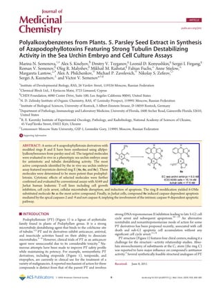
ABSTRACT: A series of 4-azapodophyllotoxin derivatives with
modified rings B and E have been synthesized using allylpo-
lyalkoxybenzenes from parsley seed oil. The targeted molecules
were evaluated in vivo in a phenotypic sea urchin embryo assay
for antimitotic and tubulin destabilizing activity. The most
active compounds identified by the in vivo sea urchin embryo
assay featured myristicin-derived ring E (4e, 6e, and 8e). These
molecules were determined to be more potent than podophyl-
lotoxin. Cytotoxic effects of selected molecules were further
confirmed and evaluated by conventional assays with A549 and
Jurkat human leukemic T-cell lines including cell growth
inhibition, cell cycle arrest, cellular microtubule disruption, and induction of apoptosis. The ring B modification yielded 6-OMe
substituted molecule 8e as the most active compound. Finally, in Jurkat cells, compound 8e induced caspase-dependent apoptosis
mediated by the apical caspases-2 and -9 and not caspase-8, implying the involvement of the intrinsic caspase-9-dependent apoptotic
pathway.
2. B dx.doi.org/10.1021/jm200737s |J. Med. Chem. XXXX, XXX, 000–000
Journal of Medicinal Chemistry ARTICLE
were designed to address epimerization at positions 1À4.3
For
example, 4-aza-PT derivatives with a chiral center C1 (Figure 1)
showed antiproliferative and pro-apoptotic activity in HeLa and
Jurkat cells (4a and 18a)15,16
and inhibited growth of human liver
cancer cells HepG2 (4a and 4i).17
Two racemic analogues of
4-aza-PTs (4a and 6a) were more cytotoxic against P388 murine
leukemia cells than the parent PT.18,19
Several 4-aza-PT deriva-
tives were reported to be potent tumor inhibitors (ex., 4a)20
and
vascular-disrupting agents.21
In addition, insecticidal (larvicidal)
activity possibly mediated by microtubule alterations was re-
ported for this class of molecules.6
However, experimental
evidence for the mechanism of action and cellular targets of
4-aza-PTs has been obtained only recently. Namely, it was shown
that some of 4-aza-PTs with hetero aryl, benzo hetero aryl, or
tetrahydronaphthalene rings A and B exhibited cytotoxicity
involving microtubule depolymerization, G2/M cell cycle arrest,
and apoptotic cell death.22,23
Considering synthetic feasibility, antimitotic, antiproliferative,
and tubulin destabilizing activity of 4-aza-PT derivatives, these
compounds continue to attract a considerable interest. In the
present study, we prepared a series of 4-aza-PT derivatives using
plant polyalkoxybenzenes easily available from parsley and dill
seed extracts.24,25
It has been suggested that rotation of the axial
E-ring in the parent PT molecule is restricted as it is almost
orthogonal to the other rings.26
Therefore a modification of the
rings B and E with additional methoxy functionality could affect
the rotation of the ring E yielding PT derivatives with promising
antimitotic potencies. Moreover, tetraalkoxybenzene pharmaco-
phore is featured in several natural products with reported
antiproliferative activity including Cactaceae tetrahydroiso-
quinolines27
and flavonoids and isoflavonoids.28À30
In designing the evaluation sequence for test molecules, we
aimed at identifying potent novel aza-analogues of PT and
confirming their specific antitubulin activity via a variety of
cell-based assays. The initial in vivo sea urchin embryo assay
allowed for the identification of hits featuring desirable antimi-
totic potency and phenotypic effect (vide infra) in comparison
with the parent molecule (PT). This primary screen based on the
simple organism model has been carried out to yield active
molecules that were likely to affect tubulin dynamics as shown by us
earlier.31À34
Tubulin targeting activity of these compounds would
be further confirmed in A549 human lung epithelial carcinoma cell
line. A detailed insight into the effects of selected active compounds
on cellular proliferation, cell cycle progression, and microtubule
distribution in cultured A549 cells was intended to confirm their
specific tubulin destabilizing mode of action. Moreover, the link
between compound-induced microtubule disruption and respective
downstream intracellular mechanisms responsible for initiation of
caspase-mediated apoptotic events was studied.
All molecules were evaluated in a phenotypic sea urchin
embryo assay in order to assess their antiproliferative and
tubulin-destabilizing activities.31
The assay includes (i) fertilized
egg test for antimitotic activity displayed by cleavage alteration/
arrest and (ii) behavioral monitoring of a free-swimming blas-
tulae treated immediately after hatching. In our earlier validation
efforts, we concluded that lack of forward movement, settlement
to the bottom of the culture vessel and rapid spinning of an
embryo around the animalÀvegetal axis suggests a tubulin-
destabilizing activity caused by a molecule (video illustrations
are available at http://www.chemblock.com). An assay setup
provides the possibility to identify a molecule with antimitotic or
nonspecific cytotoxic activity and a novel mode of action.32
Data
generated by the sea urchin embryo assay have been further
confirmed using conventional cell-based and in vitro tubulin
polymerization assays.33,34
As a result, we identified several
promising candidates for the advanced in vivo evaluation.
’ CHEMISTRY
4-Aza-PTs 4À13 and intermediate quinolines 40
À150
were
synthesized as described earlier6,15À20,35À37
via the cyclization of
4-chloro-acetoacetic acid with the corresponding amines, fol-
lowed by the addition of aldehydes 2aÀk in the presence of an
Figure 1. Structures of podophyllotoxin, etoposide, and general struc-
ture of synthesized 4-aza-analogues.
Scheme 1a
a
Reagents and conditions: (a) (i) powdered KOH, (n-Bu)4N+
BrÀ
, heat,
100 °C, 40 min; (ii) O3, CHCl3ÀMeOHÀpyridine (80:20:3 v/v), À15
°C, 1À2 h;25
(b) C6H6ÀAcOH, reflux, 8 h; (c) CF3COOH, rt, 24 h;
(d) AcOHÀNaBH3CN, rt, 15 h; (e) ref 43; (f) EtOHÀEt3N, reflux, 3 h.
Compounds 4À13 (fÀk), 17i, 18e, 19e were synthesized from the
corresponding commercial aldehydes.
3. C dx.doi.org/10.1021/jm200737s |J. Med. Chem. XXXX, XXX, 000–000
Journal of Medicinal Chemistry ARTICLE
oxidizing reagent p-chloranil (Scheme 1). Aldehydes 2aÀe were
synthesized using natural polyalkoxybenzenes extracted from
parsley seed oil.25
For the SAR study, a panel of 4-aza-PTs with
the ring E derived from commercially available aldehydes 2fÀk
has been obtained. Quinolines 40
À130
were reduced with
NaBH3CN in a glacial acetic acid to yield compounds 4À13 in
12À86% yields. 4-Aza-PTs 4À13 were stable as solids, however,
they were readily oxidized to respective quinolines 40
À130
when
dissolved in DMSO or ethanol. Dihydropyridopyrazole deriva-
tives 17À19 were obtained by a three-component condensation
of respective aminopyrazole, aldehyde 2, and tetronic acid 16 as
reported in the literature.16
Notably, compounds 4g, 40
g, 4f, and
40
f are aza-analogues of the natural lignans taiwanin C and
chinensin.36,38À42
’ RESULTS AND DISCUSSION
All synthesized aza-PTs 4À19 and 40
À150
were evaluated for
their antiproliferative, antimitotic, and microtubule destabilizing
activities using in vivo sea urchin embryo assay. Cytotoxicity,
cellular microtubule depolymerization, and apoptosis-inducing
effects of selected compounds have been further assessed using
A549 human lung epithelial carcinoma and Jurkat human
leukemic T-cell lines.
Evaluation of 4-aza-PTs in the Phenotypic Sea Urchin
Embryo Assay. Effects of 4-aza-PTs on the sea urchin embryo
are summarized in Table 1 and Table S1 of the Supporting
Information. PT and etoposide were used as references.
As evidenced from Table 1, a number of 4-aza-PTs (4a,c,eÀg,
iÀk, 5e, 6a,e, 7e, 8e,h, 9e, 11e, 110
e, 12a, and 13e) displayed
significant cleavage alteration, cleavage arrest, and embryo spinning,
suggesting their antimitotic microtubule destabilizing activity. Fig-
ure 2 illustrates typical developmental changes of an embryo when
treated with a representative compound 4e. The observed pheno-
typic changes suggest direct tubulin dynamic effects of 4-aza-PT
derivatives. Interestingly, molecules 4a, 4e, and 8h were reported as
larvicidal agents against Phaedon cochleariae beetle, whereas 4j, 4k,
Table 1. Effects of 4-Aza-podophyllotoxins on Sea Urchin Embryo and Cancer Cells
sea urchin embryo effects EC (nM)a
cytotoxicity IC50 (nM)
compd cleavage alteration cleavage arrest embryo spinning A549b
P388c
A549 cell total microtubule disruption (μM)d
PT 20 50 500 7.47 ( 0.41 10.4 0.1
etoposide 2000 >68000 >68000 7140e
1180e
NDf
4a 50 500 2000 2.71 ( 0.92 4.5 0.1
40
a >4000 >4000 >4000 11928.33 ( 5368.30 >252900 >5
4b 1000 2000 >5000 NDf
NDf
NDf
4c 100 500 4000 NDf
NDf
NDf
4d 1000 4000 >10000 299.32 ( 58.35 NDf
1
4e 5 50 50 55.18 ( 7.54 NDf
1
4f 10 50 200 NDf
85.4 NDf
4g 5 100 2000 595.58 ( 46.18 130.7 1
4i 50 200 2000 >3200 385.4 NDf
4j 5 20 100 NDf
15.7 NDf
4k 5 20 100 63.00 ( 5.21 17.2 NDf
5a 2000 >2000 >2000 291.43 ( 143.53 NDf
5
5e 50 500 2000 297.63 ( 164.54 NDf
>5
5i 2000 >2000 >2000 NDf
NDf
NDf
6a 200 1000 1000 9.20 ( 1.22 4.1 1
6e 5 50 100 64.67 ( 11.34 NDf
0.5
7e 10 50 500 282.27 ( 96.99 NDf
5
8e 0.5 5 50 15.14 ( 2.98 NDf
1
8h 2 10 200 NDf
NDf
NDf
9e 20 200 2000 533.83 ( 111.47 NDf
>5
10f >4000 >4000 >4000 NDf
NDf
NDf
11e 5 50 100 159.05 ( 8.24 NDf
NDf
110
e 200 2000 5000 NDf
NDf
NDf
12a 50 200 100 56.54 ( 6.83 NDf
0.5
13e 2 20 50 264.09 ( 52.32 NDf
5
130
e 100 1000 >5000 NDf
NDf
NDf
140
e 100 >4000 >5000 NDf
NDf
NDf
a
The sea urchin embryo assay was conducted as described previously;31
fertilized eggs and hatched blastulae were exposed to 2-fold decreasing
concentrations of compounds; duplicate measurements showed no differences in effective threshold concentration (EC) values. b
A549: human lung
epithelial carcinoma cells; data are expressed as the mean ( SD from the doseÀresponse curves of three independent experiments. c
P388: murine
leukemia cells; data from ref 19. d
A549 cells were incubated with compounds at concentrations of 0.1, 0.5, 1, and 5 μM during 8 h. Data are expressed as
compound concentration that caused total microtubule disassembly from three independent experiments (see Figure 4B,DÀF as an example). e
Data
from ref 44. f
ND: not determined.
4. D dx.doi.org/10.1021/jm200737s |J. Med. Chem. XXXX, XXX, 000–000
Journal of Medicinal Chemistry ARTICLE
and 8h were toxic against Spodoptera frugiperda larvae.6
We believe
that less potent compounds 4b,d, 18e, 60
b, and 130
e were tubulin
destabilizers as well, although they did not cause embryo spinning.
As evidenced from the detailed microscopic analysis, these com-
pounds induced formation of tuberculate eggs typical of tubulin
destabilizers (Figure 2C).31
In our hands, topoisomerase inhibitor
etoposide featured cleavage alteration but failed to induce cleavage
arrest and embryo spinning. When applied to hatched blastulae,
etoposide at 4À5 μM caused larval (pluteus) malformations. At
higher concentrations (10À20 μM), marked abnormalities of
gastrulation and morphogenesis were detected, whereas at 40À68
μM, developmental abnormalities and eventual embryo death were
observed after 2.5 h of treatment.
Because of their marginal solubility in DMSO, ethanol, and
seawater, we were unable to test 80
e, 110
a, and 120
a or reach
higher concentrations for 4b,d, 18e, 60
b, and 130
e. 140
e showed
nontubulin antiproliferative activity, as evidenced by the lack of
embryo spinning and arrested tuberculate eggs.
StructureÀActivity Studies in the Sea Urchin Embryo
Assay. 1. The Ring C: Saturated vs Unsaturated Compounds.
A number of 1,4-aza-PT analogues (4aÀg,iÀk, 5e, 6a,e, 7e,
9e, and 13e) featuring 1,4-dihydropyridine system for the ring
C exhibited potent antimitotic activity with EC50 values of
0.5 nMÀ2 μM in the sea urchin assay. These results are in
agreement with the published data.19
Conversely, the respective
aromatic pyridine derivatives 40
aÀg,iÀk, 50
e, 60
a,e, 70
e, 90
e, and
130
e) were inactive up to 4 μM concentration or exhibited
moderate cleavage alteration effect (Table 1; Table S1, Support-
ing Information), with a notable exception of 110
e, featuring
myristicin substituent for the ring E. This molecule did exhibit a
tubulin destabilizing activity although it was significantly lower
than that for the corresponding unsaturated analogue 11e. In
addition, aromatization of the ring C yielded decrease in cyto-
toxicity and microtubule effects, presumably due to the inactive
conformation of the ring E.3
Several saturated compounds were found to lose their activity
in DMSO solution at room temperature. For example, the
antimitotic effect of 4k on sea urchin embryos decreased
significantly (by ca. 5-fold) within 2 days after storage in DMSO.
According to the literature data, aromatization of 4a to yield 40
a
required harsh experimental conditions such as refluxing acetic
acid.37
We observed this conversion to take place in DMSO at
room temperature, as confirmed by the time course NMR studies
of 4k. Specifically, in 2 and 6 days, the rate of its conversion to the
respective aromatic analogue 40
k were 25% and 45%, respec-
tively. A similar activity reduction was observed for 4i and 6a,
whereas DMSO stocks of compounds 4f, 8e, 9e, and 11e
retained their activities for 2À4 days.
2. Other Substitutions: Rings A, B, and E. Introduction of a
methoxy-group to C8 position of the ring B dramatically de-
creased antiproliferative activity in sea urchin embryo assay
(Table 1, compounds 4a . 5a, 4e . 5e, and 4i . 5i).
Replacement of methylenedioxy group (the ring A) with ethy-
lenedioxy substituent did not result in a definitive outcome. For
example, the potency of respective ethylenedioxy derivative
(Table 1, 4a vs 6a) in the sea urchin embryo cleavage stages
was reduced significantly, similar to the reported insecticidal
activity.6
However, for the pair 4e vs 6e, the activity was not
affected.19
Replacement of the ring A with two methoxy substit-
uents was reported to either result in a marked reduction of
cytotoxicity19,45,46
or was negligible.16
Similar derivatives featur-
ing a single methoxy group at C7 displayed reduced potency
compared to the parent 4-aza-PT molecule (Table 1, 4e vs 7e).
On the contrary, 6-methoxy derivative 8e exhibited the strongest
antiproliferative effect of all tested 4-aza-PTs being ca. 40 times
more potent than the parent PT. Another 6-methoxy analogue
8h displayed high activity in the sea urchin assay as well as
pronounced larvicidal effect.6
Compound 9e lacking the ring A
was less active, however it did retain the antimitotic activity.
Substitution of the ring A with cyclopentane moiety was reported
to result in a moderate decrease in cytotoxicity.19
In our assay
system, this modification slightly enhanced the in vivo effect
(Table 1, 4e and 13e), however a nontubulin antiproliferative
activity of 5,6-cyclopentyl derivative 140
e was also detected. In
contrast, replacement of the ring A with a cyclohexyl group did
not influence the antimitotic potency at the sea urchin embryo
cleavage stages (Table 1, 4e and 11e; 4a and 12a). At later
developmental stages of the embryo (ex., after hatching), 12a
displayed formidable toxicity. Similarly, cyclohexyl derivative 12a
exhibited cytotoxicity against human cervical (HeLa) and breast
(MCF-7) carcinomas cells at nanomolar concentration range.23
A replacement of the AB ring system of 4-aza-PTs with
pyrazole was reported to furnish derivatives that retained both
cytotoxic and proapoptotic activities at 5 μM concentration.15,16
However, cellular target(s) or mode of action for these molecules
have not been determined. In our hands, all tested pyrazole-
containing compounds 17a,i, 18aÀc, and 19aÀc,e failed to affect
sea urchin embryo development at any stage up to 4 μM
concentration, the only exception being myristicin-derived 18e
that exhibited weak tubulin-destabilizing properties (Table S1,
Supporting Information).
As described above, 1,4-dihydropyridine derivatives (the ring C)
showed the best activities in the sea urchin embryo assay. We further
evaluated the effect of C1 substituent (the ring E) on compounds
activity. Notably, molecules featuring m-methoxybenzene- or myr-
isticin-derived substituent (Table 1, 4e,j, 5e, 6e, 7e, 8e, 9e, 11e, and
13e) consistently exhibited the highest potency. 2,3-Dimethoxy and
Figure 2. Effect of 4e on sea urchin embryo development. Time after
fertilization (21 °C): 6 h. (A) Intact embryo at early blastula stage.
Fertilized eggs were exposed continuously to 4e at 10 nM (B) and 100
nM (C). Note abnormal cleavage of cells with different size and shape
that are irregularly positioned and formation of large multinucleated
blastomeres (B). Tuberculate shape of an arrested egg seen on (C) is
typical for tubulin destabilizing agents.
Figure 3. Structures of 4e enantiomers.
5. E dx.doi.org/10.1021/jm200737s |J. Med. Chem. XXXX, XXX, 000–000
Journal of Medicinal Chemistry ARTICLE
ethylenedioxy derivatives (ex., 4f,g) were less potent, displaying
activities slightly higher than the parent PT. (see Table 1). Respec-
tive p-methoxybenzene and 3,4,5-trimethoxybenzene analogues
showed further reduced antitubulin effects in the assay (Table 1,
4a,i, 5a,i, and 6a), whereas 4-aza-PT molecules substituted with
dillapiol-, apiol-derived or tetramethoxybenzene moieties 4bÀd
were the least active. Compound 4k with unsubstituted benzene
ring E exhibited strong antimitotic tubulin destabilizing activity,
suggesting that methoxy groups in the ring E may not directly affect
antitubulin activity. Similarly, 30
,40
,50
-demethoxy PT retained sig-
nificant cytotoxicity.47
For the molecules featuring 8-methoxy
moiety, the myristicin-derived analogue 5e displayed the highest
activity(5evs5a,i).Asimilar patternwasobservedfortherespective
ethylenedioxy derivatives 6a and 6e.
In the next round of structureÀactivity studies, the effect of
stereochemistry on compound’s activity has been studied. It was
reported previously that (+)-configuration of antitubulin isoqui-
noline derivatives was essential for their cytotoxicity and inhibi-
tion of tubulin polymerization.48
For 4-aza-PTs, only R-enantiomer
of 8h was found to be cytotoxic against cultured insect cells and
larvae, showing cellular behavior consistent with its microtubule
effect.6
Figure 4. Effects of selected compounds on the interphase microtubule networks in A549 lung carcinoma cells as analyzed by indirect
immunofluorescence microscopy. Cells were treated with 0.1% DMSO (A) and with test compounds (1 μM) for 8 h: 4a (B), 40
a (C), 4e (D), 6a
(E), and 8e (F). Bar: 25 μm. Note microtubule disappearance in B, DÀF.
6. F dx.doi.org/10.1021/jm200737s |J. Med. Chem. XXXX, XXX, 000–000
Journal of Medicinal Chemistry ARTICLE
Similarly, R-enantiomer of 4-aza-PT mimetic with naphtha-
lene A,B-ring system was about 1000 times more potent in HeLa
and MCF-7 cells than S-enantiomer.23
In the present study, a
racemic 4e was separated by chiral HPLC to yield pure enantio-
mers (Figure 3 and Figure S1, Supporting Information). Both
R-isomer and a respective racemate of 4e were found to cause
cleavage alteration and arrest of the sea urchin embryo at 5 and 50
nM, respectively. The respective S-isomer was less active, ex-
hibiting cleavage abnormalities at 50 nM and cleavage arrest at
200 nM. Both enantiomers triggered embryo spinning, suggest-
ing their tubulin destabilizing properties.
In summary, the sea urchin embryo profiling of the new aza-
PT derivatives suggested that the most potent representative in
each structural group featured myristicin-derived ring E (ex., 4e,
5e, 6e, 8e, 11e, and 13e, Table 1). Notably, methylenedioxy ring
A was not required for the activity. For example, introduction of
6-methoxy or 7-methoxy groups into the ring B yielded potent
molecules (ex., 7e, 8e, and 8h).
Cell Growth Inhibitory Activity. Following outcome of the
sea urchin embryo assay, we evaluated the inhibitory effect of
selected active compounds on proliferation of A549 human lung
carcinoma cells and Jurkat leukemia T-cells using MTT49
and
trypan blue assays, respectively. Relevant A549 cellular IC50 data
for 4a,d,e,g,i,k, 5a,e, 6a,e, 7e, 8e, 9e, 11e, 12a, 13e, 40
a, and
reference compounds (PT, etoposide) are summarized in
Table 1. Reported cytotoxicity data for selected molecules
against P388 murine leukemia cells are included as well (Table 1).19
Notably, in Jurkat cells, 8e and PT exhibited IC50 values of 17.5
and 25 nM, respectively.
In general, sensitivity of sea urchin embryos to 4-aza-PTs at the
cleavage stage, when cells divide synchronously every 35À40
min, was markedly higher than that of cancer cells, probably due
to their higher frequency of mitosis. Key features of the most
active molecules identified in both assays included: (i) 1,4-
dihydropyridine template for the ring C (4a vs 40
a), (ii) a
myristicin-derived substitution in the ring E (8e), (iii) 6-methoxy
substituent replacing the ring A (4e vs 8e), and (iv) lack of
8-methoxy substitution in the ring B (4a vs 5a, 4e vs 5e). Results
of structureÀactivity relationship studies for the ring E correlated
well between both cellular (A549 and P388) and sea urchin
embryo assays. Compounds 4f and 4j exhibited high cytotoxicity
against P388 cells (Table 1).19
Molecules featuring 3,4,5-tri-
methoxybenzene substituent (4a and 6a) as well as the parent PT
were the most cytotoxic agents against both human A549 and
murine P388 cancer cell lines, 4a being more potent than PT.
Notably, derivatives endowed with the myristicin moiety showed
the greatest antimitotic activities in sea urchin embryo assay.
Similarly, in Jurkat cells a myristicin derivative 8e was more
potent than PT. Replacement of the ring A with cyclopentyl or
cyclohexyl moieties markedly decreased cytotoxicity against
A549 cells (4e vs 13e; 4a vs 12a), but this effect was almost
negligible in the sea urchin embryo test. The observed discre-
pancy could be attributed to the differences between mitotic
spindle microtubules in frequently dividing sea urchin blasto-
meres vs predominantly interphase microtubules in cultured
cancer cells targeted by tubulin destabilizing agents.
The Effect on Microtubule Distribution in Cells. Next, we
evaluated the effect of selected active compounds on microtubule
stability and distribution in cultured A549 cells. Specifically, cells
were incubated with test articles (0.1À5 μM) or PT (0.1 and
5 μM) as a positive control and 0.5% DMSO as a negative control.
Microtubules were subsequently labeled by the indirect immu-
nofluorescence method and analyzed by fluorescence micro-
scopy. Figure 4 exemplifies the microtubule destabilization effect
of selected 4-aza-PTs in A549 cells.
As shown in Table 1, compounds 4a,d,e,g, 6a, 6e, 7e, 8e, 12a,
and 13e identified as tubulin destabilizers in the sea urchin
embryo assay also affected cellular microtubule structure
(Figure 4B,DÀF). It should be noted that 5a and 5e precipitated
from 10 mM stock solutions in DMSO, therefore the data
pertinent to these molecules could not be accurately interpreted.
Compound 40
a (an aromatic analogue of 4a) featuring low cyto-
toxicity failed to induce sea urchin embryo development alterations
and to depolymerize cellular microtubules (Figure 4C). Surprisingly,
compound 9e that exhibited modest cytotoxicity without the
disruption of interphase microtubules in A549 cells did show a
considerable antimitotic tubulin destabilizing activity in the sea
urchin assay. The effect could be attributed to a somewhat selective
interaction of this molecule with spindle microtubules in the sea
urchin blastomeres vs interphase microtubules in cultured cancer
cells. Treatment with PT at 0.1 and 5 μM resulted in complete
disassembly of cellular microtubules (data not shown).
Cell Cycle Analysis. The ability of selected 4-aza-PTs to
interfere with A549 and Jurkat cell cycle progression was
estimated by propidium iodide staining followed by flow cyto-
metry analysis. In A549 cells, PT as well as its aza-analogues 4a,g
and 6e caused G2/M cell cycle arrest, exhibiting EC50 values of
16.5 ( 0.35, 16.2 ( 0.71, 230 ( 7.07, and 119 ( 1.41 nM,
respectively. Compound 40
a failed to affect the cell cycle
progression even at 50 μM concentration. Notably, all three
cell-based assays ranked the tested molecules similarly. Cyto-
toxicity and microtubule dynamics effects of 4-aza-PTs decreased
in the following order: 4a > 6e > 4g . 40
a. In Jurkat cells, both 8e
and PT were potent G2/M blockers as well. In contrast, etopo-
side caused a pronounced cell cycle arrest in G0/G1 phase
(Figure 5).
Apoptosis-Inducing Activity. It has been well established that
tubulinbindingcompounds interferewiththe dynamics andstructure
of both mitotic spindle and interphase microtubules, eventually
leading to cell death by apoptosis. However the link between
microtubule alteration and respective intracellular mechanisms
responsible for initiation of apoptotic events remains unclear.50À52
Caspases (intracellular proteases) play a key role in the apoptotic
response. Their activation by specific signals triggers proteolysis of
cellularsubstratestherebyexecutingapoptoticevents.53
Therearetwo
Figure 5. Cell cycle distribution of Jurkat cells treated with 8e (20 nM),
PT (25 nM), and etoposide (2 μM) for 24 h. The cells were stained with
propidium iodide to analyze DNA content by flow cytometry.
7. G dx.doi.org/10.1021/jm200737s |J. Med. Chem. XXXX, XXX, 000–000
Journal of Medicinal Chemistry ARTICLE
main apoptotic pathways, namely the extrinsic pathway that
involves membrane-bound death receptors and leads to activa-
tion of caspase-8 and the mitochondria-related caspase-9-depen-
dent intrinsic pathway. Both pathways converge onto the effector
caspase-3 activated by the apical (initiator) caspases-8 and -9.53,54
Caspase-2 combines both initiator and effector features.53
Its
processing requires caspase-9 and caspase-3.55
Generally, microtubule interfering agents induce caspase-
dependent apoptotic cell death through intrinsic caspase-9-
dependent pathway.50À52,56
For instance, tubulin destabilizers
combretastatin A-4 and its analogues, as well as nocodazole,
trigger apoptosis through the intrinsic pathway accompanied by
an activation of caspase-9 and caspase-3.57À59
However, other
tubulin-targeting compounds were found to induce apoptosis in
human cancer cells via the activation of caspase-8 and -2 rather
than caspase-9, suggesting the extrinsic pathway as a primary
apoptotic mechanism.60À63
Whether this difference is cell-spe-
cific or dependent on the compound remains unclear. Because
numerous malignancies are characterized by molecular defects in
the apoptotic pathways,54
the investigation of apoptotic mechan-
isms is of particular importance.
There are a few data concerning the participation of effector
caspases-3 and -7 in apoptosis caused by PT derivatives.64À66
For
4-aza-PTs, caspase-3 activation in Jurkat and A549 cells has been
described.16,22,23
Up to now there is no evidence on the involve-
ment of apical caspases-9 and -2 in cell death caused by PT
derivatives. In the present study, the details of apoptosis induc-
tion by compound 4a found to be the most cytotoxic against
Figure 6. Jurkat cells treated with PT or 8e: flow cytometry assessment of hypodyploid cells (A), cells with active form of caspase-3 (B), and with
cleaved PARP (C). (a) Control (untreated cells); (b) PT, 50 nM, 48 h; (c) 8e, 50 nM, 48 d. In (d), cells were pretreated with caspase inhibitor z-VAD-
FMK (50 μM) for 2 h, followed by the incubation with 8e as in (c). M1 comprises the hypodiploid cells (A), cells with active form of caspase-3 (B), and
cleaved PARP-positive cells (C) with corresponding percentages given in each plot.
8. H dx.doi.org/10.1021/jm200737s |J. Med. Chem. XXXX, XXX, 000–000
Journal of Medicinal Chemistry ARTICLE
A549 cells, and 8e, the most potent in the sea urchin embryo
assay, were studied in A549 and Jurkat cells, respectively.
The proapoptotic activity of compound 8e was examined in
Jurkat cells. Apoptosis induction, cell cycle distribution, expres-
sion of active effector caspase-3 in cells, and cleavage of PARP
were assessed quantitatively by flow cytometry. Procaspase-8 and
the active form of apical caspase-2 and -9 were analyzed by
Western blot. As evidenced by the flow cytometric assay, after 48
h, both 8e and PT increased the hypodiploid subpopulation peak
(sub-G1) indicative of apoptosis from 12% (control) to 55% of
the total cell population (Figure 6A). To confirm the involve-
ment of caspases in the 8e-induced Jurkat cell death, an effect of
pan-caspase inhibitor Z-VAD-fmk67
has been examined. Jurkat
cells were preincubated with Z-VAD-fmk at 50 μM for 2 h,
followed by incubation with 50 nM of 8e for additional 48 h.
Pretreatment with Z-VAD-fmk decreased the content of apop-
totic cells from 55% to 28% (Figure 6A), suggesting that cell
death induced by 8e was related to caspase activation. Approxi-
mately 30% of cells treated with 8e or PT possessed active form
of caspase-3, as compared to 7% in untreated cells (Figure 6B).
Cleaved PARP, the product resulting from caspase-3 activation,
has been identified in ca. 40% of the cells treated by 8e or PT
(Figure 6C).
Activation of caspases-9, -2, and -8 in cells treated with 8e was
further analyzed by Western blot. The exposure of Jurkat cells to
8e resulted in the processing of procaspase-9 and -2, as evidenced
by the appearance of their active forms (Figure 7A,B). The
intensities of bands corresponding to active forms of caspase-9
and -2 upon 24 h of treatment with 8e or PT were higher than
those at 48 h, while the intensity of procaspase-8 55/57 kDa
traces was unaltered (Figure 7C), suggesting lack of active
caspase-8. As expected, etoposide caused the reduction of
procaspase-8 level in Jurkat cells (Figure 7D).68
Finally, the ability of compound 4a to trigger apoptotic and
necrotic events in A549 cells was analyzed using double staining with
Annexin VÀFITC and 7-AAD (7-amino actinomycin D) dyes. The
procedure allows for the differentiation between living (intact)
(Annexin VÀFITCÀ
/7-ADDÀ
), early apoptotic (Annexin VÀ
FITC+
/7-ADDÀ
), late apoptotic (Annexin VÀFITC+
/7-ADD+
),
and necrotic cells (Annexin VÀFITCÀ
/7-ADD+
).69,70
Compound
4a as well as nocodazole (positive control) induced a significant
increase of early apoptosis and to a lesser degree necrosis without
affecting late apoptosis (Figure S2, Supporting Information). The
effects of 4a were concentration-independent, suggesting that this
compound might trigger apoptosis and necrosis even at lower
concentrations. 4a was more potent apoptotis- and necrosis-indu-
cing agent than nocodazole.
On the basis of these data, we concluded that compounds 4a and
8e caused G2/M cell cycle arrest and apoptotic response in A549
and Jurkat human cancer cells, notably, that in Jurkat cells 8e as well
as PT induced caspase-dependent apoptosis mediated by the apical
caspases-2 and -9 and not caspase-8, implying the involvement of
the intrinsic caspase-9-dependent apoptotic pathway.
PT-Related Compounds: Mode of Action. In the sea urchin
embryoassay, deathofarrested eggs exposedtotubulindestabilizing
agents was observed after 6À8 h of treatment with a test article. It
has been suggested that apoptosis never occurs during sea urchin
embryo cleavage, first appearing only at blastula stage just before
hatching.71
This explains the absence ofquick cell divisionarrest and
death at cleavage after treatment by various DNA-targeting agents
known to trigger apoptosis such as etoposide, aloe-emodin, apigen-
in, 5-fluorouracil, and 4-phenylbutyric acid.31
Correspondingly, the
formation of multinucleated cells (Figure 1B) typical of tubulin
destabilizers is an inherent morphological characteristic of mitotic
catastrophe, a cell death different from apoptosis.72
Tubulin destabilizers including colchicine, nocodazole, combre-
tastatins, phenstatin, indibulin, dolastatin 15, vinblastin, and others
interacting with different binding sites on tubulin caused embryo
spinning when applied at the swimming blastula stage. Spinning
continued for several hours, followed by slow movements near the
bottom of the vessel suggesting embryo viability.31,33,34,73
In con-
trast, PT andsomeof4-aza-PTs (4a,e,g,j, 6e, 7e, 8e,h, 12a, and13e)
induced sea urchin embryo disintegration and death after spinning
independently of compound concentration, probably owing to yet
another microtubule-unrelated effect. In addition, compound 140
e
caused cleavage alteration at 100 nM, whereas it failed to produce
cleavage arrest and embryo spinning, suggesting nontubulin antu-
proliferative activity. Similarly, 9e exerted cytotoxic effect against
A549 cells without microtubule disruption (Table 1). These results
are correlated well with the reported ability of PT derivatives to
induce cell death and sub-G1 apoptotic cell accumulation without
cell cycle arrest and interphase microtubule damage.10,14
Moreover,
in a series of 4-aza-PTs with hetero aryl or benzo hetero aryl system
instead of the A and B rings, chemical modifications of the ring E
resulted in a significant decrease of antitubulin activity and retention
of cytotoxicity.22
A combination of our results and literature data
suggests an alternative tubulin-independent mechanism of 4-aza-
PTs embryotoxicity that could be evident at postcleavage stages of
the sea urchin embryo development.
’ CONCLUSIONS
In conclusion, we synthesized a series of 4-aza-PT analogues
from the easily accessible plant-derived polyalkoxybenzenes.
A developed reaction sequence allows for an expedited synthesis
Figure 7. Western blot analysis of procaspase-8, cleaved caspase-2, and
caspase-9 in total lysates of Jurkat cells. The lysates treated with PT or 8e
were prepared in SDSÀLaemmli sample buffer. Immunoblotting was
performed using monoclonal anticaspase-9 (A), anticaspase-2 (B), and
antiprocaspase-8 (C, D) antibodies. β-Actin served as the loading
control. Lanes: (1, 6) control (untreated cells); (2) PT, 50 nM, 24 h;
(3) PT, 50 nM, 48 h; (4) 8e, 50 nM, 24 h; (5) 8e, 50 nM, 48 h; (7)
etoposide, 2 μM, 24 h.
9. I dx.doi.org/10.1021/jm200737s |J. Med. Chem. XXXX, XXX, 000–000
Journal of Medicinal Chemistry ARTICLE
of the targeted molecules in 12À86% overall yields from naturally
occurring building blocks. The resulting compounds were ex-
tensively evaluated in both phenotypic sea urchin embryo and
cellular assays linking their observed cytotoxicity to a potent
antimitotic tubulin destabilizing effect. Notably, the phenotypic
sea urchin embryo assay allowed for rapid and reproducible
identification of antitubulin molecules. These compounds were
further reconfirmed as potent antimitotic agents by a panel of
conventional cell-based assays. The most potent representative
in each structural group featured myristicin-derived ring E (4e,
6e, 8e, 11e, and 13e), as evidenced by the in vivo sea urchin
embryo assay. The activity of these new compounds compares
favorably to the reported antimitotic agents containing myristi-
cin-derived pharmacophore including corresponding phenstatin
analogue and combretastatin A-2.25,73
The ring B modification
yielded 6-methoxy substituted molecule 8e as the most active
compound. Compound 4a featuring 3,4,5- trimethoxybenzene
substituent was the most cytotoxic against A549 human lung
epithelial carcinoma cells, being more potent than the parent PT.
The activity of 4-aza-PT derivatives correlated well between the
sea urchin phenotypic and conventional cell-based assays. In
Jurkat cells, both 8e and PT induced caspase-dependent apop-
tosis mediated by the apical caspases-2 and -9 and not caspase-8,
implying the involvement of the intrinsic caspase-9-dependent
apoptotic pathway. The structureÀactivity data reported in this
paper warrant further synthetic studies and biological evaluation
of novel heterocyclic myristicine derivatives. Four specific mol-
ecules (4e, 7e, 8e, and 8h) have been selected for advanced
NCI60 human tumor cell line anticancer drug screen. Their
antiproliferative activity will be reported separately.
’ EXPERIMENTAL SECTION
Chemistry. Materials and Methods. NMR spectra were col-
lected on a Bruker DR-500 instrument [working frequencies of 500.13
MHz (1
H) and 75.47 (13
C)]. Mass spectra were obtained on a Finnigan
MAT/INCOS 50 instrument (70 eV) using direct probe injection.
Elemental analysis was accomplished with the automated Perkin-Elmer
2400 CHN microanalyzer. HPLC was performed using a Laboratorni
Pristroje (Praha, Czech Republic) chromatograph. Ozonolysis was
conducted using a custom-designed apparatus (Science and Technology
Park, St. Petersburg State Polytechnic University, Russia) equipped with
an IR detector of O3 concentration (Japan) and an automated shut-
down circuit. The device allowed for the controlled generation of ozone,
with a maximal capacity of 10 g of O3 per h from O2. The compound
purity has been determined by NMR, HPLC, and elemental analyses.
Purity of compounds 40
À150
, 4À13, and 17À19 was determined to be
g95%.
Isolation of Plant Allylpolyalkoxybenzenes24
. Liquid CO2
extraction of parsley and dill seeds was carried out earlier by Company
Karavan Ltd. (Krasnodar, Russia). Allylpolymethoxybenzenes 1bÀe
with 98À99% purity were obtained by high-efficiency distillation using
a pilot plant device at N. D. Zelinsky Institute of Organic Chemistry,
RAS (Moscow, Russia). The seed essential oils of parsley varieties
cultivated in Russia contained 70À75% of 1b (var. Sakharnaya), 21% of
1d (var. Slavyanovskaya), and 40À46% of 1e (var. Astra). Indian dill
seeds were purchased from Vremya & Co. (St. Petersburg, Russia). The
dill seed essential oil contained 30À33% of 1c.
General Procedure A for the Synthesis of Quinolines
40
À15036
. A solution of aromatic amine (50 mmol), ethyl 4-chloroa-
cetoacetate (50 mmol) and acetic acid (3 mL) in 100 mL of benzene was
refluxed for 8 h. The reaction mixture was cooled to room temperature,
and the precipitated product was collected by vacuum filtration and
washed with EtOH (30 mL). In most cases, the resulting products 3
were obtained with >85% purity as judged by NMR analysis. A solution
of corresponding anilinolactone 3 (2 mmol), appropriate aromatic
aldehyde (2aÀk) (2.2 mmol), and p-chloranil (2 mmol) in TFA
(3 mL) was stirred at room temperature for 24 h. The reaction mixture
was quenched with water (50 mL) and extracted with CH2Cl2 (3 Â
20 mL). The organic layers were combined, washed with 5% NaHCO3
(2 Â 20 mL), water (2 Â 20 mL), dried over anhydrous sodium sulfate,
and concentrated in vacuum to obtain the crude product. Flash
chromatography purification (petroleum ether/EtOAc, 4:1À1:1) af-
forded targeted quinolines 40
À150
as a white solid.
General Procedure B for the Synthesis of 4-Aza-podo-
phyllotoxin Derivatives 4À13. A suspension of quinoline 40
À130
(0.26 mmol) in a glacial AcOH (3.96 mmol, 3 mL) was treated with
NaBH3CN (0.78 mmol), and the resulting mixture was stirred for 3 h at
room temperature and treated with the ice water (15 mL). The
precipitate was filtered, washed with ethanol (2 Â 1 mL), and the
product was purified by column chromatography (petroleum ether/
EtOAc, 2:1) to afford the targeted 4-aza-podophyllotoxins 4À13 as a
white solid. NMR analysis confirmed the absence of intermediates
40
À130
.
General Procedure C for the Synthesis of Dihydropyrido-
pyrazole Derivatives 17À19. A mixture of of corresponding
pyrazole (1 mmol), tetronic acid 16 (1 mmol), triethylamine
(0.05 mL), and appropriate aldehyde (2aÀk) (1 mmol) in EtOH
(5 mL) was refluxed for 3 h. The reaction mixture was cooled to
+4 °C, and the precipitated product was collected by vacuum filtration
and washed with EtOH (2 mL) at 0 °C. Purification by flash chroma-
tography (petroleum ether/EtOAc, 4:1À2:1) afforded the targeted
4-aza-dihydropodophylltoxins 17À19 as a white solid.
Synthetic and Analytical Data for Selected 4-Aza-podo-
phyllotoxins. Data for the other compounds are presented in
Supporting Information.
9-(6,7-Dimethoxy-1,3-benzodioxol-5-yl)[1,3]dioxolo[4,5-
g]furo[3,4-b]quinolin-8(6H)-one (40
c). The title compound was
obtained according to the general procedure A. Yield 27%, mp
241À244 °C. 1
H NMR (DMSO-d6): δ 3.35 (s, 3H, OCH3-60
), 4.02
(s, 3H, OCH3-70
), 5.42 (d, J = 15.4 Hz, 1H, OCH2-6), 5.49 (d, J = 15.4
Hz, 1H, OCH2-6), 6.11 and 6.13 (s, 2H, OCH2O-20
), 6.28 (s, 2H,
OCH2O-2), 6.51 (s, 1H, H-40
), 6.89 (s, 1H, H-10), 7.52 (s, 1H, H-4).
EIMS m/z 409 [M]+
(25), 394 (1), 351 (15), 350 (68), 224 (23), 167
(100). Anal. Calcd for C21H15NO8: C, 61.62; H, 3.69; N, 3.42. Found:
C, 61.69; H, 3.72; N, 3.36.
9-(6,7-Dimethoxy-1,3-benzodioxol-5-yl)-6,9-dihydro-
[1,3]dioxolo[4,5-g]furo[3,4-b]quinolin-8(5H)-one (4c). The ti-
tle compound was obtained according to the general procedure B. Yield
26%, mp 295À298 °C. 1
H NMR (DMSO-d6): δ 3.73 (s, 3H, OCH3-60
),
3.94 (s, 3H, OCH3-70
), 4.83 (d, J = 15.6 Hz, 1H, OCH2-6), 4.95 (d, J =
15.7 Hz, 1H, OCH2-6), 5.13 (s, 1H, H-9), 5.88 and 5.94 (s, 2H,
OCH2O-2), 5.89 and 5.91 (s, 2H, OCH2O-20
), 6.24 (s, 2H, H-40
),
6.42 (s, 1H, H-10), 6.49 (s, 1H, H-4), 9.81 (s, 1H, NH-5). EIMS m/z
411 [M]+
(20), 380 (100), 366 (17), 230 (39), 172 (7). Anal. Calcd for
C21H17NO8: C, 61.32; H, 4.17; N, 3.40. Found: C, 61.29; H, 4.14;
N, 3.44.
9-(7-Methoxy-1,3-benzodioxol-5-yl)-6,9-dihydro[1,3]
dioxolo[4,5-g]furo[3,4-b]quinolin-8(5H)-one (4e). The title
compound was obtained according to the general procedure B. Yield
55%, mp 308À310 °C. 1
H NMR (DMSO-d6): δ 3.80 (s, 3H, OCH3-70
),
4.83 (s, 1H, H-9), 4.84 (d, J = 15.7 Hz, 1H, OCH2-6), 4.97 (d, J = 15.7
Hz, 1H, OCH2-6), 5.90 and 5.91 (s, 2H, OCH2O-20
), 5.90 and 5.96 (s,
2H, OCH2O-2), 6.33 (d, J = 1.4 Hz, 1H, H-40
), 6.52 (s, 1H, H-10), 6.56
(d, J = 1.4 Hz, 1H, H-60
), 6.64 (s, 1H, H-4), 9.87 (s, 1H, NH-5). EIMS
m/z 381 [M]+
(5), 231 (14), 230 (100), 152 (24), 151 (7). Anal. Calcd
10. J dx.doi.org/10.1021/jm200737s |J. Med. Chem. XXXX, XXX, 000–000
Journal of Medicinal Chemistry ARTICLE
for C20H15NO7: C, 62.99; H, 3.96; N, 3.67. Found: C, 63.06; H, 4.03;
N, 3.60.
9-Phenyl-6,9-dihydro[1,3]dioxolo[4,5-g]furo[3,4-b]quin-
olin-8(5H)-one (4k). The title compound was obtained according to
the general procedure B. Yield 73%, mp 290À293 °C (lit.6
mp
295À298 °C). 1
H NMR (DMSO-d6): δ 4.85 (d, J = 15.7 Hz, 1H,
OCH2-6), 4.91 (s, 1H, H-9), 4.94 (d, J = 15.7 Hz, 1H, OCH2-6), 5.89
and 5.95 (s, 2H, OCH2O-2), 6.53 (s, 1H, H-10), 6.56 (s, 1H, H-4), 7.15
(t, J = 7.2 Hz, 1H, H-40
), 7.19 (d, J = 7.2 Hz, 2H, H-20
,60
), 7.26 (d, J = 7.2
Hz, 2H, H-30
,50
), 9.87 (s, 1H, NH-5). EIMS m/z 307 [M]+
(20), 230
(21), 78 (12), 77 (100). Anal. Calcd for C18H13NO4: C, 70.35; H, 4.26;
N, 4.56. Found: C, 70.29; H, 4.22; N, 4.63.
10-Methoxy-9-(7-methoxy-1,3-benzodioxol-5-yl)-6,9-
dihydro[1,3]dioxolo[4,5-g]furo[3,4-b]quinolin-8(5H)-one
(5e). The title compound was obtained according to the general
procedure B. Yield 26%, mp 321À326 °C. 1
H NMR (DMSO-d6): δ
3.66 (s, 3H, OCH3-10), 3.77 (s, 3H, OCH3-70
), 4.78 (d, J = 15.7 Hz, 1H,
OCH2-6), 4.89 (d, J = 15.7 Hz, 1H, OCH2-6), 4.89 (s, 1H, H-9), 5.89
and 5.96 (s, 2H, OCH2O-2), 5.89 and 5.90 (s, 2H, OCH2O-20
), 6.22 (d,
J = 1.4 Hz, 1H, H-40
), 6.33 (s, 1H, H-4), 6.42 (d, J = 1.4 Hz, 1H, H-60
),
9.93 (s, 1H, NH-5). EIMS m/z 411 [M]+
(3), 384 (0.5), 260 (100), 245
(25), 152 (31), 151 (9). Anal. Calcd for C21H17NO8: C, 61.32; H, 4.17;
N, 3.40. Found: C, 61.36; H, 4.18; N, 3.38.
6-Methoxy-9-(7-methoxy-1,3-benzodioxol-5-yl)-4,9-dih-
ydrofuro[3,4-b]quinolin-1(3H)-one (8e). The title compound
was obtained according to the general procedure B. Yield 60%, mp
278À280 °C. 1
H NMR (DMSO-d6): δ 3.70 (s, 3H, OCH3-6), 3.80
(s, 3H, OCH3-70
), 4.84 (d, J = 15.5 Hz, 1H, OCH2-3), 4.86 (s, 1H, H-9),
4.97 (d, J = 15.5 Hz, 1H, OCH2-3), 5.89 and 5.90 (s, 2H, OCH2O-20
),
6.31 (d, J = 1.4 Hz, 1H, H-40
), 6.44 (d, J = 2.5 Hz, H-5), 6.52 (dd, J = 8.6,
2.5 Hz, 1H, H-7), 6.53 (d, J = 1.4 Hz, 1H, H-60
), 6.99 (d, J = 8.6 Hz, H-8),
9.94 (s, 1H, NH-4). EIMS m/z 367 [M]+
(14), 217 (15), 216 (100), 152
(16), 151 (7). Anal. Calcd for C20H17NO6: C, 65.39; H, 4.66; N, 3.81.
Found: C, 65.42; H, 4.69; N, 3.74.
9-(3,5-Dimethoxyphenyl)-6-methoxy-4,9-dihydrofuro-
[3,4-b]quinolin-1(3H)-one (8h). The title compound was obtained
according to the general procedure B. Yield 43%, mp 229À233 °C (lit.6
mp 254 °C). 1
H NMR (DMSO-d6): δ 3.68 (s, 6H, 2 Â OCH3-30
, 50
),
3.70 (s, 3H, OCH3-6), 4.85 (d, J = 15.8 Hz, 1H, OCH2-3), 4.85 (s, 1H,
H-9), 4.98 (d, J = 15.8 Hz, 1H, OCH2-3), 6.30 (t, J = 2.2 Hz, 1H, H-40
),
6.32 (d, J = 2.2 Hz, 2H, H-20
,60
), 6.45 (d, J = 2.6 Hz, H-5), 6.52 (dd, J =
8.5, 2.6 Hz, 1H, H-7), 6.99 (d, J = 8.5 Hz, H-8), 9.94 (s, 1H, NH-4).
EIMS m/z 353 [M]+
(12), 217 (15), 216 (100), 138 (16). Anal. Calcd
for C20H19NO5: C, 67.98; H, 5.42; N, 3.96. Found: C, 67.91; H, 5.38;
N, 4.04.
4-(4,7-Dimethoxy-1,3-benzodioxol-5-yl)-3-methyl-1,4,7,
8-tetrahydro-5H-furo[3,4-b]pyrazolo[4,3-e]pyridin-5-one
(18b). The title compound was obtained according to the general
procedure C. Yield 47%, mp 295À297 °C. 1
H NMR (DMSO-d6): δ 1.83
(s, 3H, CH3-3), 3.69 (s, 3H, OCH3-40
), 3.71 (s, 3H, OCH3-70
), 4.78
(d, J = 15.7 Hz, 1H, OCH2-7), 4.85 (d, J = 15.7 Hz, 1H, OCH2-7), 4.99
(s, 1H, H-4), 5.98 and 5.99 (s, 2H, OCH2O-20
), 6.23 (s, 1H, H-60
), 10.03
(s, 1H, NH-8), 11.79 (s, 1H, NH-1). EIMS m/z 371 [M]+
(31), 341
(14), 340 (74), 327 (5), 326 (21), 312 (16), 296 (14), 191 (11), 190
(100), 182 (10). Anal. Calcd for C18H17N3O6: C, 58.22; H, 4.61; N,
11.32. Found: C, 58.30; H, 4.68; N, 11.23.
Biology. Materials and Methods. Sea Urchin Embryo
Assay. Adult sea urchins Paracentrotus lividus were collected from the
Mediterranean Sea at the Cyprus coast and kept in an aerated seawater
tank. Gametes were obtained by intracoelomic injection of 0.5 M KCl.
Eggs were washed with filtered seawater and fertilized by adding drops of
a diluted sperm. Embryos were cultured at room temperature under
gentle agitation with a motor-driven plastic paddle (60 rpm) in filtered
seawater. The embryos were observed with a light microscope Biolam
(LOMO, S.-Petersburg, Russia). For treatment with the test com-
pounds, 5 mL aliquots of embryo suspension were transferred to 6-well
plates and incubated as a monolayer at a concentration up to 2000
embryos/mL. Stock solutions of compounds were prepared in DMSO at
5À10 mM concentrations, followed by a 10-fold dilution with 95%
EtOH. This procedure enhanced solubility of the test compounds in the
salt-containing medium (seawater), as evidenced by microscopic exam-
ination of the samples. The maximal tolerated concentrations of DMSO
and EtOH in the in vivo assay were determined to be 0.05% and 1%
respectively. Higher concentrations of either DMSO (g0.1%) or EtOH
(>1%) caused nonspecific alteration and retardation of the sea urchin
embryo development independent of the treatment stage. Podophyllo-
toxin (Aldrich) and topoiomerase II inhibitor etoposide (LANS, Ver-
opharm, Russia, 20 mg/mL in 96% EtOH) served as reference com-
pounds.
The antiproliferative activity was assessed by exposing fertilized eggs
(8À20 min after fertilization, 43À55 min before the first mitotic cycle
completion) to 2-fold decreasing concentrations of the compound.
Cleavage alteration and arrest were clearly detected at 2.5À5.5 h after
fertilization. The effects were quantitatively estimated as a threshold
concentration resulting in cleavage alteration and embryo death before
hatching or full mitotic arrest. At these concentrations, all tested tubulin
destabilizers caused 100% cleavage alteration and embryo death before
hatching, whereas at 2-fold lower concentrations, the compounds failed
to produce any effect. For tubulin destabilizing activity, the compounds
were tested on free-swimming blastulae just after hatching (9À10 h after
fertilization), originated from the same embryo culture. Embryo spin-
ning was observed after 15 min to 20 h of treatment, depending on the
nature and concentration of the compound. Both spinning and lack of
forward movement were interpreted to be the result of the tubulin
destabilizing activity of a molecule according to previous studies.31
Video illustrations are available at http://www.chemblock.com). Com-
pound substructure search in sea urchin embryo screening database is
available free online at http://www.zelinsky.ru.
’ ASSOCIATED CONTENT
bS Supporting Information. Effects of compounds 40
À150
and 17À19 on the sea urchin embryo development, experimen-
tal details regarding syntheses and analytical data, biological assay
methods. This material is available free of charge via the Internet
at http://pubs.acs.org.
’ AUTHOR INFORMATION
Corresponding Author
*Phone: +7 (916) 620-9584. Fax: +7 (499) 137-2966. E-mail:
vs@zelinsky.ru.
’ ACKNOWLEDGMENT
This work was supported by Alexander von Humboldt
Foundation (no. 3.4-Fokoop-Rus/1015567, Germany) and a
grant from Chemical Block Ltd.
’ ABBREVIATIONS USED
PT, podophyllotoxin;SAR, structureÀactivity relationship;7-AAD,
7-amino actinomycin D;PARP, poly ADP-ribose polymerase
’ REFERENCES
(1) Ravelli, R. B. G.; Gigant, B.; Curmi, P. A.; Jourdain, I.; Lachkar, S.;
Sobel, A.; Knossow, M. Insight into tubulin regulation from a complex with
colchicine and a stathmin-like domain. Nature 2004, 428, 198–202.
11. K dx.doi.org/10.1021/jm200737s |J. Med. Chem. XXXX, XXX, 000–000
Journal of Medicinal Chemistry ARTICLE
(2) Gupta, S.; Das, L.; Datta, A. B.; Poddar, A.; Janik, M. E.;
Bhattacharyya, B. Oxalone and lactone moieties of podophyllotoxin
exhibit properties of both the B and C rings of colchicine in its binding
with tubulin. Biochemistry 2006, 45, 6467–6475.
(3) Desbene, S.; Giorgi-Renault, S. Drugs that inhibit tubulin
polymerization: The particular case of podophyllotoxin and analogues.
Curr. Med. Chem.: Anti-Cancer Agents 2002, 2, 71–90.
(4) Chen, S.-W.; Wang, Y.-H.; Jin, Y.; Tian, X.; Zheng, Y.-T.; Luo,
D.-Q.; Tu, Y.-Q. Synthesis and anti-HIV-1 activities of novel podophyl-
lotoxin derivatives. Bioorg. Med. Chem. Lett. 2007, 17, 2091–2095.
(5) Saitoh, T.; Kuramochi, K.; Imai, T.; Takata, K.; Takehara, M.;
Kobayashi, S.; Sakaguchia, K.; Sugawara, F. Podophyllotoxin directly
binds a hinge domain in E2 of HPV and inhibits an E2/E7 interaction in
vitro. Bioorg. Med. Chem. 2008, 16, 5815–5825.
(6) Frackenpohl, J.; Adelt, I.; Antonicek, H.; Arnold, C.; Behrmann,
P.; Blaha, N.; B€ohmer, J.; Gutbrod, O.; Hanke, R.; Hohmann, S.; van
Houtdreve, M.; Losel, P.; Malsam, O.; Melchers, M.; Neufert, V.;
Peschel, E.; Reckmann, U.; Schenke, T.; Thiesen, H.-P.; Velten, R.;
Vogelsang, K.; Weiss, H.-C. Insecticidal heterolignans—tubuline po-
lymerization inhibitors with activity against chewing pests. Bioorg. Med.
Chem. 2009, 17, 4160–4184.
(7) Xu, H.; Lv, M.; Tian, X. A review on hemisynthesis, biosynthesis,
biological activities, mode of action, and structureÀactivity relationship
of podophyllotoxins: 2003À2007. Curr. Med. Chem. 2009, 16, 327–349.
(8) Xu, H.; Xiao, X. Natural products-based insecticidal agents 4.
Semisynthesis and insecticidal activity of novel esters of 2-chloropodo-
phyllotoxin against Mythimna separata Walker in vivo. Bioorg. Med.
Chem. Lett. 2009, 19, 5415–5418.
(9) Xu, H.; He, X.-Q. Natural products-based insecticidal agents 6.
Design, semisynthesis, and insecticidal activity of novel monomethyl
phthalate derivatives of podophyllotoxin against Mythimna separata
Walker in vivo. Bioorg. Med. Chem. Lett. 2010, 20, 4503–4506.
(10) Damayanthi, Y.; Lown, J. W. Podophyllotoxins: current status
and recent developments. Curr. Med. Chem. 1998, 5, 205–252.
(11) Canel, C.; Moraes, R. M.; Dayan, F. E.; Ferreira, D. Podophyl-
lotoxin. Phytochemistry 2000, 54, 115–120.
(12) Meresse,P.;Dechaux,E.;Monneret,C.;Bertounesque,E.Etoposide:
discovery and medicinal chemistry. Curr. Med. Chem. 2004, 11, 2443–2466.
(13) Duca, M.; Guianvarc’h, D.; Meresse, P.; Bertounesque, E.;
Dauzonne, D.; Kraus-Berthier, L.; Thirot, S.; Leonce, S.; Pierre, A.;
Pfeiffer, B.; Renard, P.; Arimondo, P. B.; Monneret, C. Synthesis and
biological study of a new series of 40
-demethylepipodophyllotoxin
derivatives. J. Med. Chem. 2005, 48, 593–603.
(14) Castro, M. A.; del Corral, J. M. M.; Garcia, P. A.; Rojo, M. V.; de la
Iglesia-Vicente, J.; Mollinedo, F.; Cuevas, C.; San Feliciano, A. Synthesis and
biological evaluation of new podophyllic aldehyde derivatives with cytotoxic
and apoptosis-inducing activities. J. Med. Chem. 2010, 53, 983–993.
(15) Magedov, I. V.; Manpadi, M.; Rozhkova, E.; Przheval’skii,
N. M.; Rogelj, S.; Shors, S. T.; Steelant, W. F.; Van Slambrouck, S.;
Kornienko, A. Structural simplification of bioactive natural products
with multicomponent synthesis: dihydropyridopyrazole analogues of
podophyllotoxin. Bioorg. Med. Chem. Lett. 2007, 17, 1381–1385.
(16) Magedov, I. V.; Manpadi, M.; Slambrouck, S. V.; Steelant, W. F.;
Rozhkova, E.; Przheval’skii, N. M.; Rogelj, S.; Kornienko, A. Discovery and
investigation of antiproliferative and apoptosis-inducing properties of new
heterocyclic podophyllotoxin analogues accessible by a one-step multi-
component synthesis. J. Med. Chem. 2007, 50, 5183–5192.
(17) Shi, C.; Wang, J.; Chen, H.; Shi, D. Regioselective synthesis and
in vitro anticancer activity of 4-aza-podophyllotoxin derivatives cata-
lyzed by L-proline. J. Comb. Chem. 2010, 12, 430–434.
(18) Hitotsuyanagi, Y.; Kobayashi, M.; Morita, H.; Itokawa, H.;
Takeya, K. Synthesis of (À)-4-aza-4-deoxypodophyllotoxin from (À)-
podophyllotoxin. Tetrahedron Lett. 1999, 40, 9107–9110.
(19) Hitotsuyanagi, Y.; Fukuyo, M.; Tsuda, K.; Kobayashi, M.; Ozeki,
A.; Itokawa, H.; Takeya, K. 4-Aza-2,3-dehydro-4-deoxypodophyllotoxins:
simple aza-podophyllotoxin analogues possessing potent cytotoxicity.
Bioorg. Med. Chem. Lett. 2000, 10, 315–317.
(20) Husson, H.-P.; Giorgi-Renault, S.; Tratrat, C.; Atassi, G.; Pierre,
A.; Renard, P.; Pfeiffer, B. Dihydrofuro[3,4-b]quinolin-1-one com-
pounds. U.S. Patent 6,548,515, 2003.
(21) Labruere, R.; Gautier, B.; Testud, M.; Seguin, J.; Lenoir, C.;
Desbene-Finck, S.; Helissey, P.; Garbay, C.; Chabot, G. G.; Vidal, M.;
Giorgi-Renault, S. Design, synthesis, and biological evaluation of the first
podophyllotoxin analogues as potential vascular-disrupting agents.
ChemMedChem. 2010, 5, 2016–2025.
(22) Kamal, A.; Suresh, P.; Mallareddy, A.; Kumar, B. A.; Reddy, P. V.;
Raju, P.; Tamboli, J. R.; Shaik, T. B.; Jain, N.; Kalivendi, S. V. Synthesis of a
new 4-aza-2,3-didehydropodophyllotoxin analogues as potent cytotoxicand
antimitotic agents. Bioorg. Med. Chem. 2011, 19, 2349–2358.
(23) Magedov, I. V.; Frolova, L.; Manpadi, M.; Bhoga, U. D.; Tang,
H.; Evdokimov, N. M.; George, O.; Georgiou, K. H.; Renner, S.; Getlik,
M.; Kinnibrugh, T. L.; Fernandes, M. A.; van Slambrouck, S.; Steelant,
W. F. A.; Shuster, C. B.; Rogelj, S.; van Otterlo, W. A. L.; Kornienko, A.
Anticancer properties of an important drug lead podophyllotoxin can be
efficiently mimicked by diverse heterocyclic scaffolds accessible via one-
step synthesis. J. Med. Chem. 2011, 54, 4234–4246.
(24) Semenov, V. V.; Rusak, V. A.; Chartov, E. M.; Zaretsky, M. I.;
Konyushkin, L. D.; Firgang, S. I.; Chizhov, A. O.; Elkin, V. V.; Latin,
N. N.; Bonashek, V. M.; Stas’eva, O. N. Polyalkoxybenzenes from plant
raw materials 1. Isolation of polyalkoxybenzenes from CO2 extracts of
Umbelliferae plant seeds. Russ. Chem. Bull. 2007, 56, 2448–2455.
(25) Semenov, V. V.; Kiselyov, A. S.; Titov, I. Y.; Sagamanova, I. K.;
Ikizalp, N. N.; Chernysheva, N. B.; Tsyganov, D. V.; Konyushkin, L. D.;
Firgang, S. I.; Semenov, R. V.; Karmanova, I. B.; Raihstat, M. M.; Semenova,
M. N. Synthesis of antimitotic polyalkoxyphenyl derivatives of combretas-
tatinusing plant allylpolyalkoxybenzenes.J.Nat.Prod.2010, 73, 1796–1802.
(26) (a) Petcher, T. J.; Weber, H. P.; Kuhn, M.; Von Wartburg, A.
Crystal structure and absolute configuration of 20
-bromopodophyllo-
toxin-0.5 ethyl acetate. J. Chem. Soc., Perkin Trans. 2 1973, 2, 288–292.
(b) Rithner, C. D.; Bushweller, C. H.; Gender, W. J.; Hoogasian, S.
Dynamic nuclear magnetic resonance and empirical force field studies of
podophyllotoxin. J. Org. Chem. 1983, 48, 1491–1495.
(27) Shulgin, A. T. Tinkal: The Continuation; Transform Press:
Berkeley, CA, 1977; p 646.
(28) Lichius, J. J.; Thoison, O.; Montagnac, A.; Pais, M.; Gueritte-
Voegelein, F.; Sevenet, T.; Cosson, J. P.; Hadi, A. H. Antimitotic and
cytotoxic flavonols from Zieridium pseudobtusifolium and Acronychia
porteri. J. Nat. Prod. 1994, 57, 1012–1016.
(29) Walle, T. Methoxylated flavones, a superior cancer chemopre-
ventive flavonoid subclass? Semin Cancer Biol. 2007, 17, 354–362.
(30) Quintin, J.; Buisson, D.; Thoret, S.; Cresteil, T.; Lewin, G.
Semisynthesis and antiproliferative evaluation of a series of 30
-amino-
flavones. Bioorg. Med. Chem. Lett. 2009, 19, 3502–3506.
(31) Semenova, M. N; Kiselyov, A. S.; Semenov, V. V. Sea urchin
embryo as a model organism for the rapid functional screening of tubulin
modulators. BioTechniques 2006, 40, 765–774.
(32) Semenova, M. N.; Tsyganov, D. V.; Yakubov, A. P.; Kiselyov,
A. S.; Semenov, V. V. A synthetic derivative of plant allylpolyalkox-
ybenzenes induces selective loss of motile cilia in sea urchin embryos.
ACS Chem. Biol. 2008, 3, 95À100.
(33) Semenova, M. N.; Kiselyov, A. S.; Titov, I. Y.; Raihstat, M. M.;
Molodtsov, M.; Grishchuk, E.; Spiridonov, I.; Semenov, V. V. In vivo
evaluation of indolyl glyoxamides in the sea urchin embryo model:
correlation with in vitro tubulin dynamics effects. Chem. Biol. Drug Des.
2007, 70, 485–490.
(34) Kiselyov, A. S.; Semenova, M. N.; Chernyshova, N. B.; Leitao,
A.; Samet, A. V.; Kislyi, K. A.; Raihstat, M. M.; Oprea, T.; Lemcke, T.;
Lantowe, M.; Weiss, D. G.; Ikizalp, N. N.; Kuznetsov, S. A.; Semenov, V. V.
Novel derivatives of 1,3,4-oxadiazoles are potent mitostatic agents featuring
strong microtubule depolymerizing activity in the sea urchin embryo and
cell culture assays. Eur. J. Med. Chem. 2010, 45, 1683–1697.
(35) Schmidt, D. G.; Seemuth, P. D.; Zimmer, H. Substituted
.gamma.-butyrolactones.Part31.2,4(3H,5H)-Furandione:heteroannulations
with aromatic o-amino carbonyl compounds and condensations with some
vic-polyones. J. Org. Chem. 1983, 48, 1914–1916.
12. L dx.doi.org/10.1021/jm200737s |J. Med. Chem. XXXX, XXX, 000–000
Journal of Medicinal Chemistry ARTICLE
(36) Hitotsuyanagi, Y.; Kobayashi, M.; Fukuyo, M.; Takeya, K.;
Itokawa, H. A facile synthesis of the 4-aza-analogs of 1-arylnaphthalene
lignans chinensin, justicidin B, and taiwanin C. Tetrahedron Lett. 1997,
38, 8295–8296.
(37) Tratrat, C.; Giorgi-Renault, S.; Husson, H.-P. A multicompo-
nent reaction for the one-pot synthesis of 4-aza-2,3-didehydropodo-
phyllotoxin and derivatives. Org. Lett. 2002, 4, 3187–3189.
(38) Ogiku, T.; Yoshida, S.; Ohmizu, H.; Iwasaki, T. Efficient
syntheses of 1-arylnaphthalene lignan lactones and related compounds
from cyanohydrins. J. Org. Chem. 1995, 60, 4585–4590.
(39) Cow, C.; Leung, C.; Charlton, J. L. Antiviral activity of aryln-
aphthalene and aryldihydronaphthalene lignans. Can. J. Chem. 2000, 78,
553–561.
(40) Ban, H. S.; Lee, S.; Kim, Y. P.; Yamaki, K.; Shin, K. H.; Ohuchi,
K. Inhibition of prostaglandin E(2) production by taiwanin C isolated
from the root of Acanthopanax chiisanensis and the mechanism of action.
Biochem. Pharmacol. 2002, 64, 1345–1354.
(41) Chang, S.-T.; Sheng-Yang Wang, S.-Y.; Yueh-Hsiung Kuo,
Y.-H. Resources and bioactive substances from Taiwania (Taiwania
cryptomerioides). J. Wood Sci. 2003, 49, 1–4.
(42) Foley, P.; Eghbali, N.; Anastas, P. T. Silver-catalyzed one-pot
synthesis of arylnaphthalene lactone natural products. J. Nat. Prod. 2010,
73, 811–813.
(43) Momose, T.; Toyooka, N.; Takeuchi, Y. A laboratory synthesis
of 4-hydroxy-2(5H)-furanone (β-tetronic acid). Heterocycles 1986,
24, 1429–1431.
(44) Jin, Y.; Chen, S. W.; Tian, X. Synthesis and biological evaluation
of new spin-labeled derivatives of podophyllotoxin. Bioorg. Med. Chem.
2006, 14, 3062–3068.
(45) Cisney, M. E.; Shilling, W. L.; Hearon, W. M.; Goheen, D. W.
Conidendrin. II. The stereochemistry and reactions of the lactone ring.
J. Am. Chem. Soc. 1954, 76, 5083–5087.
(46) Dantzig, A.; LaLonde, R. T.; Ramdayal, F.; Shepard, R. L.;
Yanai, K.; Zhang, M. Cytotoxic responses to aromatic ring and config-
urational variations in alpha-conidendrin, podophyllotoxin, and sikki-
motoxin derivatives. J. Med. Chem. 2001, 44, 180–185.
(47) Berkowitz, D. B.; Maeng, J.-H.; Dantzig, A. H.; Shepard, R. L.;
Norman, B. H. Chemoenzymatic and ring E-modular approach to the
(À)-podophyllotoxin skeleton. Synthesis of 30
,40
,50
-tridemethoxy-(À)-
podophyllotoxin. J. Am. Chem. Soc. 1996, 118, 9426–9427.
(48) Goldbrunner, M.; Loidl, G.; Polossek, T.; Mannschreck, A.; von
Angerer, E. Inhibition of tubulin polymerization by 5,6-dihydroindolo-
[2,1-alpha]isoquinoline derivatives. J. Med. Chem. 1997, 40, 3524–3533.
(49) Mosmann, T. Rapid colorimetric assay for cellular growth and
survival: application to proliferation and cytotoxicity assays. J. Immunol.
Methods 1983, 65, 55–63.
(50) Bhalla, K. N. Microtubule-targeted anticancer agents and
apoptosis. Oncogene 2003, 22, 9075–9086.
(51) Mollinedo, F.; Gajate, C. Microtubules, microtubule-interfering
agents and apoptosis. Apoptosis 2003, 8, 413–450.
(52) Gascoigne, K. E.; Taylor, S. S. How do anti-mitotic drugs kill
cancer cells? J Cell Sci. 2009, 122, 2579–2585.
(53) Pop, C.; Salvesen, G. S. Human caspases: activation, specificity,
and regulation. J. Biol. Chem. 2009, 284, 21777–21781.
(54) Ghobrial, I. M.; Witzig, T. E.; Adjei, A. A. Targeting apoptosis
pathways in cancer therapy. Cancer J. Clin. 2005, 55, 178–194.
(55) Inoue, S.; Browne, G.; Melino, G.; Cohen, G. M. Ordering of
caspases in cells undergoing apoptosis by the intrinsic pathway. Cell
Death Differ. 2009, 16, 1053–1061.
(56) Viola, G.; Cecconet, L.; Leszl, A.; Basso, G.; Brun, P.; Salvador,
A.; Dall’Acqua, F.; Diana, P.; Barraja, P.; Cirrincione, G. Pyrrolotetra-
zinones deazaanalogues of Temozolomide induce apoptosis in Jurkat
cell line: involvement of tubulin polymerization inhibition. Cancer
Chemother. Pharm. 2009, 64, 1235–1251.
(57) Beswick, R. W.; Ambrose, H. E.; Wagner, S. D. Nocodazole, a
microtubule depolymerising agent, induces apoptosis of chronic lym-
phocytic leukaemia cells associated with changes in Bcl-2 phosphoryla-
tion and expression. Leukemia Res. 2006, 30, 427–436.
(58) Vitale, I.; Antoccia, A.; Cenciarelli, C.; Crateri, P.; Meschini, S.;
Arancia, G.; Pisano, C.; Tanzarella, C. Combretastatin CA-4 and
combretastatin derivative induce mitotic catastrophe dependent on
spindle checkpoint and caspase-3 activation in non-small cell lung
cancer cells. Apoptosis 2007, 12, 155–166.
(59) Romagnoli, R.; Baraldi, P. G.; Cruz-Lopez, O.; Cara, C. L.;
Carrion, M. D.; Brancale, A.; Hamel, E.; Chen, L.; Bortolozzi, R.; Basso,
G.; Viola, G. Synthesis and antitumor ativity of 1,5-disubstituted 1,2,4-
triazoles as cis-restricted combretastatin analogues. J. Med. Chem. 2010,
53, 4248–4258.
(60) Chen, Y.-C.; Lu, P.-H.; Pan, S. L.; Teng, C.-M.; Kuo, S.-C.; Lin,
T.-P.; Ho, Y.-F.; Huang, Y.-C.; Guh, J.-H. Quinolone analogue inhibits
tubulin polymerization and induces apoptosis via Cdk1-involved signal-
ing pathways. Biochem. Pharmacol. 2007, 74, 10–19.
(61) McElligott, A. M.; Maginn, E. N.; Greene, L. M.; Siobhan
McGuckin, S.; Hayat, A.; Browne, P. V.; Butini, S.; Campiani, G.;
Catherwood, M. A.; Vandenberghe, E.; Williams, D. C.; Zisterer,
D. M.; Mark Lawler, M. The novel tubulin-targeting agent pyrrolo-
1,5-benzoxazepine-15 induces apoptosis in poor prognostic subgroups
of chronic lymphocytic leukemia. Cancer Res. 2009, 69, 8366–8375.
(62) Hsieh, C.-C.; Kuo, Y.-H.; Kuo, C.-C.; Chen, L.-T.; Cheung,
C.-H. A.; Chao, T.-Y.; Lin, C.-H.; Pan, W.-Y.; Chang, C.-Y.; Chien, S.-C.;
Chen, T.-W.; Lung, C.-C.; Chang, J.-Y. Chamaecypanone C, a novel
skeleton microtubule inhibitor, with anticancer activity by trigger
caspase 8-Fas/FasL dependent apoptotic pathway in human cancer
cells. Biochem. Pharmacol. 2010, 79, 1261–1271.
(63) Maginn, E. N.; Browne, P. V.; Hayden, P.; Vandenberghe, E.;
MacDonagh, B.; Evans, P.; Goodyer, M.; Tewari, P.; Campiani, G.;
Butini, S.; Williams, D. C.; Zisterer, D. M.; Lawler, M. P.; McElligott,
A. M. PBOX-15, a novel microtubule targeting agent, induces apoptosis,
upregulates death receptors, and potentiates TRAIL-mediated apoptosis
in multiple myeloma cells. Br. J. Cancer 2011, 104, 281–289.
(64) Qi, Y.; Liao, F.; Zhao, C.; Lin, Y.; Zuo, M. Cytotoxicity,
apoptosis induction, and mitotic arrest by a novel podophyllotoxin
glucoside, 4DPG, in tumor cells. Acta Pharmacol. Sin. 2005, 26,
1000–1008.
(65) Yong, Y.; Shin, S. Y.; Lee, Y. H.; Lim, Y. Antitumor activity of
deoxypodophyllotoxin isolated from Anthriscus sylvestris: Induction of
G2/M cell cycle arrest and caspase-dependent apoptosis. Bioorg. Med.
Chem. Lett. 2009, 19, 4367–4371.
(66) Shin, S. Y.; Yong, Y.; Kim, C. G.; Lee, Y. H.; Lim, Y.
Deoxypodophyllotoxin induces G2/M cell cycle arrest and apoptosis
in HeLa cells. Cancer Lett. 2010, 287, 231–239.
(67) Ko, S. C.; Johnson, V. L.; Chow, S. C. Functional characterization of
Jurkat T cells rescued from CD95/Fas-induced apoptosis through the
inhibitionofcaspases.Biochem.Biophys.Res.Commun.2000,270,1009–1015.
(68) Sohn, D.; Schulze-Setoff, K.; J€anicke, R. U. Caspase-8 can be
activated by interchain proteolysis without receptor-triggered dimerization
during drug-induced apoptosis. J. Biol. Chem. 2005, 280, 5267–5273.
(69) Vermes, I.; Haanen, C.; Steffens-Nakken, H.; Reutelingsperger,
C. A novel assay for apoptosis. Flow cytometric detection of phospha-
tidylserine expression on early apoptotic cells using fluorescein labelled
Annexin V. J. Immunol. Methods 1995, 184, 39–51.
(70) Koopman, G.; Reutelingsperger, C. P.; Kuijten, G. A.; Keehnen,
R. M.; Pals, S. T.; van Oers, M. H. Annexin V for flow cytometric
detection of phosphatidylserine expression on B cells undergoing
apoptosis. Blood 1994, 84, 1415–1420.
(71) Thurber, R. V.; Epel, D. Apoptosis in early development of the
sea urchin, Strongylocentrotus purpuratus. Dev. Biol. 2007, 303, 336–346.
(72) Castedo, M.; Perfettini, J. L.; Roumier, T.; Andreau, K.;
Medema, R.; Kroemer, G. Cell death by mitotic catastrophe: a molecular
definition. Oncogene 2004, 23, 2825–2837.
(73) Titov, I. Y.; Sagamanova, I. K.; Gritsenko, R. T.; Karmanova,
I. B.; Atamanenko, O. P.; Semenova, M. N.; Semenov, V. V. Bioorg. Med.
Chem. Lett. 2011, 21, 1578–1581.